



PKC beta 2 Antibody (YA129)
(Synonyms: PKCA, PRKACA, PRKCA, Protein kinase C alpha type, PKC-A, PKC-alpha)
Based on 1 Customer Validation
PKC beta 2 Antibody (YA129) is a Rabbit-derived and non-conjugated IgG monoclonal antibody, targeting to PKC beta 2.
-
Host:
Rabbit
-
Isotype:
IgG
-
Application:
WB, ICC/IF, IHC-P, IP, FC
-
Reactivity :
Human, Mouse
-
Formulation:
Supplied in 1*TBS (pH7.4), 0.05% BSA and 40% Glycerol. Preservative: 0.05% Sodium Azide.
-
Conjugation:
Non-conjugated


To place orders, for customer services and technical support, please contact: MedChemExpress USA
Tel: 609-228-6898 E-mail: [email protected] [email protected]





Applications
| Application |
WB

WB: Western Blot
|
ICC/IF
ICC/IF: Immunocytochemistry/
Immunofluorescence |
IHC-P
IHC-P: Immunohistochemistry-Paraffin
|
FC
FC: Flow Cytometry
|
IP
IP: Immunoprecipitation
|
|---|---|---|---|---|---|
| Dilution Ratio | 1:1000-1:2000 | 1:50-1:200 | 1:50-1:200 | 1:50-1:100 | Use at an assay dependent concentration. |


Product Details
PKC beta 2 Antibody (YA129) is a Rabbit-derived and non-conjugated IgG monoclonal antibody, targeting to PKC beta 2.
-
Host Rabbit

-
Clonality Monoclonal,Recombinant

-
Species ReactivityHuman, Mouse
-
Observed Molecular WeightObserved band size: 77 kDa
Note: Due to possible protein modifications or aggregation, the molecular weight should be confirmed by actual measurement, and the predicted value is for reference only.

-
Calculated Molecular Weight Predicted band size: 77 kDa

Synthetic peptide corresponding to Human PKC beta 2.AA range:615-660.
Endogenous
Protein A affinity purified.
Non-conjugated
Unmodified
IgG


Product Properties
-
Appearance
Solution
-
Formulation
Supplied in 1*TBS (pH7.4), 0.05% BSA and 40% Glycerol. Preservative: 0.05% Sodium Azide.
-
Concentration
Batch-dependent, Please check the COA for the concentration of each lot. Check Lot Concentration
-
Storage & Stability
Stored at -20°C for 1 year. Avoid repeated freeze / thaw cycles.
-
Shipping
Shipping with blue ice.


Verification Images
-
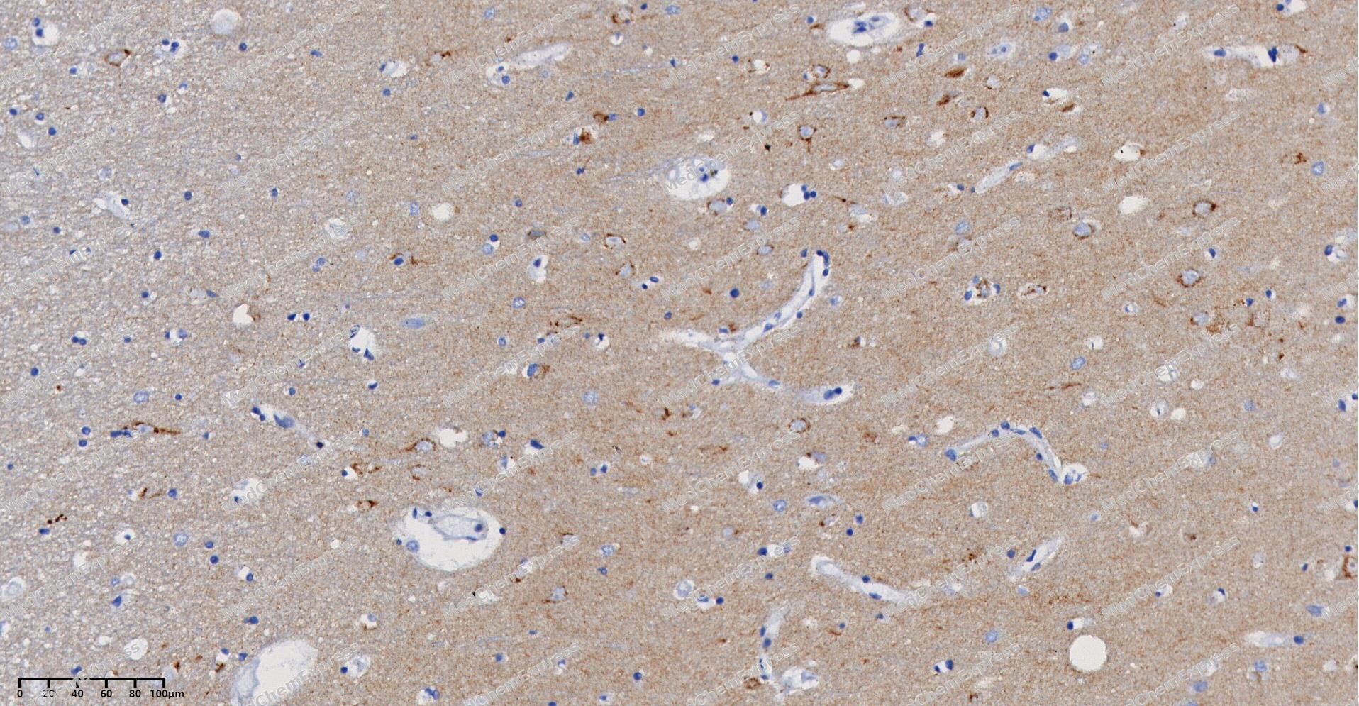 Immunohistochemical analysis of paraffin-embedded human Brain tissue using PKC beta 2 antibody was performed. The section was pretreated using high-temperature and high-pressure mediated EDTA antigen retrieval buffer (pH 9.0), for 5 minutes. The tissues were incubated with primary antibody (HY-P80432, 1:100 dilution) at room temperature for 60 minutes. Detection was performed using an HRP conjugated compact polymer system. DAB was used as the chromogen. The tissues were counterstained with hematoxylin and mounted with neutral balsam mounting medium.
 -
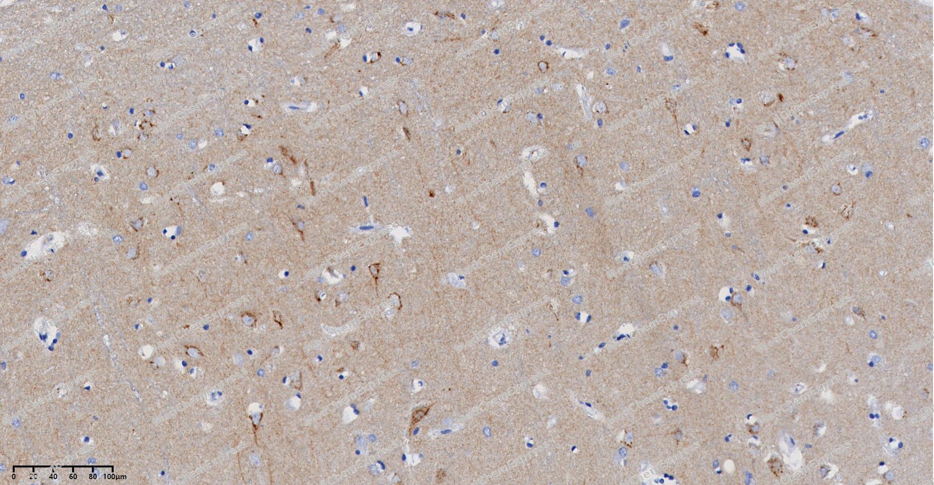 Immunohistochemical analysis of paraffin-embedded human Brain tissue using PKC beta 2 antibody was performed. The section was pretreated using high-temperature and high-pressure mediated EDTA antigen retrieval buffer (pH 9.0), for 5 minutes. The tissues were incubated with primary antibody (HY-P80432, 1:100 dilution) at room temperature for 60 minutes. Detection was performed using an HRP conjugated compact polymer system. DAB was used as the chromogen. The tissues were counterstained with hematoxylin and mounted with neutral balsam mounting medium.
 -
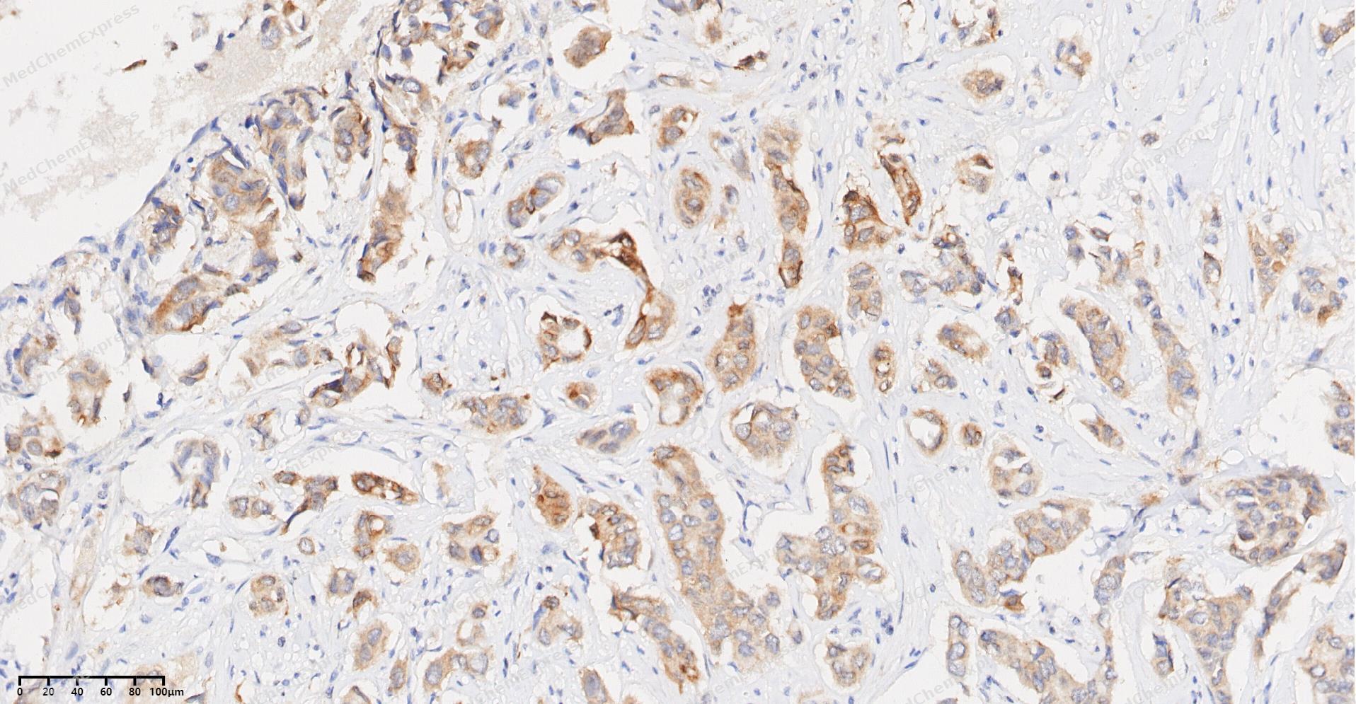 Immunohistochemical analysis of paraffin-embedded human Breast Cancer tissue using PKC beta 2 antibody was performed. The section was pretreated using high-temperature and high-pressure mediated EDTA antigen retrieval buffer (pH 9.0), for 5 minutes. The tissues were incubated with primary antibody (HY-P80432, 1:100 dilution) at room temperature for 60 minutes. Detection was performed using an HRP conjugated compact polymer system. DAB was used as the chromogen. The tissues were counterstained with hematoxylin and mounted with neutral balsam mounting medium.
 -
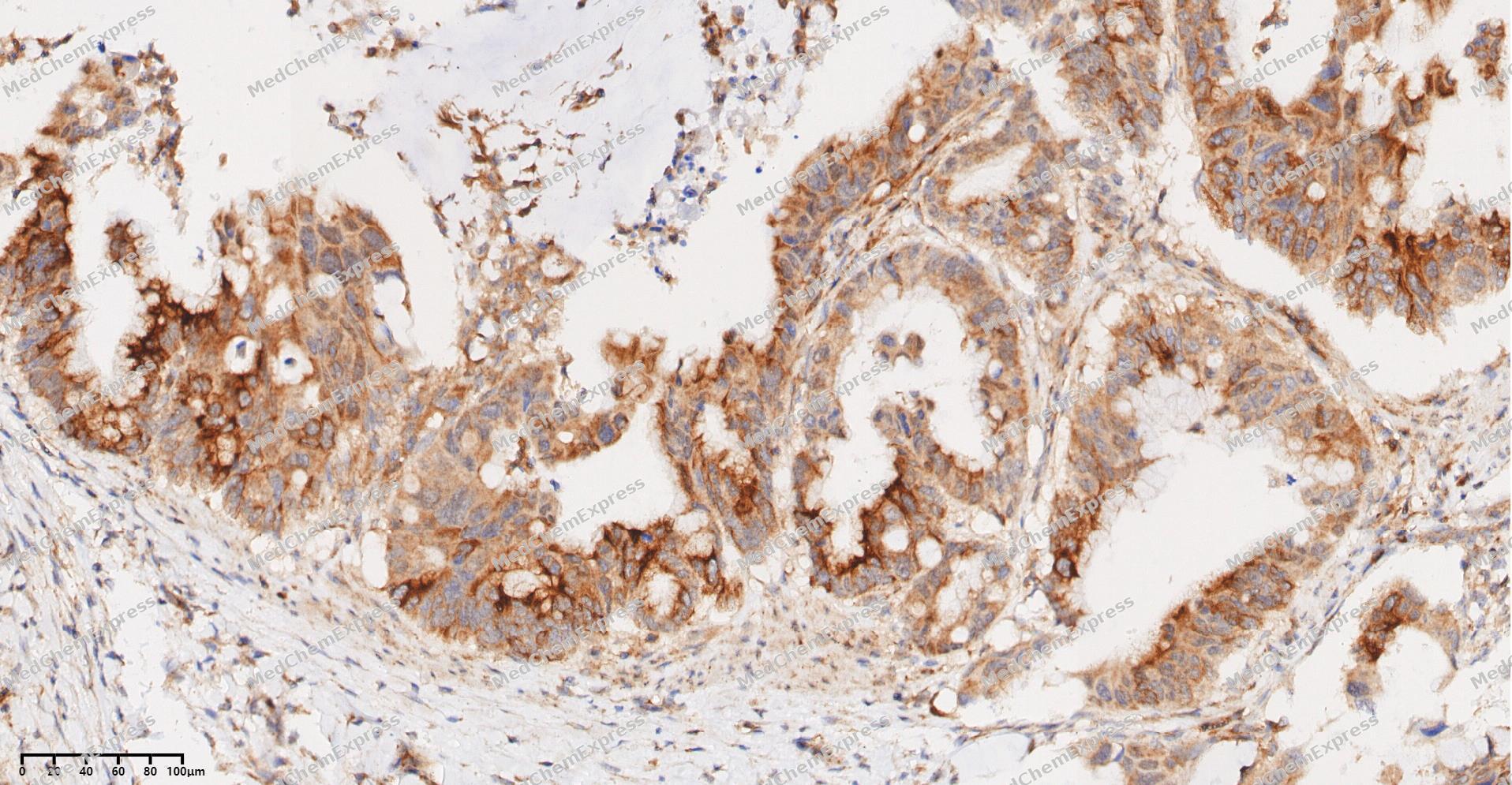 Immunohistochemical analysis of paraffin-embedded human Gastric Cancer tissue using PKC beta 2 antibody was performed. The section was pretreated using high-temperature and high-pressure mediated EDTA antigen retrieval buffer (pH 9.0), for 5 minutes. The tissues were incubated with primary antibody (HY-P80432, 1:100 dilution) at room temperature for 60 minutes. Detection was performed using an HRP conjugated compact polymer system. DAB was used as the chromogen. The tissues were counterstained with hematoxylin and mounted with neutral balsam mounting medium.
 -
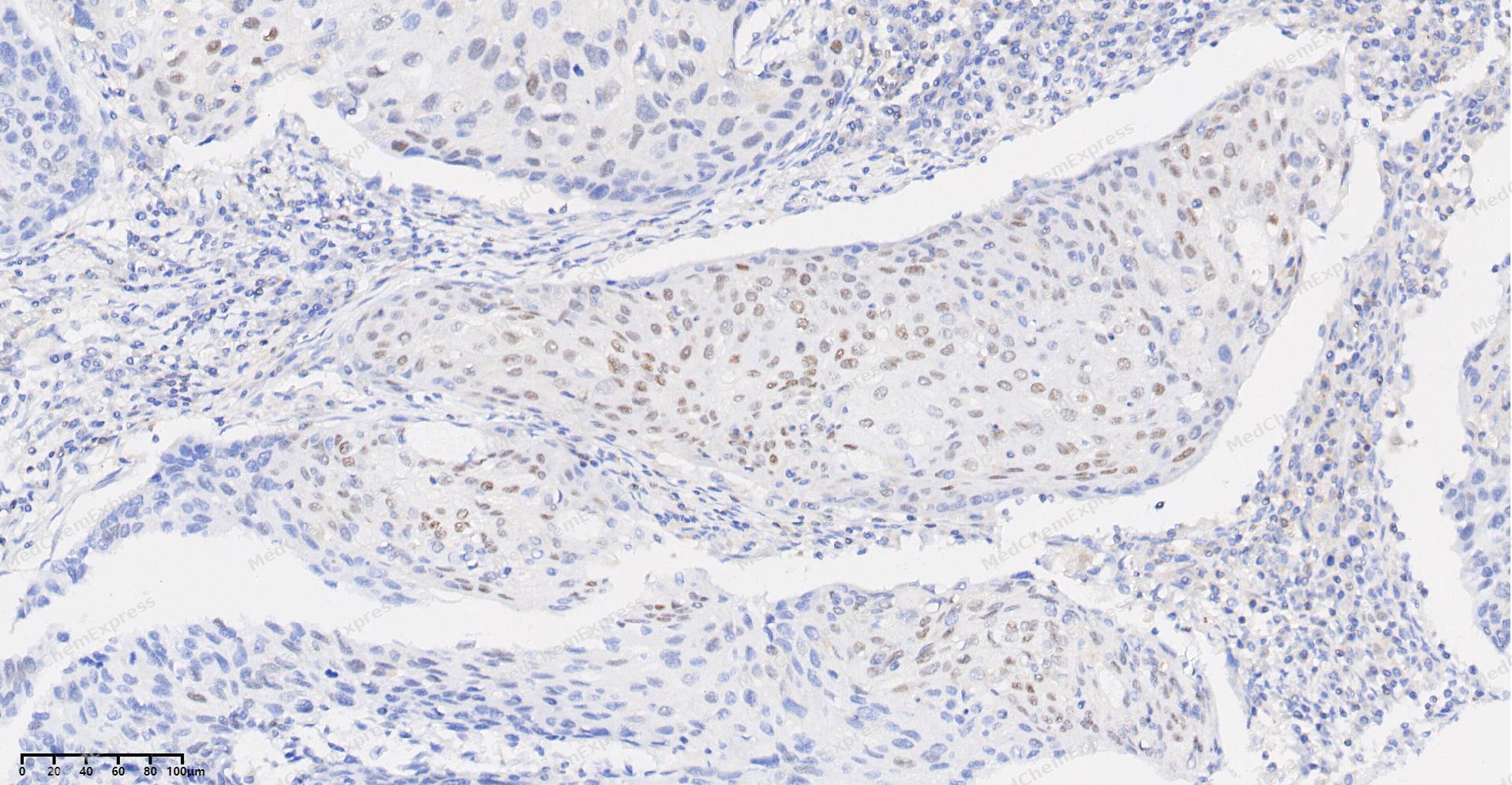 Immunohistochemical analysis of paraffin-embedded human Esophageal Carcinoma tissue using PKC beta 2 antibody was performed. The section was pretreated using high-temperature and high-pressure mediated EDTA antigen retrieval buffer (pH 9.0), for 5 minutes. The tissues were incubated with primary antibody (HY-P80432, 1:100 dilution) at room temperature for 60 minutes. Detection was performed using an HRP conjugated compact polymer system. DAB was used as the chromogen. The tissues were counterstained with hematoxylin and mounted with neutral balsam mounting medium.
 -
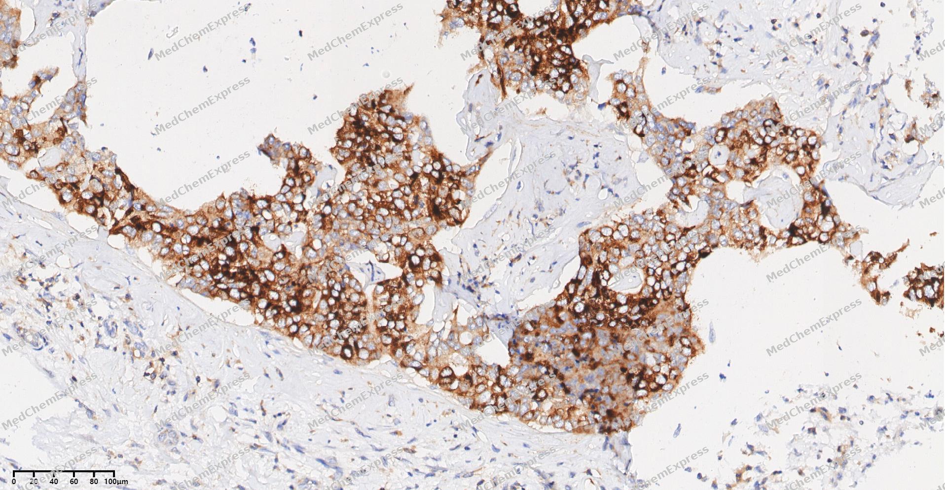 Immunohistochemical analysis of paraffin-embedded human cholangiocarcinoma tissue using PKC beta 2 antibody was performed. The section was pretreated using high-temperature and high-pressure mediated EDTA antigen retrieval buffer (pH 9.0), for 5 minutes. The tissues were incubated with primary antibody (HY-P80432, 1:100 dilution) at room temperature for 60 minutes. Detection was performed using an HRP conjugated compact polymer system. DAB was used as the chromogen. The tissues were counterstained with hematoxylin and mounted with neutral balsam mounting medium.
 -
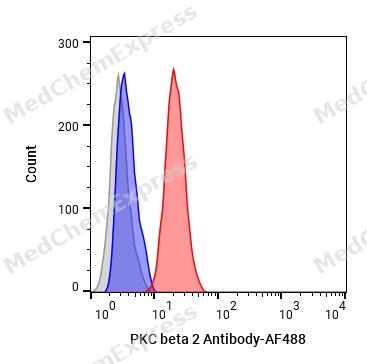 Flow cytometric analysis of 1X10⁶ HeLa cells labeling PKC beta 2 Antibody (HY-P80432, red). Cells were fixed with 4% paraformaldehyde and permeabilised with 90% methanol. Then stained with the primary antibody at 1/50 dilution for an hour at 4℃. AF488-conjugated Goat Anti-Rabbit IgG H&L (HY-P8002) was used as the secondary antibody at 1/1,000 dilution for 30 minutes at 4℃. Rabbit IgG Isotype Control (HY-P80879, blue) was used as the isotype control, cells without incubation with primary antibody were used as the unlabeled control (black).
 -
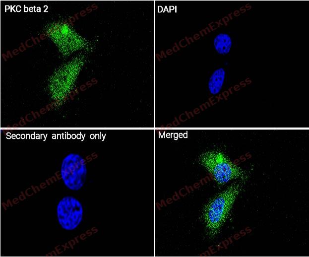 Immunocytochemistry analysis of Hela cells labeling PKC beta 2 with PKC beta 2 Antibody (HY-P80432) at 1/100 dilution. Cells were fixed in 4% paraformaldehyde for 15 minutes at room temperature, permeabilized with 0.1% Triton X-100 in PBS for 15 minutes at room temperature, then blocked with quick block buffer for 10 minutes at room temperature. Cells were then incubated with PKC beta 2 Antibody (HY-P80432) at 1/100 dilution in quick block buffer overnight at 4 ℃. AF488-conjugated Goat Anti-Rabbit IgG H&L(HY-P8002, Green) was used as the secondary antibody at 1/1,000 dilution. PBS instead of the primary antibody was used as the secondary antibody only control. The Nuclear counterstain was DAPI (Blue).
 -
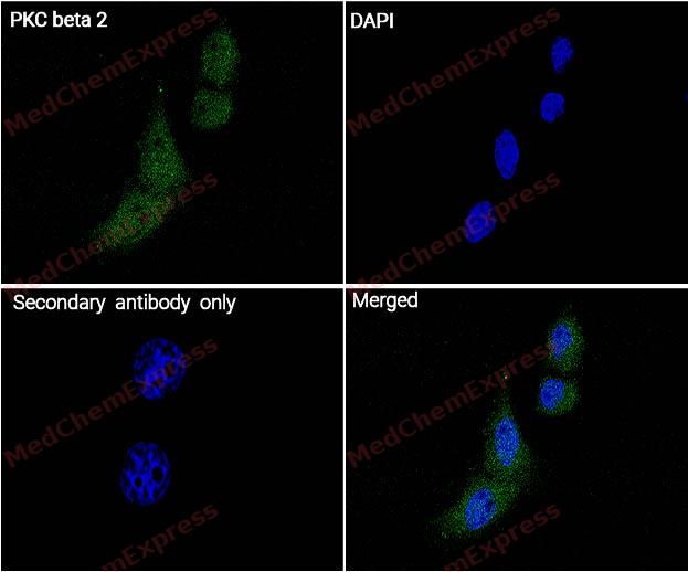 Immunocytochemistry analysis of Hela cells labeling PKC beta 2 with PKC beta 2 Antibody (HY-P80432) at 1/200 dilution. Cells were fixed in 4% paraformaldehyde for 15 minutes at room temperature, permeabilized with 0.1% Triton X-100 in PBS for 15 minutes at room temperature, then blocked with quick block buffer for 10 minutes at room temperature. Cells were then incubated with PKC beta 2 Antibody (HY-P80432) at 1/200 dilution in quick block buffer overnight at 4 ℃. AF488-conjugated Goat Anti-Rabbit IgG H&L(HY-P8002, Green) was used as the secondary antibody at 1/1,000 dilution. PBS instead of the primary antibody was used as the secondary antibody only control. The Nuclear counterstain was DAPI (Blue).


Background
-
Function
PKC beta 2 is a Calcium-activated, phospholipid- and diacylglycerol (DAG)-dependent serine/threonine-protein kinase involved in various cellular processes such as regulation of the B-cell receptor (BCR) signalosome, oxidative stress-induced apoptosis, androgen receptor-dependent transcription regulation, insulin signaling and endothelial cells proliferation. Plays a key role in B-cell activation by regulating BCR-induced NF-kappa-B activation. Mediates the activation of the canonical NF-kappa-B pathway (NFKB1) by direct phosphorylation of CARD11/CARMA1 at 'Ser-559', 'Ser-644' and 'Ser-652'. Phosphorylation induces CARD11/CARMA1 association with lipid rafts and recruitment of the BCL10-MALT1 complex as well as MAP3K7/TAK1, which then activates IKK complex, resulting in nuclear translocation and activation of NFKB1. Plays a direct role in the negative feedback regulation of the BCR signaling, by down-modulating BTK function via direct phosphorylation of BTK at 'Ser-180', which results in the alteration of BTK plasma membrane localization and in turn inhibition of BTK activity. Involved in apoptosis following oxidative damage: in case of oxidative conditions, specifically phosphorylates 'Ser-36' of isoform p66Shc of SHC1, leading to mitochondrial accumulation of p66Shc, where p66Shc acts as a reactive oxygen species producer. Acts as a coactivator of androgen receptor (AR)-dependent transcription, by being recruited to AR target genes and specifically mediating phosphorylation of 'Thr-6' of histone H3 (H3T6ph), a specific tag for epigenetic transcriptional activation that prevents demethylation of histone H3 'Lys-4' (H3K4me) by LSD1/KDM1A. In insulin signaling, may function downstream of IRS1 in muscle cells and mediate insulin-dependent DNA synthesis through the RAF1-MAPK/ERK signaling cascade. Participates in the regulation of glucose transport in adipocytes by negatively modulating the insulin-stimulated translocation of the glucose transporter SLC2A4/GLUT4. Phosphorylates SLC2A1/GLUT1, promoting glucose uptake by SLC2A1/GLUT1. Under high glucose in pancreatic beta-cells, is probably involved in the inhibition of the insulin gene transcription, via regulation of MYC expression. In endothelial cells, activation of PRKCB induces increased phosphorylation of RB1, increased VEGFA-induced cell proliferation, and inhibits PI3K/AKT-dependent nitric oxide synthase (NOS3/eNOS) regulation by insulin, which causes endothelial dysfunction. Also involved in triglyceride homeostasis. Phosphorylates ATF2 which promotes cooperation between ATF2 and JUN, activating transcription. Phosphorylates KLHL3 in response to angiotensin II signaling, decreasing the interaction between KLHL3 and WNK4. Phosphorylates and activates LRRK1, which phosphorylates RAB proteins involved in intracellular trafficking[1][2][3][4][5][6].
-
Subcellular Localization
Cytoplasm; Nucleus; Membrane; Peripheral membrane protein
-
Subunit
Interacts with PDPK1/PDK1 (By similarity). Interacts in vitro with PRKCBP1. Interacts with PHLPP1 and PHLPP2; both proteins mediate its dephosphorylation. Interacts with KDM1A/LSD1, PKN1 and AR
-
SwissProt ID
-
Synonyms
PKCA, PRKACA, PRKCA, Protein kinase C alpha type, PKC-A, PKC-alpha
-
Research Field
Signal Transduction


Documentation
-
Data Sheet (236 KB)
-
SDS (251 KB)
- English - EN (251 KB)
- Français - FR (251 KB)
- Deutsch - DE (251 KB)
- Norwegian - NO (251 KB)
- Español - ES (251 KB)
- Swedish - SV (251 KB)
- Italian - IT (251 KB)
- Korean - KR (251 KB)
- Portuguese - PT (251 KB)
-
User Guide for Antibodies (1077 KB)

 Powered by Bioz
