



Syntenin 1 Antibody (YA1051)
(Synonyms: SDCBP; MDA9; SYCL; Syntenin-1; Melanoma differentiation-associated protein 9; MDA-9; Pro-TGF-alpha cytoplasmic domain-interacting protein 18; TACIP18; Scaffold protein Pbp1; Syndecan-binding protein 1)
Based on 1 Customer Validation
Syntenin 1 Antibody (YA1051) is a Rabbit-derived and non-conjugated IgG monoclonal antibody, targeting to Syntenin 1.
-
Host:
Rabbit
-
Isotype:
IgG
-
Application:
WB, IHC-P, ICC/IF, IP, FC
-
Reactivity :
Human
-
Formulation:
Supplied in Rabbit IgG in 10mM phosphate buffered saline , pH 7.4, 150mM sodium chloride, 0.05% BSA, 0.02% sodium azide and 50% glycerol.
-
Conjugation:
Non-conjugated


To place orders, for customer services and technical support, please contact: MedChemExpress USA
Tel: 609-228-6898 E-mail: [email protected] [email protected]





Applications
| Application |
WB

WB: Western Blot
|
IHC-P
IHC-P: Immunohistochemistry-Paraffin
|
ICC/IF
ICC/IF: Immunocytochemistry/
Immunofluorescence |
IP
IP: Immunoprecipitation
|
FC
FC: Flow Cytometry
|
|---|---|---|---|---|---|
| Dilution Ratio | 1:500-1:1000 | 1:50-1:100 | 1:50-1:200 | 1:50 | 1:50-1:100 |


Product Details
Syntenin 1 Antibody (YA1051) is a Rabbit-derived and non-conjugated IgG monoclonal antibody, targeting to Syntenin 1.
-
Host Rabbit

-
Clonality Recombinant,Monoclonal

-
Species ReactivityHuman
-
Observed Molecular WeightObserved band size: 32 kDa
Note: Due to possible protein modifications or aggregation, the molecular weight should be confirmed by actual measurement, and the predicted value is for reference only.

-
Calculated Molecular Weight Predicted band size: 32 kDa

A synthesized peptide derived from human Syntenin aa2-79.
Endogenous
Affinity Chromatography
Non-conjugated
Unmodified
IgG


Product Properties
-
Appearance
Solution
-
Formulation
Supplied in Rabbit IgG in 10mM phosphate buffered saline , pH 7.4, 150mM sodium chloride, 0.05% BSA, 0.02% sodium azide and 50% glycerol.
-
Concentration
Batch-dependent, Please check the COA for the concentration of each lot. Check Lot Concentration
-
Storage & Stability
Stored at -20°C for 1 year. Avoid repeated freeze / thaw cycles.
-
Shipping
Shipping with blue ice.


Verification Images
-
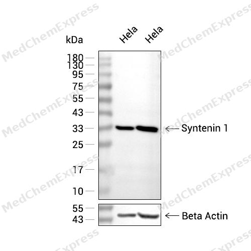 Western blot analysis of extracts from Hela (lane 2(20μg) , Hela (lane 3(40μg),using Syntenin 1 Antibody. Proteins were transferred to a PVDF membrane and blocked with 5% BSA in TBST for 2 hour at room temperature. The primary antibody and Loading control antibody (Beta Actin, HY-P80438, 1/3000) was used in 5% BSA in TBST at 4°C overnight. Goat Anti-Mouse/Rabbit IgG-HRP Secondary Antibody (HY-P8004/HY-P8001, 1/10,000) was used for 1 hour at room temperature.
 -
 Immunohistochemical analysis of paraffin-embedded human liver tissue using Syntenin 1 Antibody (HY-P81310A, 1/100). The section was pretreated using heat mediated antigen retrieval with sodium citrate buffer (pH 6.0) for 8 minutes. The tissues were blocked with quick block buffer for 0.5 hours at room temperature, washed with PBS and PBST, and then incubated with the primary antibody overnight at 4℃. The detection was performed using an HRP conjugated compact polymer system. DAB was used as the chromogen. Tissues were counterstained with hematoxylin and mounted with DPX.
 -
 Immunohistochemical analysis of paraffin-embedded human cerebral cortex tissue using Syntenin 1 Antibody (HY-P81310A, 1/100). The section was pretreated using heat mediated antigen retrieval with sodium citrate buffer (pH 6.0) for 8 minutes. The tissues were blocked with quick block buffer for 0.5 hours at room temperature, washed with PBS and PBST, and then incubated with the primary antibody overnight at 4℃. The detection was performed using an HRP conjugated compact polymer system. DAB was used as the chromogen. Tissues were counterstained with hematoxylin and mounted with DPX.
 -
 Immunohistochemical analysis of paraffin-embedded human cerebellum tissue using Syntenin 1 Antibody (HY-P81310A, 1/100). The section was pretreated using heat mediated antigen retrieval with sodium citrate buffer (pH 6.0) for 8 minutes. The tissues were blocked with quick block buffer for 0.5 hours at room temperature, washed with PBS and PBST, and then incubated with the primary antibody overnight at 4℃. The detection was performed using an HRP conjugated compact polymer system. DAB was used as the chromogen. Tissues were counterstained with hematoxylin and mounted with DPX.
 -
 Immunohistochemical analysis of paraffin-embedded human spleen tissue using Syntenin 1 Antibody (HY-P81310A, 1/100). The section was pretreated using heat mediated antigen retrieval with sodium citrate buffer (pH 6.0) for 8 minutes. The tissues were blocked with quick block buffer for 0.5 hours at room temperature, washed with PBS and PBST, and then incubated with the primary antibody overnight at 4℃. The detection was performed using an HRP conjugated compact polymer system. DAB was used as the chromogen. Tissues were counterstained with hematoxylin and mounted with DPX.
 -
 Immunohistochemical analysis of paraffin-embedded human lymph node tissue using Syntenin 1 Antibody (HY-P81310A, 1/100). The section was pretreated using heat mediated antigen retrieval with sodium citrate buffer (pH 6.0) for 8 minutes. The tissues were blocked with quick block buffer for 0.5 hours at room temperature, washed with PBS and PBST, and then incubated with the primary antibody overnight at 4℃. The detection was performed using an HRP conjugated compact polymer system. DAB was used as the chromogen. Tissues were counterstained with hematoxylin and mounted with DPX.
 -
 Immunohistochemical analysis of paraffin-embedded human tonsil tissue using Syntenin 1 Antibody (HY-P81310A, 1/100). The section was pretreated using heat mediated antigen retrieval with sodium citrate buffer (pH 6.0) for 8 minutes. The tissues were blocked with quick block buffer for 0.5 hours at room temperature, washed with PBS and PBST, and then incubated with the primary antibody overnight at 4℃. The detection was performed using an HRP conjugated compact polymer system. DAB was used as the chromogen. Tissues were counterstained with hematoxylin and mounted with DPX.
 -
 Immunohistochemical analysis of paraffin-embedded human adrenal gland tissue using Syntenin 1 Antibody (HY-P81310A, 1/100). The section was pretreated using heat mediated antigen retrieval with sodium citrate buffer (pH 6.0) for 8 minutes. The tissues were blocked with quick block buffer for 0.5 hours at room temperature, washed with PBS and PBST, and then incubated with the primary antibody overnight at 4℃. The detection was performed using an HRP conjugated compact polymer system. DAB was used as the chromogen. Tissues were counterstained with hematoxylin and mounted with DPX.
 -
 Immunohistochemical analysis of paraffin-embedded human salivary gland tissue using Syntenin 1 Antibody (HY-P81310A, 1/100). The section was pretreated using heat mediated antigen retrieval with sodium citrate buffer (pH 6.0) for 8 minutes. The tissues were blocked with quick block buffer for 0.5 hours at room temperature, washed with PBS and PBST, and then incubated with the primary antibody overnight at 4℃. The detection was performed using an HRP conjugated compact polymer system. DAB was used as the chromogen. Tissues were counterstained with hematoxylin and mounted with DPX.
 -
 Immunohistochemical analysis of paraffin-embedded human epididymis tissue using Syntenin 1 Antibody (HY-P81310A, 1/100). The section was pretreated using heat mediated antigen retrieval with sodium citrate buffer (pH 6.0) for 8 minutes. The tissues were blocked with quick block buffer for 0.5 hours at room temperature, washed with PBS and PBST, and then incubated with the primary antibody overnight at 4℃. The detection was performed using an HRP conjugated compact polymer system. DAB was used as the chromogen. Tissues were counterstained with hematoxylin and mounted with DPX.
 -
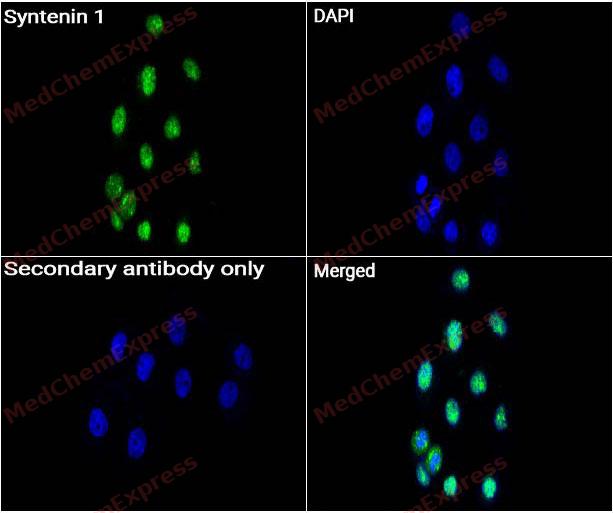 Immunocytochemistry analysis of Hela cells labeling Syntenin 1 with Syntenin 1 antibody (HY-P81310A) at 1/50 dilution. Cells were fixed in 4% paraformaldehyde for 15 minutes at room temperature, permeabilized with 0.1% Triton X-100 for 10 minutes at room temperature, then blocked with QuickBlock™ Blocking Buffer for Immunol Staining for 10 min at room temperature. Cells were then incubated with Syntenin 1 antibody (HY-P81310A) at 1/50 dilution in QuickBlock™ Blocking Buffer for Immunol Staining at 4 ℃. Alexa Fluor® 488-conjugated AffiniPure Goat Anti-Rabbit IgG H&L(HY-P8002, Green) was used as the secondary antibody at 1/1,000 dilution. PBS instead of the primary antibody was used as the secondary antibody only control. The Nuclear counterstain was DAPI (Blue).
 -
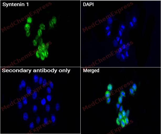 Immunocytochemistry analysis of Hela cells labeling Syntenin 1 with Syntenin 1 antibody (HY-P81310A) at 1/200 dilution. Cells were fixed in 4% paraformaldehyde for 15 minutes at room temperature, permeabilized with 0.1% Triton X-100 for 10 minutes at room temperature, then blocked with QuickBlock™ Blocking Buffer for Immunol Staining for 10 min at room temperature. Cells were then incubated with Syntenin 1 antibody (HY-P81310A) at 1/200 dilution in QuickBlock™ Blocking Buffer for Immunol Staining at 4 ℃. Alexa Fluor® 488-conjugated AffiniPure Goat Anti-Rabbit IgG H&L(HY-P8002, Green) was used as the secondary antibody at 1/1,000 dilution. PBS instead of the primary antibody was used as the secondary antibody only control. The Nuclear counterstain was DAPI (Blue).
 -
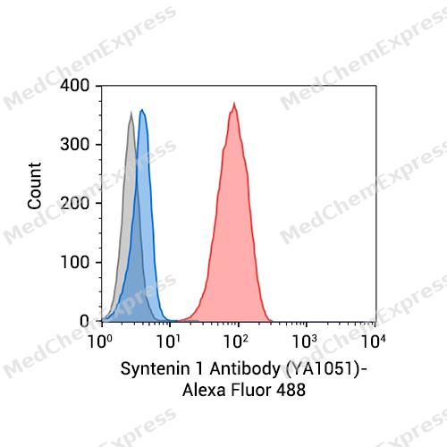 Flow cytometric analysis of 1X10^6 Hela cells labeling Syntenin 1 Antibody (YA1051) (HY-P81310A, red). Cells were fixed with 4% paraformaldehyde and permeabilised with 90% methanol. Then stained with the primary antibody at 1/50 dilution for an hour at 4℃. Alexa Fluor® 488-conjugated AffiniPure Goat Anti-Rabbit IgG H&L (HY-P8002) was used as the secondary antibody at 1/1,000 dilution for 30 minutes at 4℃. Rabbit IgG Isotype Control (HY-P80879, blue) was used as the isotype control, cells without incubation with primary antibody were used as the unlabeled control (black).


Background
-
Function
Syntenin 1 multifunctional adapter protein involved in diverse array of functions including trafficking of transmembrane proteins, neuro and immunomodulation, exosome biogenesis, and tumorigenesis. Positively regulates TGFB1-mediated SMAD2/3 activation and TGFB1-induced epithelial-to-mesenchymal transition (EMT) and cell migration in various cell types. May increase TGFB1 signaling by enhancing cell-surface expression of TGFR1 by preventing the interaction between TGFR1 and CAV1 and subsequent CAV1-dependent internalization and degradation of TGFR1. In concert with SDC1/4 and PDCD6IP, regulates exosome biogenesis. Regulates migration, growth, proliferation, and cell cycle progression in a variety of cancer types. In adherens junctions may function to couple syndecans to cytoskeletal proteins or signaling components. Seems to couple transcription factor SOX4 to the IL-5 receptor (IL5RA). May also play a role in vesicular trafficking. Seems to be required for the targeting of TGFA to the cell surface in the early secretory pathway[1][2][3][4][5][6][7].
-
Subcellular Localization
Cell junction, focal adhesion; Cell junction, adherens junction; Cell membrane; Peripheral membrane protein; Endoplasmic reticulum membrane; Peripheral membrane protein; Nucleus; Melanosome; Cytoplasm, cytosol; Cytoplasm, cytoskeleton; Secreted, extracellular exosome; Membrane raft
-
Expression
Tissue_specificity:Expressed in lung cancer, including adenocarcinoma, squamous cell carcinoma, and small cell carcinoma (protein level) (PubMed: 25893292) . Widely expressed. Expressed in fetal kidney, liver, lung, and brain. In humans, the highest expression is found in the heart and placenta.
Induction:By IFNG/IFN-gamma in melanoma cells -
Isoforms & Post-Translational Modification
O00560 has 3 isomers: O00560-1: 32444 Da (predicted); O00560-2: 32316 Da (predicted); O00560-3: 31762 Da (predicted).
Phosphorylated on tyrosine residues -
Subunit
Monomer and homodimer (By similarity). Interacts with SDC1, SDC2, SDC3, SDC4, NRXN2, EPHA7, EPHB1, NF2 isoform 1, TGFA and IL5RA. Interacts with NFASC and PTPRJ (By similarity). Interacts with SDCBP2 (PubMed:11152476). Interacts with PDCD6IP (PubMed:22660413). Forms a complex with PDCD6IP and SDC2 (PubMed:22660413). Interacts (via C-terminus) with TGFBR1 (PubMed:25893292). Binds to FZD7; this interaction is increased by inositol trisphosphate (IP3) (PubMed:27386966). Interacts with SMO (By similarity)
-
SwissProt ID
-
Synonyms
SDCBP; MDA9; SYCL; Syntenin-1; Melanoma differentiation-associated protein 9; MDA-9; Pro-TGF-alpha cytoplasmic domain-interacting protein 18; TACIP18; Scaffold protein Pbp1; Syndecan-binding protein 1
-
Research Field
Neuroscience


Documentation
-
Data Sheet (262 KB)
-
SDS (252 KB)
- English - EN (252 KB)
- Français - FR (252 KB)
- Deutsch - DE (252 KB)
- Norwegian - NO (252 KB)
- Español - ES (252 KB)
- Swedish - SV (252 KB)
- Italian - IT (252 KB)
- Korean - KR (252 KB)
- Portuguese - PT (252 KB)
-
User Guide for Antibodies (1077 KB)
References
[1]. Philley JV, et al. MDA-9/Syntenin Control. J Cell Physiol. 2016 Mar;231(3):545-50. [Content Brief]
[2]. Hwangbo C, et al. Syntenin regulates TGF-β1-induced Smad activation and the epithelial-to-mesenchymal transition by inhibiting caveolin-mediated TGF-β type I receptor internalization. Oncogene. 2016 Jan 21;35(3):389-401. [Content Brief]
[3]. Baietti MF, et al. Syndecan-syntenin-ALIX regulates the biogenesis of exosomes. Nat Cell Biol. 2012 Jun 3;14(7):677-85. [Content Brief]
[4]. Kashyap R, et al. Syntenin controls migration, growth, proliferation, and cell cycle progression in cancer cells. Front Pharmacol. 2015;6:241. [Content Brief]
[5]. Geijsen N, et al. Cytokine-specific transcriptional regulation through an IL-5Ralpha interacting protein. Science. 2001 Aug 10;293(5532):1136-8. [Content Brief]
[6]. Zimmermann P, et al. Characterization of syntenin, a syndecan-binding PDZ protein, as a component of cell adhesion sites and microfilaments. Mol Biol Cell. 2001 Feb;12(2):339-50. [Content Brief]
[7]. Fernández-Larrea J, et al. A role for a PDZ protein in the early secretory pathway for the targeting of proTGF-alpha to the cell surface. Mol Cell. 1999 Apr;3(4):423-33. [Content Brief]

 Powered by Bioz
