



TNFAIP3 Antibody (YA2231)
(Synonyms: TNFAIP3; OTUD7C; Tumor necrosis factor alpha-induced protein 3; TNF alpha-induced protein 3; OTU domain-containing protein 7C; Putative DNA-binding protein A20; Zinc finger protein A20)
Based on 1 Customer Validation
TNFAIP3 Antibody (YA2231) is a Rabbit-derived and non-conjugated IgG monoclonal antibody, targeting to TNFAIP3.
-
Host:
Rabbit
-
Isotype:
IgG
-
Application:
WB, IHC-F, IHC-P, ICC/IF
-
Reactivity :
Human
-
Formulation:
Supplied in 50mM Tris-Glycine(pH 7.4), 0.15M NaCl, 40% Glycerol, 0.01% Sodium azide and 0.05% BSA
-
Conjugation:
Non-conjugated


To place orders, for customer services and technical support, please contact: MedChemExpress USA
Tel: 609-228-6898 E-mail: [email protected] [email protected]





Applications
| Application |
WB

WB: Western Blot
|
IHC-P
IHC-P: Immunohistochemistry-Paraffin
|
IHC-F
IHC-F: Immunohistochemistry-Frozen
|
ICC/IF
ICC/IF: Immunocytochemistry/
Immunofluorescence |
|---|---|---|---|---|
| Dilution Ratio | 1:500-1:1000 | 1:50-1:100 | 1:50-1:100 | 1:50-1:200 |


Product Details
TNFAIP3 Antibody (YA2231) is a Rabbit-derived and non-conjugated IgG monoclonal antibody, targeting to TNFAIP3.
-
Host Rabbit

-
Clonality Recombinant,Monoclonal

-
Species ReactivityHuman
-
Observed Molecular WeightObserved band size: 90 kDa
Note: Due to possible protein modifications or aggregation, the molecular weight should be confirmed by actual measurement, and the predicted value is for reference only.

-
Calculated Molecular Weight Predicted band size: 90 kDa

A synthetic peptide of human TNFAIP3
Endogenous
Affinity Purified
Non-conjugated
Unmodified
IgG


Product Properties
-
Appearance
Liquid
-
Formulation
Supplied in 50mM Tris-Glycine(pH 7.4), 0.15M NaCl, 40% Glycerol, 0.01% Sodium azide and 0.05% BSA
-
Concentration
Batch-dependent, Please check the COA for the concentration of each lot. Check Lot Concentration
-
Storage & Stability
Stored at -20°C for 1 year. Avoid repeated freeze / thaw cycles.
-
Shipping
Shipping with blue ice.


Verification Images
-
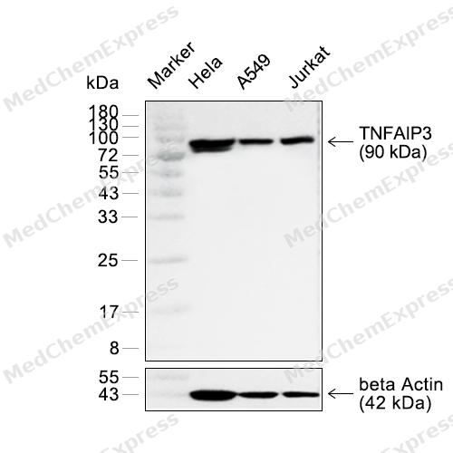 Western blot analysis of extracts from HeLa (lane 2(20μg)) , A549 (lane 3(20μg)) and Jurkat(lane 4(20μg)) using TNFAIP3 Rabbit mAb. Proteins were transferred to a PVDF membrane and blocked with 5% non-fat milk in TBST for 2 hour at room temperature. The primary antibody (1/1000) and Loading control antibody (beta Actin, HY-P83730, 1/5000) was used in 5% non-fat milk in TBST at 4°C overnight. Goat Anti-Rabbit/Mouse IgG-HRP Secondary Antibody (1/10000) was used for 1 hour at room temperature.
 -
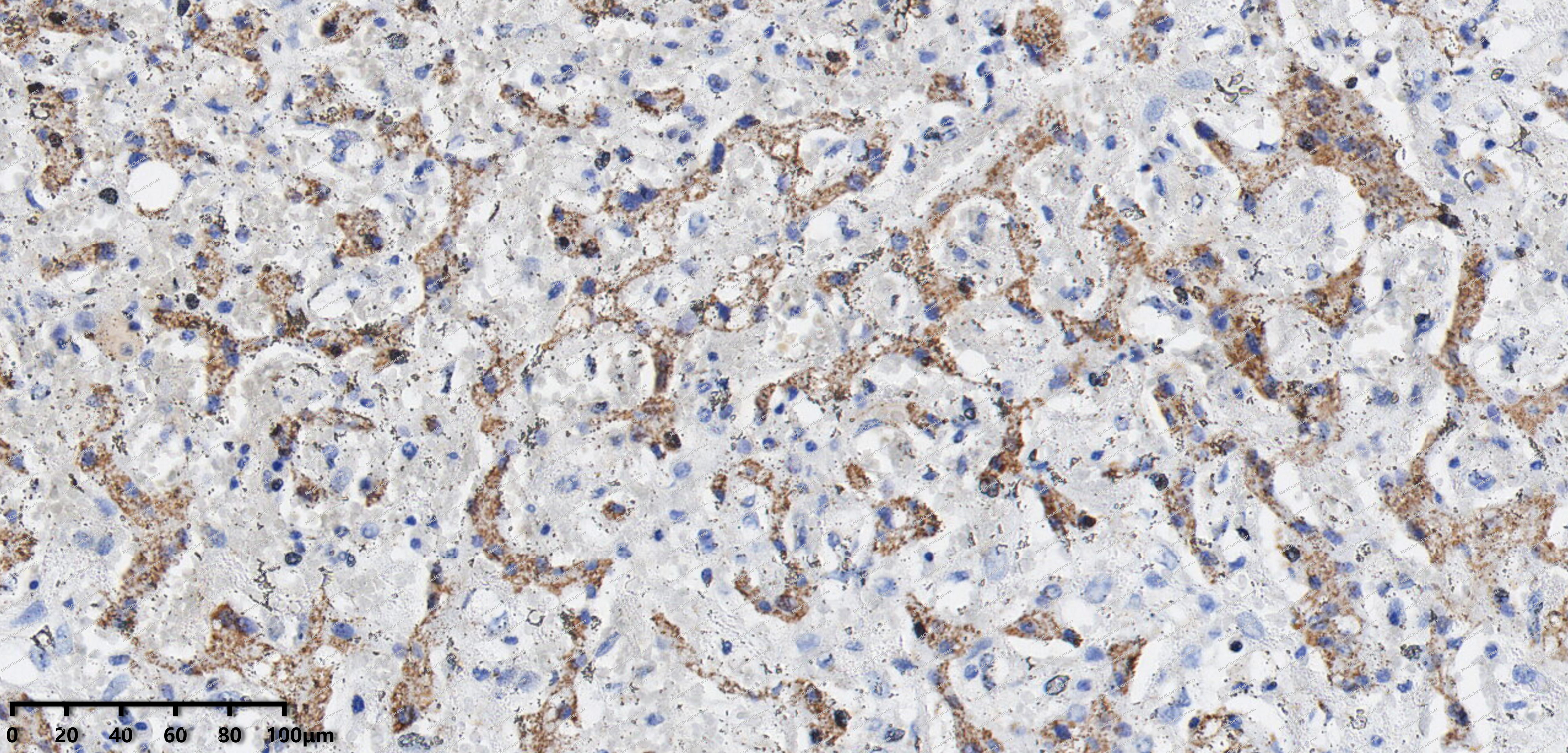 Immunohistochemical analysis of paraffin-embedded human Liver cyst tissue using TNFAIP3 antibody was performed. The section was pretreated using high-temperature mediated EDTA antigen retrieval buffer (pH 9.0), for 20 minutes. The tissues were incubated with primary antibody (HY-P82486, 1:200 dilution) at room temperature for 20 minutes. Detection was performed using an HRP conjugated compact polymer system. DAB was used as the chromogen. The tissues were counterstained with hematoxylin and mounted with neutral balsam mounting medium.
 -
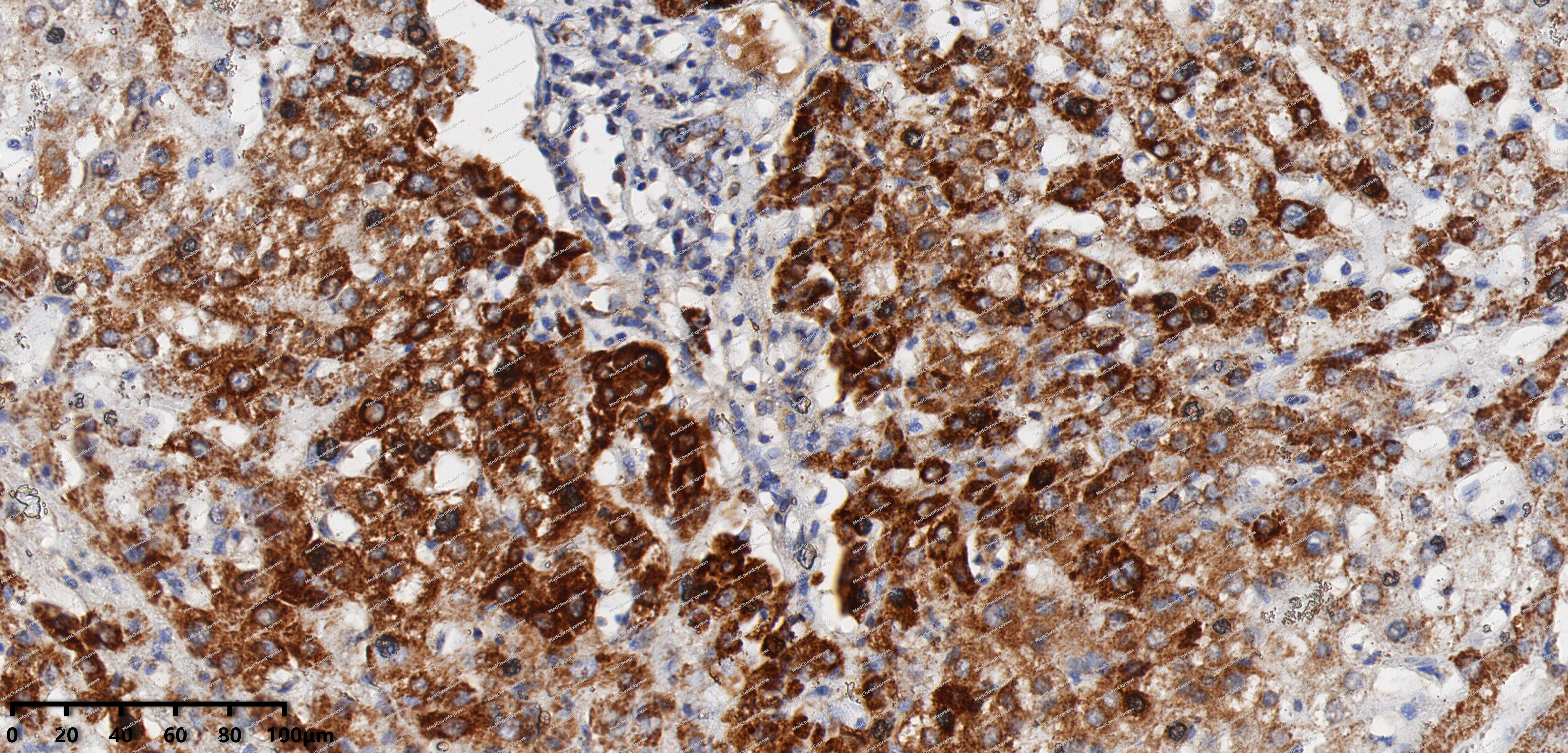 Immunohistochemical analysis of paraffin-embedded human Hepatocellular Carcinoma tissue using TNFAIP3 antibody was performed. The section was pretreated using high-temperature mediated EDTA antigen retrieval buffer (pH 9.0), for 20 minutes. The tissues were incubated with primary antibody (HY-P82486, 1:200 dilution) at room temperature for 20 minutes. Detection was performed using an HRP conjugated compact polymer system. DAB was used as the chromogen. The tissues were counterstained with hematoxylin and mounted with neutral balsam mounting medium.
 -
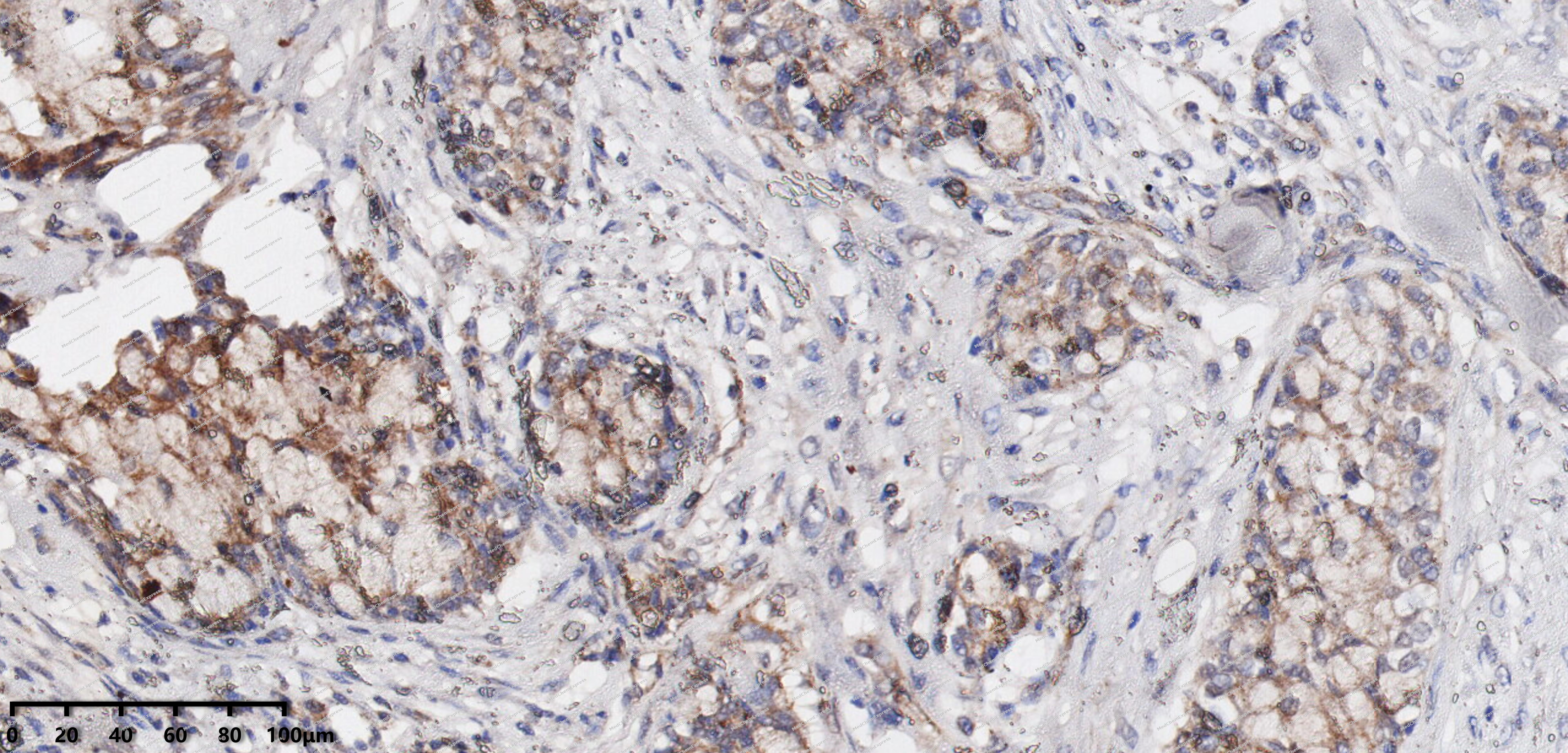 Immunohistochemical analysis of paraffin-embedded human Mucoepidermoid carcinoma tissue using TNFAIP3 antibody was performed. The section was pretreated using high-temperature mediated EDTA antigen retrieval buffer (pH 9.0), for 20 minutes. The tissues were incubated with primary antibody (HY-P82486, 1:200 dilution) at room temperature for 20 minutes. Detection was performed using an HRP conjugated compact polymer system. DAB was used as the chromogen. The tissues were counterstained with hematoxylin and mounted with neutral balsam mounting medium.
 -
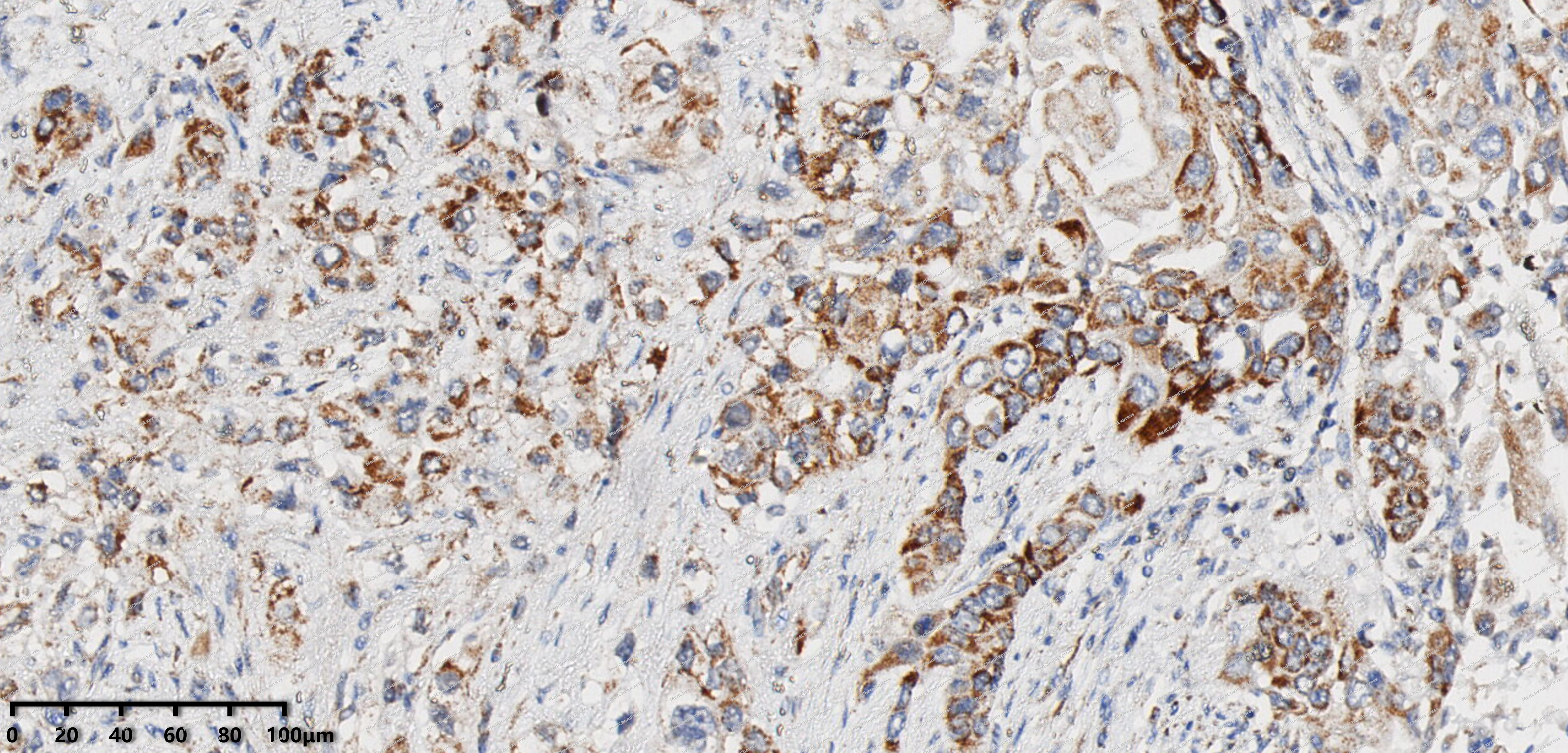 Immunohistochemical analysis of paraffin-embedded human Pancreatic cancer tissue using TNFAIP3 antibody was performed. The section was pretreated using high-temperature mediated EDTA antigen retrieval buffer (pH 9.0), for 20 minutes. The tissues were incubated with primary antibody (HY-P82486, 1:200 dilution) at room temperature for 20 minutes. Detection was performed using an HRP conjugated compact polymer system. DAB was used as the chromogen. The tissues were counterstained with hematoxylin and mounted with neutral balsam mounting medium.
 -
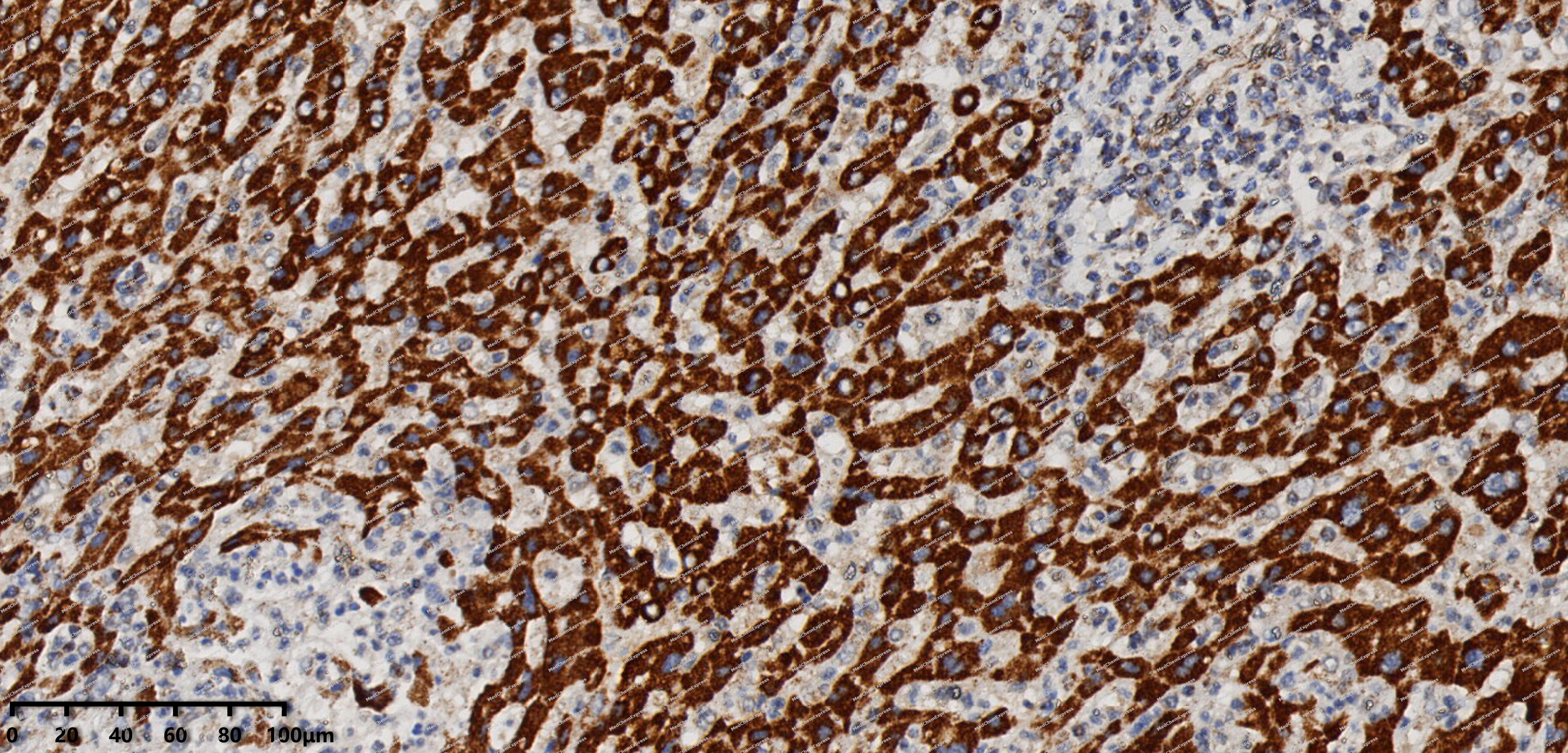 Immunohistochemical analysis of paraffin-embedded human Intrahepatic cholangiocarcinoma tissue using TNFAIP3 antibody was performed. The section was pretreated using high-temperature mediated EDTA antigen retrieval buffer (pH 9.0), for 20 minutes. The tissues were incubated with primary antibody (HY-P82486, 1:200 dilution) at room temperature for 20 minutes. Detection was performed using an HRP conjugated compact polymer system. DAB was used as the chromogen. The tissues were counterstained with hematoxylin and mounted with neutral balsam mounting medium.
 -
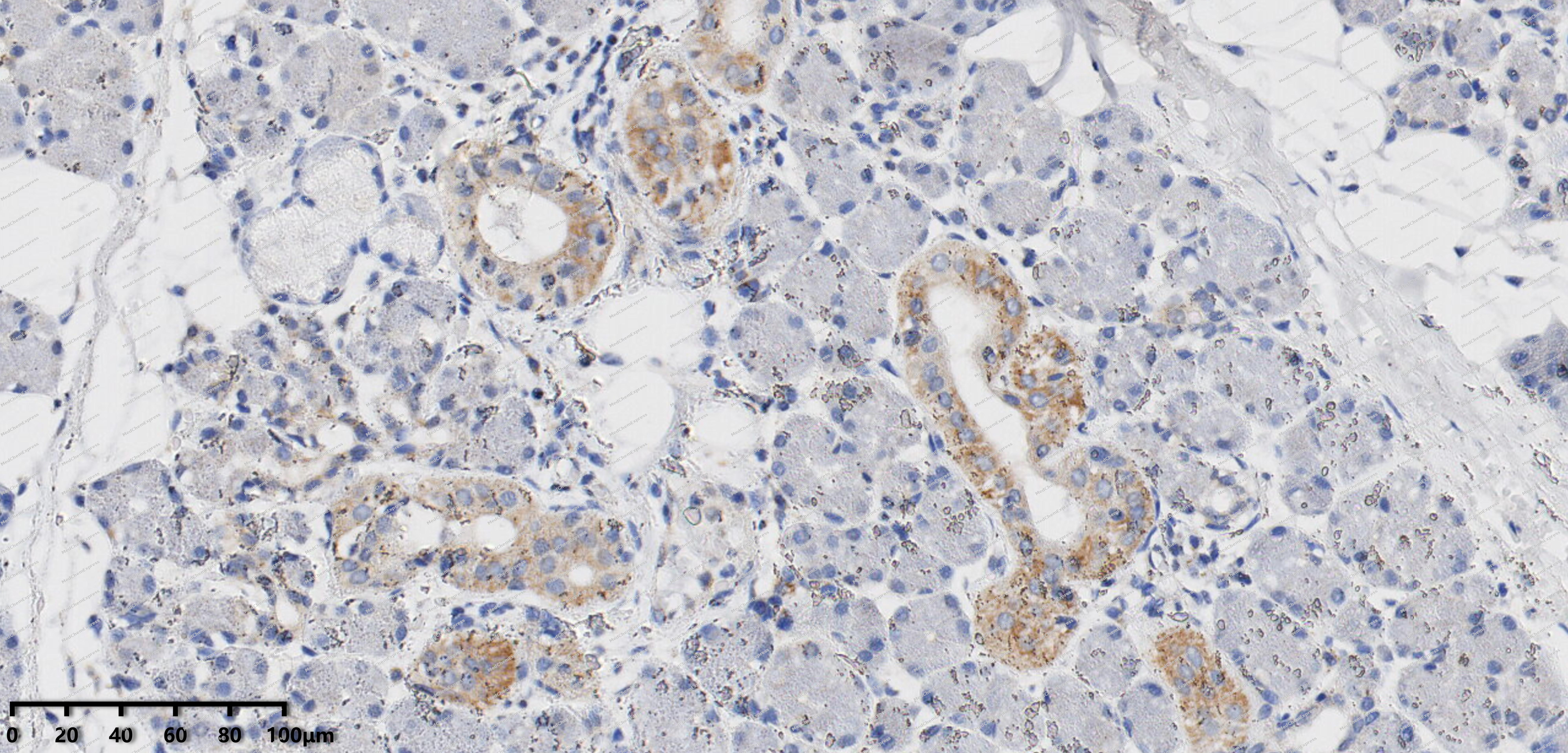 Immunohistochemical analysis of paraffin-embedded human pleomorphic adenoma tissue using TNFAIP3 antibody was performed. The section was pretreated using high-temperature mediated EDTA antigen retrieval buffer (pH 9.0), for 20 minutes. The tissues were incubated with primary antibody (HY-P82486, 1:200 dilution) at room temperature for 20 minutes. Detection was performed using an HRP conjugated compact polymer system. DAB was used as the chromogen. The tissues were counterstained with hematoxylin and mounted with neutral balsam mounting medium.
 -
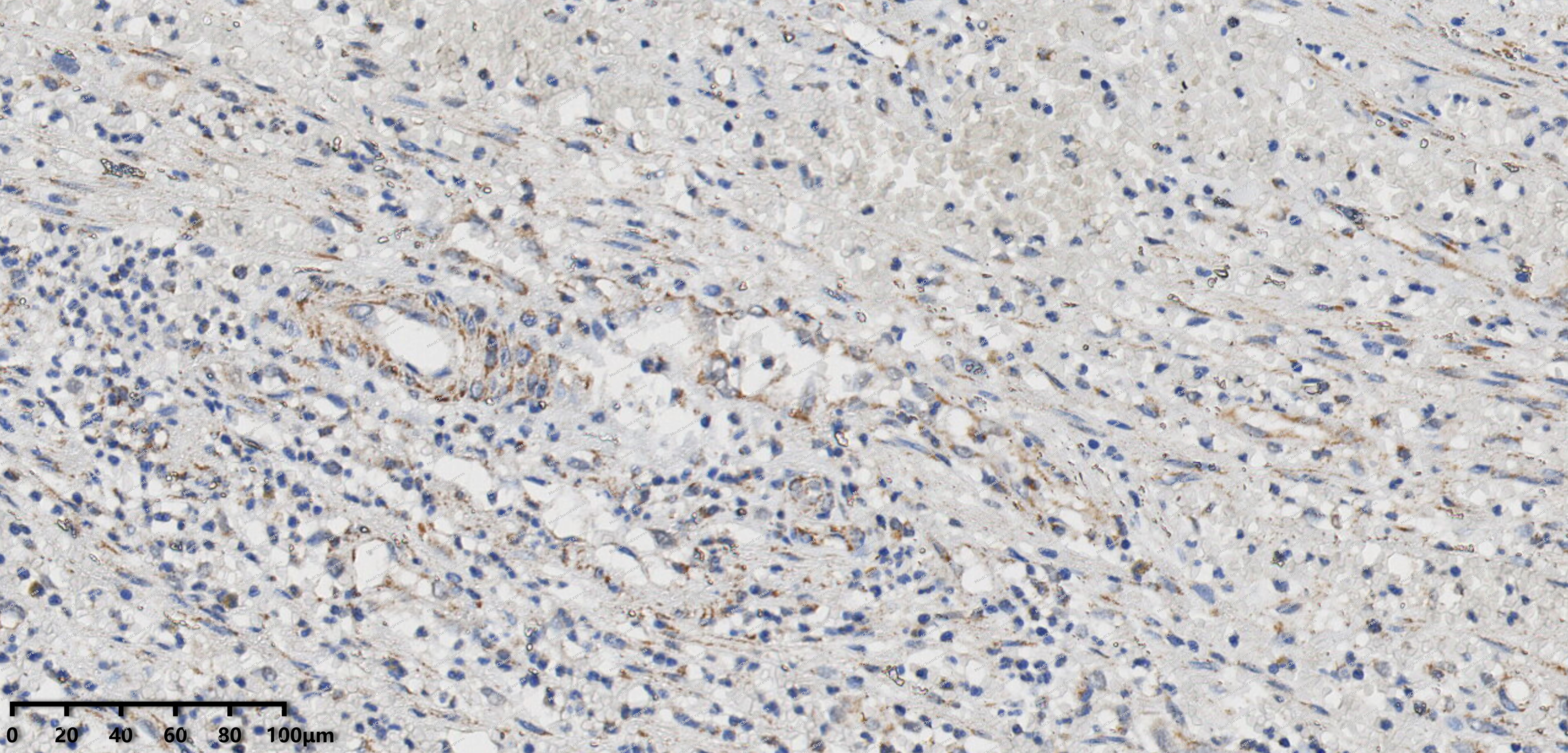 Immunohistochemical analysis of paraffin-embedded human appendix tissue using TNFAIP3 antibody was performed. The section was pretreated using high-temperature mediated EDTA antigen retrieval buffer (pH 9.0), for 20 minutes. The tissues were incubated with primary antibody (HY-P82486, 1:200 dilution) at room temperature for 20 minutes. Detection was performed using an HRP conjugated compact polymer system. DAB was used as the chromogen. The tissues were counterstained with hematoxylin and mounted with neutral balsam mounting medium.
 -
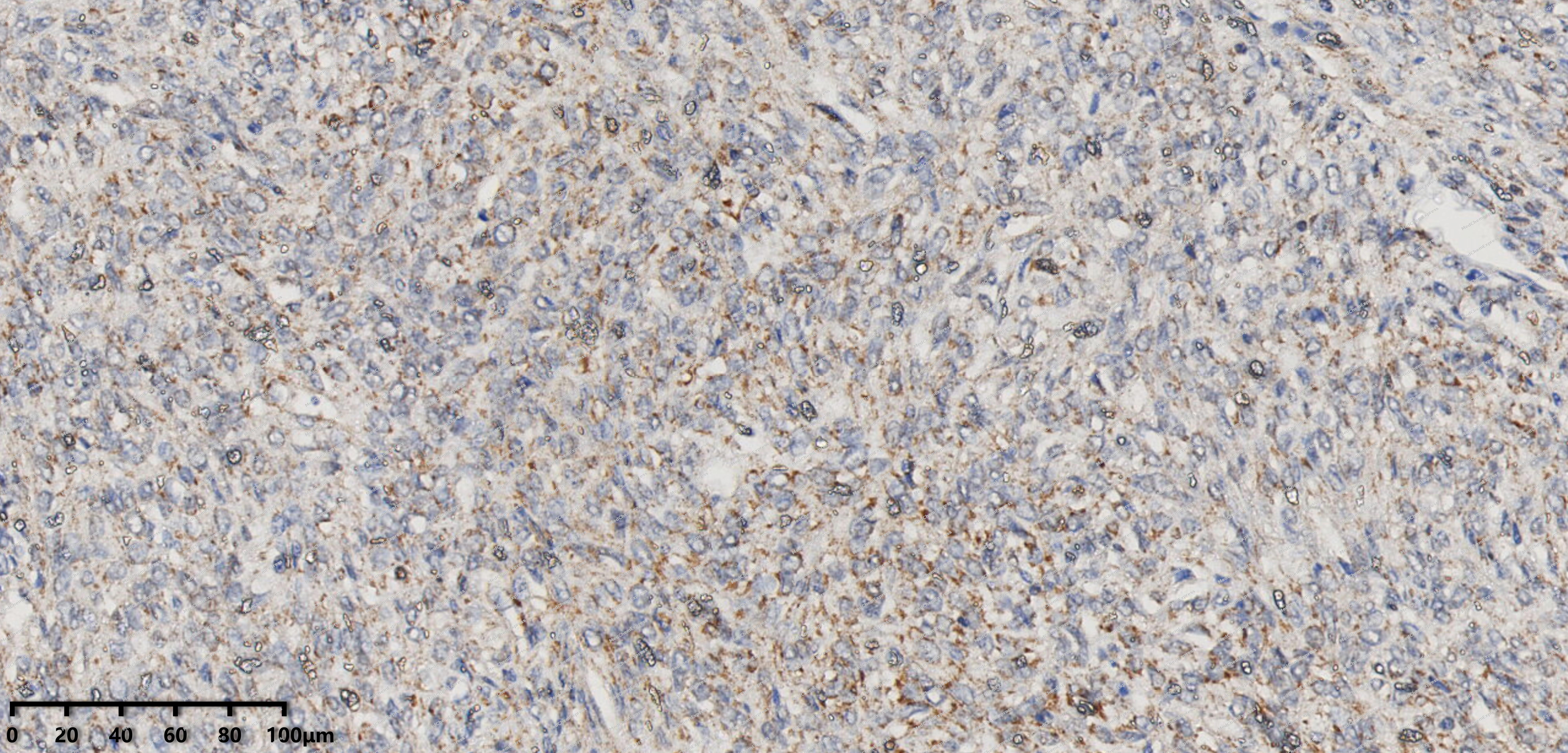 Immunohistochemical analysis of paraffin-embedded human solitary fibrous tumor tissue using TNFAIP3 antibody was performed. The section was pretreated using high-temperature mediated EDTA antigen retrieval buffer (pH 9.0), for 20 minutes. The tissues were incubated with primary antibody (HY-P82486, 1:200 dilution) at room temperature for 20 minutes. Detection was performed using an HRP conjugated compact polymer system. DAB was used as the chromogen. The tissues were counterstained with hematoxylin and mounted with neutral balsam mounting medium.
 -
 Immunocytochemistry analysis of HepG2 cells labeling TNFAIP3 with TNFAIP3 Antibody (HY-P82486) at 1/100 dilution. Cells were fixed in 4% paraformaldehyde for 15 minutes at room temperature, permeabilized with 0.1% Triton X-100 in PBS for 15 minutes at room temperature, then blocked with quick block buffer for 10 minutes at room temperature. Cells were then incubated with TNFAIP3 Antibody (HY-P82486) at 1/100 dilution in quick block buffer overnight at 4 ℃. AF488-conjugated Goat Anti-Rabbit IgG H&L (HY-P8002, Green) was used as the secondary antibody at 1/1,000 dilution. PBS instead of the primary antibody was used as the secondary antibody only control. The Nuclear counterstain was DAPI (Blue).
 -
 Immunocytochemistry analysis of HepG2 cells labeling TNFAIP3 with TNFAIP3 Antibody (HY-P82486) at 1/200 dilution. Cells were fixed in 4% paraformaldehyde for 15 minutes at room temperature, permeabilized with 0.1% Triton X-100 in PBS for 15 minutes at room temperature, then blocked with quick block buffer for 10 minutes at room temperature. Cells were then incubated with TNFAIP3 Antibody (HY-P82486) at 1/200 dilution in quick block buffer overnight at 4 ℃. AF488-conjugated Goat Anti-Rabbit IgG H&L (HY-P8002, Green) was used as the secondary antibody at 1/1,000 dilution. PBS instead of the primary antibody was used as the secondary antibody only control. The Nuclear counterstain was DAPI (Blue).


Background
-
Function
TNFAIP3 is an Ubiquitin-editing enzyme that contains both ubiquitin ligase and deubiquitinase activities. Involved in immune and inflammatory responses signaled by cytokines, such as TNF-alpha and IL-1 beta, or pathogens via Toll-like receptors (TLRs) through terminating NF-kappa-B activity. Essential component of a ubiquitin-editing protein complex, comprising also RNF11, ITCH and TAX1BP1, that ensures the transient nature of inflammatory signaling pathways. In cooperation with TAX1BP1 promotes disassembly of E2-E3 ubiquitin protein ligase complexes in IL-1R and TNFR-1 pathways; affected are at least E3 ligases TRAF6, TRAF2 and BIRC2, and E2 ubiquitin-conjugating enzymes UBE2N and UBE2D3. In cooperation with TAX1BP1 promotes ubiquitination of UBE2N and proteasomal degradation of UBE2N and UBE2D3. Upon TNF stimulation, deubiquitinates 'Lys-63'-polyubiquitin chains on RIPK1 and catalyzes the formation of 'Lys-48'-polyubiquitin chains. This leads to RIPK1 proteasomal degradation and consequently termination of the TNF- or LPS-mediated activation of NF-kappa-B. Deubiquitinates TRAF6 probably acting on 'Lys-63'-linked polyubiquitin. Upon T-cell receptor (TCR)-mediated T-cell activation, deubiquitinates 'Lys-63'-polyubiquitin chains on MALT1 thereby mediating disassociation of the CBM (CARD11:BCL10:MALT1) and IKK complexes and preventing sustained IKK activation. Deubiquitinates NEMO/IKBKG; the function is facilitated by TNIP1 and leads to inhibition of NF-kappa-B activation. Upon stimulation by bacterial peptidoglycans, probably deubiquitinates RIPK2. Can also inhibit I-kappa-B-kinase (IKK) through a non-catalytic mechanism which involves polyubiquitin; polyubiquitin promotes association with IKBKG and prevents IKK MAP3K7-mediated phosphorylation. Targets TRAF2 for lysosomal degradation. In vitro able to deubiquitinate 'Lys-11'-, 'Lys-48'- and 'Lys-63' polyubiquitin chains. Inhibitor of programmed cell death. Has a role in the function of the lymphoid system. Required for LPS-induced production of pro-inflammatory cytokines and IFN beta in LPS-tolerized macrophages
-
Subcellular Localization
Cytoplasm; Nucleus; Lysosome; Cytoplasm
-
Expression
Induction:By TNF -
Subunit
Homodimer. Interacts with TNIP1, TAX1BP1 and TRAF2. Interacts with RNF11, ITCH and TAX1BP1 only after TNF stimulation; these interaction are transient and they are lost after 1 hour of stimulation with TNF (By similarity). Interacts with YWHAZ and YWHAH. Interacts with IKBKG; the interaction is induced by TNF stimulation and by polyubiquitin. Interacts with RIPK1. Interacts with UBE2N; the interaction requires TAX1BP1. Interacts with TRAF6; the interaction is inhibited by HTLV-1 protein Tax
-
SwissProt ID
-
Synonyms
TNFAIP3; OTUD7C; Tumor necrosis factor alpha-induced protein 3; TNF alpha-induced protein 3; OTU domain-containing protein 7C; Putative DNA-binding protein A20; Zinc finger protein A20
-
Research Field
Immunology


Documentation
-
Data Sheet (234 KB)
-
SDS (252 KB)
- English - EN (252 KB)
- Français - FR (252 KB)
- Deutsch - DE (252 KB)
- Norwegian - NO (252 KB)
- Español - ES (252 KB)
- Swedish - SV (252 KB)
- Italian - IT (252 KB)
- Korean - KR (252 KB)
- Portuguese - PT (252 KB)
-
User Guide for Antibodies (1077 KB)

 Powered by Bioz
