



TREM2 Antibody
(Synonyms: TREM2; TREM-2; Trem2a; Trem2b; Trem2c; triggering receptor expressed on myeloid cells 2)Based on 6 publication(s) in Google Scholar
TREM2 Antibody is a Rabbit-derived and non-conjugated IgG polyclonal antibody, targeting to TREM2.
-
Host:
Rabbit
-
Isotype:
IgG
-
Application:
WB, IHC-P, ICC/IF
-
Reactivity :
Human, Mouse, Rat
-
Formulation:
Supplied in 1*PBS (pH 7.3), 50% glycerol and 0.5% BSA. Preservative: 0.02% sodium azide.
-
Conjugation:
Non-conjugated


To place orders, for customer services and technical support, please contact: MedChemExpress USA
Tel: 609-228-6898 E-mail: [email protected] [email protected]



Publications Citing Use of MedChemExpress (MCE) TREM2 Antibody
More

Applications
| Application |
WB

WB: Western Blot
|
IHC-P
IHC-P: Immunohistochemistry-Paraffin
|
ICC/IF
ICC/IF: Immunocytochemistry/
Immunofluorescence |
|---|---|---|---|
| Dilution Ratio | 1:500-1:1000 | 1:50-1:100 | 1:50-1:200 |


Product Details
TREM2 Antibody is a Rabbit-derived and non-conjugated IgG polyclonal antibody, targeting to TREM2.
-
Host Rabbit

-
Clonality Polyclonal

-
Species ReactivityHuman, Mouse, Rat
-
Observed Molecular WeightObserved band size:25,30-60 kDa
Note: Due to possible protein modifications or aggregation, the molecular weight should be confirmed by actual measurement, and the predicted value is for reference only.

-
Calculated Molecular Weight Predicted band size: 25 kDa

Entrez Gene: 54209 Human ; 83433 Mouse ; 301227 Rat
SwissProt: Q9NZC2 Human ; Q99NH8 Mouse ;
OMIM: 618193 Human
Synthetic peptide corresponding to Human TREM2.AA range:19-160.
Endogenous
affinity purified
Non-conjugated
Unmodified
IgG


Product Properties
-
Appearance
Solution
-
Formulation
Supplied in 1*PBS (pH 7.3), 50% glycerol and 0.5% BSA. Preservative: 0.02% sodium azide.
-
Concentration
Batch-dependent, Please check the COA for the concentration of each lot. Check Lot Concentration
-
Storage & Stability
Stored at -20°C for 1 year. Avoid repeated freeze / thaw cycles.
-
Shipping
Shipping with blue ice.


Publications (6)
-
Journal Impact Factor
-
Most Recent
-
Phytomedicine
Gualou-Xiebai-Banxia-Tang regulates liver-gut axis to ameliorate Metabolic Syndrome in HFD-fed mice. [Abstract]2024 Sep:132:155320. PMID: 38901285 -
Commun Biol
Microgravity-cultured glioblastoma organoids integrated with microfluidic chip for CAR-γδ T evaluation. [Abstract]2025 Dec 18;8(1):1791. PMID: 41407922


Verification Images
-
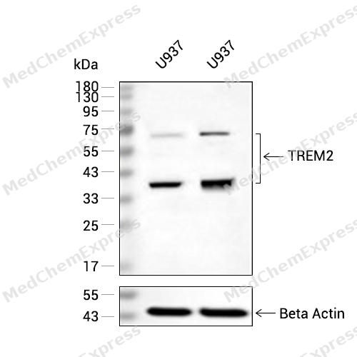 Western blot analysis of extracts from U937 (lane 2(20μg) and U937(lane 3(20μg) using TREM2 (HY-P80920) Rabbit mAb. Proteins were transferred to a PVDF membrane and blocked with 5% non-fat milk in TBST for 2 hour at room temperature. The primary antibody (1/1000) and Loading control antibody (Beta Actin, HY-P80438, 1/10000) was used in 5% non-fat milk in TBST at 4°C overnight. Goat Anti-Mouse/Rabbit IgG-HRP Secondary Antibody (1/10000) was used for 1 hour at room temperature.
 -
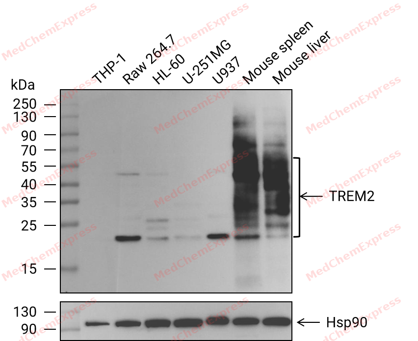 Western blot analysis of extracts from THP-1 (lane 1(20μg)) 、Raw 264.7 (lane 2(20μg)) 、HL-60 (lane 3(20μg)) 、U-251MG (lane 4(20μg)) 、U937 (lane 5(20μg)) 、Mouse spleen (lane 6(20μg)) and Mouse liver (lane 7(20μg)) using TREM2 Antibody (HY-P80920) . Proteins were transferred to a PVDF membrane and blocked with 5% nonfat dry milk in TBST for 1.5 hour at room temperature. The primary antibody (1/1000) and Loading control antibody (Hsp90, 1/10000) was used in 5% nonfat dry milk in TBST at 4℃ overnight. Goat Anti-Rabbit IgG-HRP Secondary Antibody (1/10,000) was used for 1 hour at room temperature.
 -
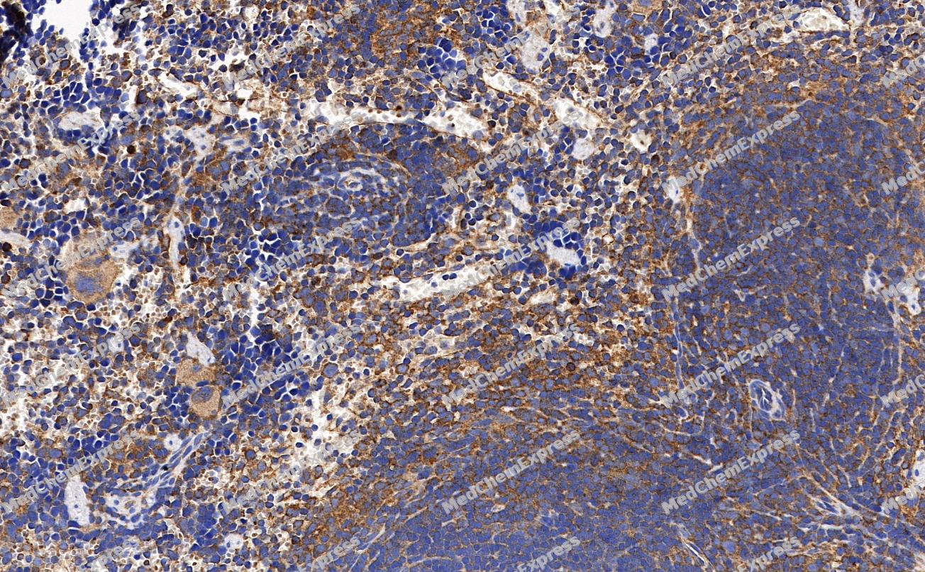 Immunohistochemical analysis of paraffin-embedded Mouse spleen tissue using TREM2 Antibody. The section was pre-treated using heat mediated antigen retrieval with sodium citrate buffer (pH 6.0) for 8 minutes. The tissues were blocked in QuickBlock for 20 minutes at room temperature, washed with ddH2O and PBS, and then probed with the primary antibody (HY-P80920, 1/100) in 4℃ overnight. The detection was performed using an HRP conjugated compact polymer system. DAB was used as the chromogen. Tissues were counterstained with hematoxylin and mounted with DPX.
 -
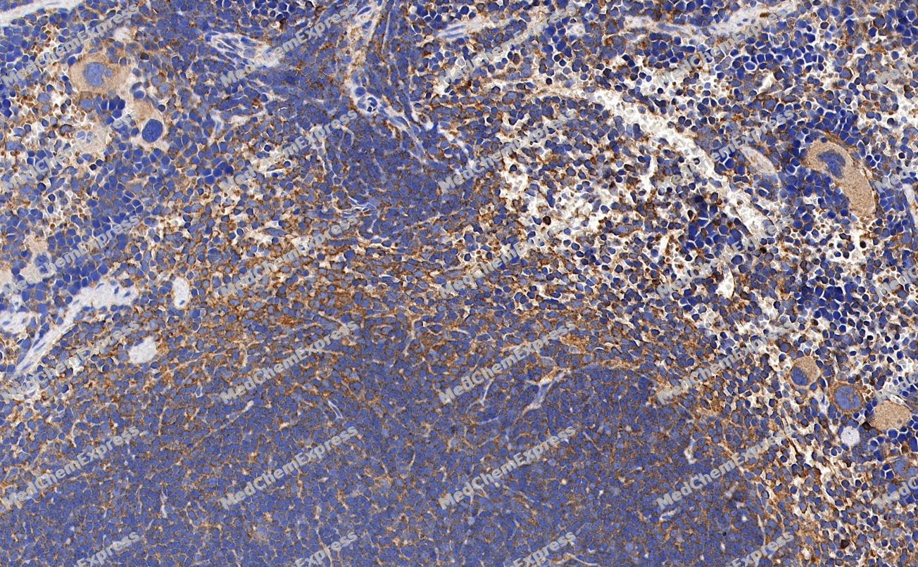 Immunohistochemical analysis of paraffin-embedded Mouse spleen tissue using TREM2 Antibody. The section was pre-treated using heat mediated antigen retrieval with sodium citrate buffer (pH 6.0) for 8 minutes. The tissues were blocked in QuickBlock for 20 minutes at room temperature, washed with ddH2O and PBS, and then probed with the primary antibody (HY-P80920, 1/100) in 4℃ overnight. The detection was performed using an HRP conjugated compact polymer system. DAB was used as the chromogen. Tissues were counterstained with hematoxylin and mounted with DPX.
 -
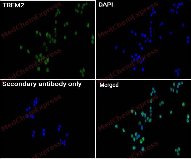 Immunocytochemistry analysis of RAW264.7 cells labeling TREM2 with TREM2 antibody (HY-P80920) at 1/50 dilution. Cells were fixed in 4% paraformaldehyde for 15 minutes at room temperature, permeabilized with 0.1% Triton X-100 for 10 minutes at room temperature, then blocked with QuickBlock™ Blocking Buffer for Immunol Staining for 10 min at room temperature. Cells were then incubated with TREM2 antibody (HY-P80920) at 1/50 dilution in QuickBlock™ Blocking Buffer for Immunol Staining at 4 ℃. Alexa Fluor® 488-conjugated AffiniPure Goat Anti-Rabbit IgG H&L(HY-P8002, Green) was used as the secondary antibody at 1/1,000 dilution. PBS instead of the primary antibody was used as the secondary antibody only control. The Nuclear counterstain was DAPI (Blue).
 -
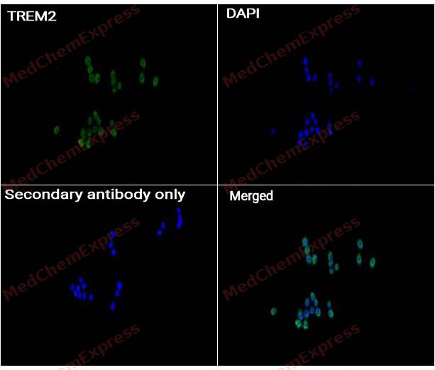 Immunocytochemistry analysis of RAW264.7 cells labeling TREM2 with TREM2 antibody (HY-P80920) at 1/200 dilution. Cells were fixed in 4% paraformaldehyde for 15 minutes at room temperature, permeabilized with 0.1% Triton X-100 for 10 minutes at room temperature, then blocked with QuickBlock™ Blocking Buffer for Immunol Staining for 10 min at room temperature. Cells were then incubated with TREM2 antibody (HY-P80920) at 1/200 dilution in QuickBlock™ Blocking Buffer for Immunol Staining at 4 ℃. Alexa Fluor® 488-conjugated AffiniPure Goat Anti-Rabbit IgG H&L(HY-P8002, Green) was used as the secondary antibody at 1/1,000 dilution. PBS instead of the primary antibody was used as the secondary antibody only control. The Nuclear counterstain was DAPI (Blue).


Background
-
Function
TREM2 (Triggering Receptor Expressed on Myeloid Cells 2) is an innate immune receptor predominantly expressed by microglia and functions as a critical regulator of microglial activation, phagocytosis, metabolic fitness, and tissue surveillance in the central nervous system[1][2]. Mechanistically, TREM2 coordinates microglial responses to lipid-associated and neurodegenerative cues and supports the transition of microglia toward disease-associated functional states that maintain tissue homeostasis during stress and injury[1][3]. The TREM2-APOE pathway has emerged as a key regulatory axis controlling disease-associated microglia, linking lipid metabolism, immune signaling, and neurodegenerative responses[1][4]. In neurodegenerative disease models, particularly Alzheimer’s disease, TREM2 regulates microglial responses to amyloid pathology, promotes plaque-associated microglial activation, and supports protective barrier formation around amyloid deposits[5][6]. Genetic studies further demonstrate that loss-of-function variants, including the R47H variant, impair ligand binding, phagocytosis, and downstream transcriptional responses while increasing susceptibility to Alzheimer’s disease[5]. Compared with other members of the TREM receptor family, TREM2 is distinguished by its central role in microglial biology and neurodegeneration-associated immune adaptation rather than primarily amplifying acute inflammatory responses[1]. TREM2 also regulates cellular metabolism, including lipid handling and bioenergetic capacity, which are required for sustained microglial function under pathological conditions[2][7]. For experimental applications, TREM2-targeting agonistic antibodies have been shown to enhance microglial metabolism, proliferation, and protective responses in preclinical neurodegeneration models, supporting their utility for mechanistic studies of microglial immunobiology[1][7].
-
Subcellular Localization
Cell membrane; Single-pass type I membrane protein; Secreted; Secreted
-
Expression
Tissue_specificity:Expressed in the brain, specifically in microglia and in the fusiform gyrus (at protein level) (PubMed:27477018, PubMed:28802038, PubMed:28855300, PubMed:29752066) . Expressed on macrophages and dendritic cells but not on granulocytes or monocytes (PubMed:10799849, PubMed:28855301) . In the CNS strongest expression seen in the basal ganglia, corpus callosum, medulla oblongata and spinal cord (PubMed:12080485) -
Isoforms & Post-Translational Modification
Q9NZC2 has 3 isomers: Q9NZC2-1: 25447 Da (predicted); Q9NZC2-2: 24328 Da (predicted); Q9NZC2-3: 24669 Da (predicted).
Undergoes ectodomain shedding through proteolytic cleavage by ADAM10 and ADAM17 to produce a transmembrane segment, the TREM2 C-terminal fragment (TREM2-CTF), which is subsequently cleaved by gamma-secretase -
Subunit
Monomer (PubMed:27995897). After ectodomain shedding, the extracellular domain oligomerizes, which is enhanced and stabilized by binding of phosphatidylserine (PubMed:29794134).
-
SwissProt ID
-
Synonyms
TREM2; TREM-2; Trem2a; Trem2b; Trem2c; triggering receptor expressed on myeloid cells 2
-
Research Field
Immunology
Documentation
-
Data Sheet (262 KB)
-
SDS (251 KB)
- English - EN (251 KB)
- Français - FR (251 KB)
- Deutsch - DE (251 KB)
- Norwegian - NO (251 KB)
- Español - ES (251 KB)
- Swedish - SV (251 KB)
- Italian - IT (251 KB)
- Korean - KR (251 KB)
- Portuguese - PT (251 KB)
-
User Guide for Antibodies (1077 KB)
References
[1]. Colonna M. The biology of TREM receptors. Nat Rev Immunol. 2023 Sep;23(9):580-594. doi: 10.1038/s41577-023-00837-1. Epub 2023 Feb 7. PMID: 36750615; PMCID: PMC9904274. et al. The biology of TREM receptors. Nat Rev Immunol. 2023 Sep;23(9):580-594. [Content Brief]
[2]. Ulland TK, et al. TREM2 - a key player in microglial biology and Alzheimer disease. Nat Rev Neurol. 2018 Nov;14(11):667-675. [Content Brief]
[3]. Kot A, et al. Exosomal miRNA Profiling in Vitreous Humor in Proliferative Diabetic Retinopathy. Cells. 2022 Dec 28;12(1):123. [Content Brief]
[4]. Kiros M, et al. Trends in HIV-1 pretreatment drug resistance and HIV-1 variant dynamics among antiretroviral therapy-naive Ethiopians from 2003 to 2018: a pooled sequence analysis. Virol J. 2023 Oct 25;20(1):243. [Content Brief]
[5]. Rabezanahary H, et al. Live virus neutralizing antibodies against pre and post Omicron strains in food and retail workers in Québec, Canada. Heliyon. 2024 May 21;10(10):e31026. [Content Brief]
[6]. Qin Q, et al. TREM2, microglia, and Alzheimer's disease. Mech Ageing Dev. 2021 Apr;195:111438. [Content Brief]
[7]. Feiten AF, et al. TREM2 expression level is critical for microglial state, metabolic capacity and efficacy of TREM2 agonism. Nat Commun. 2026;17(1):2002. [Content Brief]

 Powered by Bioz
