



Ubiquitin D Antibody (YA021)
(Synonyms: Diubiquitin, FAT10, GABBR1, UBD, UBD 3, ubiquitin D, Ubiquitin like protein FAT10)Based on 1 publication(s) in Google Scholar
Ubiquitin D Antibody (YA021) is a Rabbit-derived and non-conjugated IgG monoclonal antibody, targeting to Ubiquitin D.
-
Host:
Rabbit
-
Isotype:
IgG
-
Application:
WB, ICC/IF, IHC-P
-
Reactivity :
Human, Mouse, Rat
-
Formulation:
Supplied in 1*TBS (pH7.4), 0.05% BSA and 40% Glycerol. Preservative: 0.05% Sodium Azide.
-
Conjugation:
Non-conjugated


To place orders, for customer services and technical support, please contact: MedChemExpress USA
Tel: 609-228-6898 E-mail: [email protected] [email protected]



Publications Citing Use of MedChemExpress (MCE) Ubiquitin D Antibody (YA021)
More

Applications
| Application |
WB

WB: Western Blot
|
ICC/IF
ICC/IF: Immunocytochemistry/
Immunofluorescence |
IHC-P
IHC-P: Immunohistochemistry-Paraffin
|
|---|---|---|---|
| Dilution Ratio | 1:1000-1:2000 | 1:100-1:500 | 1:50-1:200 |


Product Details
Ubiquitin D Antibody (YA021) is a Rabbit-derived and non-conjugated IgG monoclonal antibody, targeting to Ubiquitin D.
-
Host Rabbit

-
Clonality Recombinant,Monoclonal

-
Species ReactivityHuman, Mouse, Rat
-
Observed Molecular WeightObserved band size: 23 kDa
Note: Due to possible protein modifications or aggregation, the molecular weight should be confirmed by actual measurement, and the predicted value is for reference only.

-
Calculated Molecular Weight Predicted band size: 18 kDa

Entrez Gene: 10537 Human ; 24108 Mouse ; 29168 Rat
SwissProt: O15205 Human ; P63072 Mouse ; Q921A3 Rat
OMIM: 606050 Human
Synthetic peptide corresponding to Human Ubiquitin D.AA range:114-158.
Endogenous
Protein A affinity purified.
Non-conjugated
Unmodified
IgG


Product Properties
-
Appearance
Solution
-
Formulation
Supplied in 1*TBS (pH7.4), 0.05% BSA and 40% Glycerol. Preservative: 0.05% Sodium Azide.
-
Concentration
Batch-dependent, Please check the COA for the concentration of each lot. Check Lot Concentration
-
Storage & Stability
Stored at -20°C for 1 year. Avoid repeated freeze / thaw cycles.
-
Shipping
Shipping with blue ice.


Publications (1)
-
Journal Impact Factor
-
Most Recent
-
J Cell Commun Signal
UBD-mediated glycolytic reprogramming promotes M2 macrophage polarization in ovarian cancer immune evasion. [Abstract]2025 Jul 21;19(3):e70034. PMID: 40692714


Verification Images
-
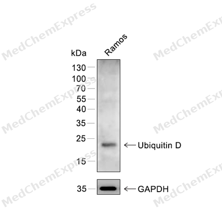 Western blot analysis of extracts from Ramos using Ubiquitin D antibody. Proteins were transferred to a PVDF membrane and blocked with 5% nonfat powdered milk in PBST for 2 hour at room temperature. The primary antibody (1/1000) and loading control antibody (GAPDH, 1/3000) was diluted with 5% nonfat powdered milk in PBST at 4°C overnight. Goat Anti-Rabbit IgG-HRP Secondary Antibody (1/8,000) was incubated for 45min at room temperature.
 -
 Immunocytochemistry analysis of Hela cells labeling Ubiquitin D with Ubiquitin D Antibody (HY-P80364) at 1/100 dilution. Cells were fixed in 4% paraformaldehyde for 15 minutes at room temperature, permeabilized with 0.1% Triton X-100 in PBS for 15 minutes at room temperature, then blocked with quick block buffer for 10 minutes at room temperature. Cells were then incubated with Ubiquitin D Antibody (HY-P80364) at 1/100 dilution in quick block buffer overnight at 4 ℃. AF488-conjugated Goat Anti-Rabbit IgG H&L(HY-P8002, Green) was used as the secondary antibody at 1/1,000 dilution. PBS instead of the primary antibody was used as the secondary antibody only control. The Nuclear counterstain was DAPI (Blue).
 -
 Immunocytochemistry analysis of Hela cells labeling Ubiquitin D with Ubiquitin D Antibody (HY-P80364) at 1/200 dilution. Cells were fixed in 4% paraformaldehyde for 15 minutes at room temperature, permeabilized with 0.1% Triton X-100 in PBS for 15 minutes at room temperature, then blocked with quick block buffer for 10 minutes at room temperature. Cells were then incubated with Ubiquitin D Antibody (HY-P80364) at 1/200 dilution in quick block buffer overnight at 4 ℃. AF488-conjugated Goat Anti-Rabbit IgG H&L(HY-P8002, Green) was used as the secondary antibody at 1/1,000 dilution. PBS instead of the primary antibody was used as the secondary antibody only control. The Nuclear counterstain was DAPI (Blue).


Background
-
Function
Ubiquitin D is an Ubiquitin-like protein modifier which can be covalently attached to target proteins and subsequently leads to their degradation by the 26S proteasome, in a NUB1-dependent manner. Conjugation to the target protein is activated by UBA6 via adenylation of its C-terminal glycine. Promotes the expression of the proteasome subunit beta type-9 (PSMB9/LMP2). Regulates TNF-alpha-induced and LPS-mediated activation of the central mediator of innate immunity NF-kappa-B by promoting TNF-alpha-mediated proteasomal degradation of ubiquitinated-I-kappa-B-alpha. Required for TNF-alpha-induced p65 nuclear translocation in renal tubular epithelial cells (RTECs). May be involved in dendritic cell (DC) maturation, the process by which immature dendritic cells differentiate into fully competent antigen-presenting cells that initiate T-cell responses. Mediates mitotic non-disjunction and chromosome instability, in long-term in vitro culture and cancers, by abbreviating mitotic phase and impairing the kinetochore localization of MAD2L1 during the prometaphase stage of the cell cycle. May be involved in the formation of aggresomes when proteasome is saturated or impaired. Mediates apoptosis in a caspase-dependent manner, especially in renal epithelium and tubular cells during renal diseases such as polycystic kidney disease and Human immunodeficiency virus (HIV)-associated nephropathy (HIVAN)[1][2][3][4][5][6][7][8][9][10].
-
Subcellular Localization
Nucleus; Cytoplasm
-
Expression
Tissue_specificity:Constitutively expressed in mature dendritic cells and B-cells. Mostly expressed in the reticuloendothelial system (e.g. thymus, spleen) , the gastrointestinal system, kidney, lung and prostate gland
Induction:Rapidly degraded by the proteasome. Cell-cycle regulation with highest expression during the S-phase (at protein level) . Induced during dendritic cell maturation. Negatively regulated by p53/TP53. High levels in various gastrointestinal and gynecological cancer cells. Induced in RTECs in common renal diseases including diabetic nephropathy (DN) , IgA nephropathy (IgAN) , and hypertensive nephrosclerosis (HN) , as well as in hepatocellular carcinoma (HCC) and during HIVAN. Inducible by the pro-inflammatory cytokines IFNG/IFN-gamma and TNF in cancers of liver and colon. Repressed by NUB1 (at protein level) -
Subunit
Interacts directly with the 26S proteasome (PubMed:16707496). Interacts with NUB1; this interaction facilitates the linking of UBD-conjugated target protein to the proteasome complex and accelerates its own degradation and that of its conjugates (PubMed:14757770, PubMed:16707496, PubMed:25422469). Interacts (via ubiquitin-like 1 domain) with the spindle checkpoint protein MAD2L1 during mitosis (PubMed:10200259, PubMed:16495226, PubMed:25422469). Present in aggresomes of proteasome inhibited cells. Interacts with HDAC6 under proteasome impairment conditions (PubMed:19033385, PubMed:25422469). Forms a thioester with UBA6 in cells stimulated with tumor necrosis factor-alpha (TNFa) and interferon-gamma (IFNg) (PubMed:17889673, PubMed:25422469). Interacts with SQSTM1 and TP53/p53 (PubMed:25422469)
-
SwissProt ID
-
Synonyms
Diubiquitin, FAT10, GABBR1, UBD, UBD 3, ubiquitin D, Ubiquitin like protein FAT10
-
Research Field
Microbiology
Documentation
-
Data Sheet (235 KB)
-
SDS (251 KB)
- English - EN (251 KB)
- Français - FR (251 KB)
- Deutsch - DE (251 KB)
- Norwegian - NO (251 KB)
- Español - ES (251 KB)
- Swedish - SV (251 KB)
- Italian - IT (251 KB)
- Korean - KR (251 KB)
- Portuguese - PT (251 KB)
-
User Guide for Antibodies (1077 KB)

 Powered by Bioz
