



ARL 17477
Based on 1 publication(s) in Google Scholar
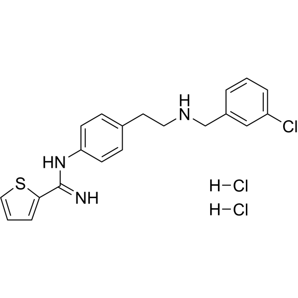
ARL 17477 is a blood-brain barrier-penetrant selective neuronal nitric oxide synthase (nNOS) inhibitor with an IC50 of 1 μM. ARL 17477 reduces nitric oxide production and activates TFEB and lysosomal biogenesis, induces lysosomal membrane permeabilization, but inhibits autophagy-lysosomal function, autophagic flux, protein aggregate clearance, and proliferation. ARL 17477 induces ERK hyperphosphorylation and slight AKT inhibition. ARL 17477 exhibits NOS1 -independent anticancer activity against multiple cancer cells. ARL 17477 inhibits xenograft tumor growth in vivo. ARL 17477 can be used in research on colon cancer, non-small cell lung cancer, osteosarcoma, pancreatic cancer, arterial occlusion, and pain.
For research use only. We do not sell to patients.
- Purity : 98.88%
- CAS No.: 866914-87-6
- Formula: C20H22Cl3N3S
- Molecular Weight:442.83
-
Storage:
4°C, sealed storage, away from moisture
* In solvent : -80°C, 6 months; -20°C, 1 month (sealed storage, away from moisture)

To place orders, for customer services and technical support, please contact: MedChemExpress USA
Tel: 609-228-6898 E-mail: [email protected] [email protected]


-
 Biological Activity
-
Chemical Information
-
Solvent & Solubility
- Protocols
- Purity & Documentation
- References
-
 Help & FAQs


-
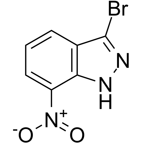
HY-101175
3-Bromo-7-nitroindazolenNOS Inhibitor -
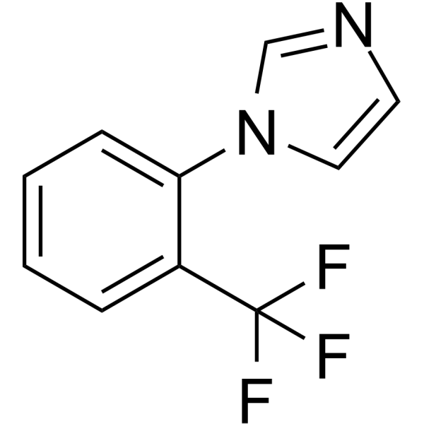
HY-101316
TRIMNOS Inhibitor -
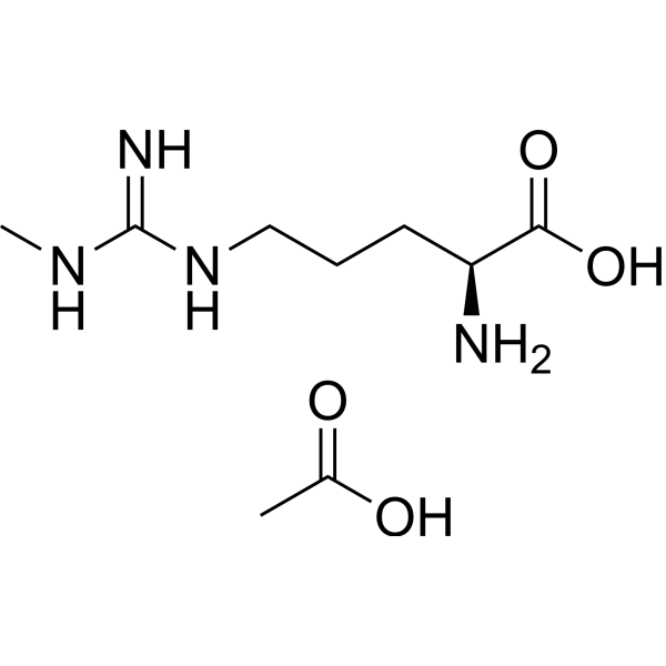
HY-18732A
L-NMMA acetateEndogenous Metabolite -
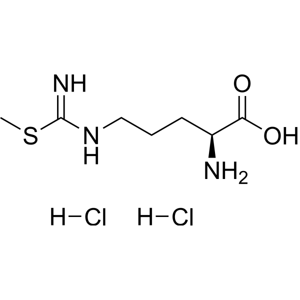
HY-U00432A
S-MTC dihydrochlorideNOS Inhibitor -
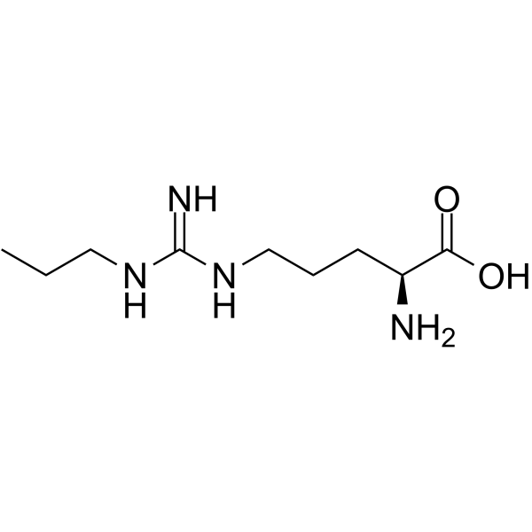
HY-102062
Nω-Propyl-L-argininenNOS Inhibitor -
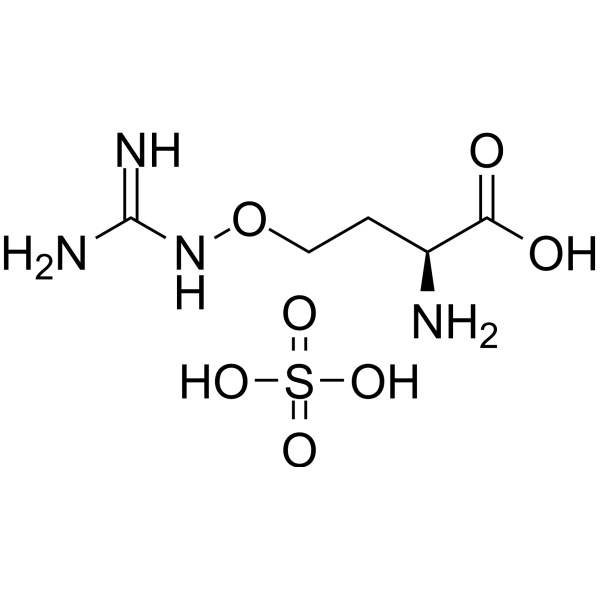
HY-B1581A
L-Canavanine sulfateNO Synthase Inhibitor -

HY-107720R
ARL 17477 (Standard)Reference Standard -
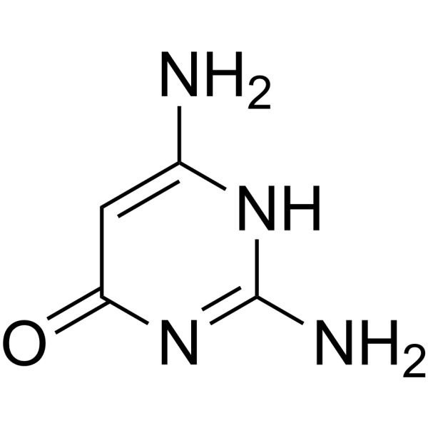
HY-100954
2,4-Diamino-6-hydroxypyrimidineGTP Cyclohydrolase I Inhibitor -
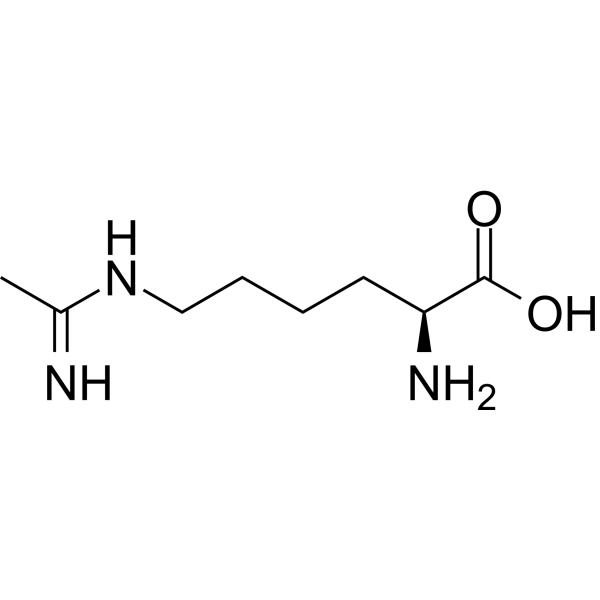
HY-12116
L-NILNO Synthase Inhibitor -
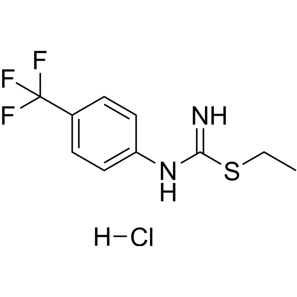
HY-W484263
hnNOS-IN-3nNOS Inhibitor -
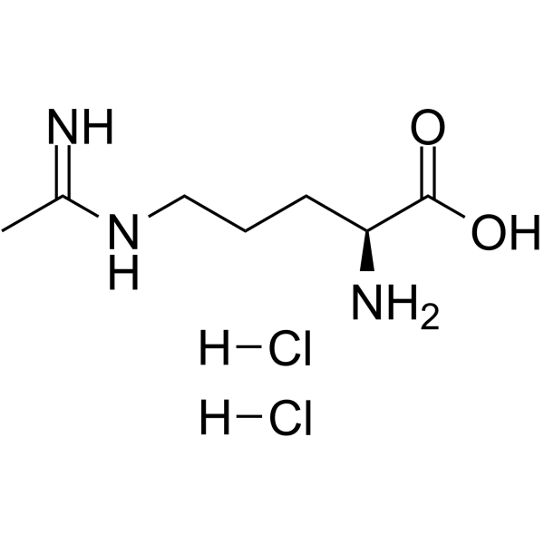
HY-100986
L-NIO dihydrochlorideNOS Inhibitor -
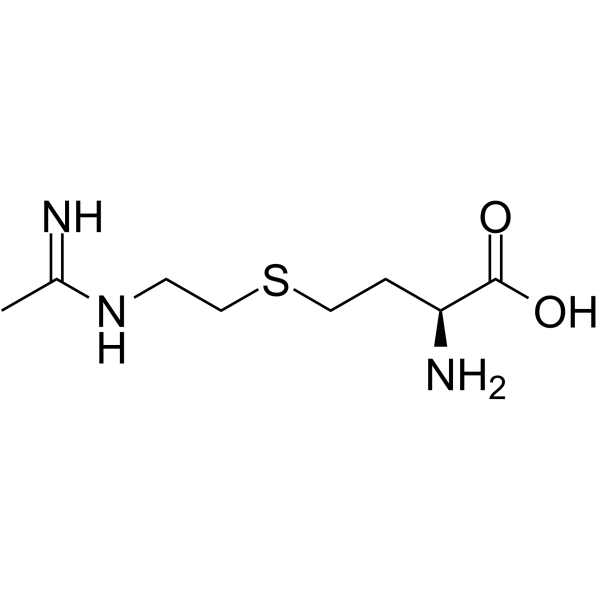
HY-12119
GW274150iNOS Inhibitor
Publications Citing Use of MedChemExpress (MCE) ARL 17477
More

Biological Activity
Description
IC50 & Target
[1]|
nNOS 1 μM (IC50) |
eNOS 17 μM (IC50) |
Cellular Effect
|
Cell Line

|
Type | Value | Description | References |
|---|---|---|---|---|
| SW48 | IC50 |
15.0 μM
|
Cytotoxicity against human colon cancer SW48 cells assessed as viability reduction incubated 2 days by Cell Counting Kit-8 assay.
Cytotoxicity against human colon cancer SW48 cells assessed as viability reduction incubated 2 days by Cell Counting Kit-8 assay.
|
37402770 |
| U-251 | IC50 |
13.0 μM
|
Cytotoxicity against human neuroblastoma U251-MG cells assessed as viability reduction incubated 2 days by Cell Counting Kit-8 assay.
Cytotoxicity against human neuroblastoma U251-MG cells assessed as viability reduction incubated 2 days by Cell Counting Kit-8 assay.
|
37402770 |
| HCT-116 | IC50 |
7.4 μM
|
Cytotoxicity against human colon cancer HCT116 cells assessed as viability reduction incubated 2 days by Cell Counting Kit-8 assay.
Cytotoxicity against human colon cancer HCT116 cells assessed as viability reduction incubated 2 days by Cell Counting Kit-8 assay.
|
37402770 |
| A549 | IC50 |
6.8 μM
|
Cytotoxicity against human lung cancer A549 cells assessed as viability reduction incubated 2 days by Cell Counting Kit-8 assay.
Cytotoxicity against human lung cancer A549 cells assessed as viability reduction incubated 2 days by Cell Counting Kit-8 assay.
|
37402770 |
| 143B | IC50 |
4.4 μM
|
Cytotoxicity against human osteosarcoma 143B cells assessed as viability reduction incubated 2 days by Cell Counting Kit-8 assay.
Cytotoxicity against human osteosarcoma 143B cells assessed as viability reduction incubated 2 days by Cell Counting Kit-8 assay.
|
37402770 |
| MIA PaCa-2 | IC50 |
4.3 μM
|
Cytotoxicity against human pancreatic cancer MIA PaCa-2 cells assessed as viability reduction incubated 2 days by Cell Counting Kit-8 assay.
Cytotoxicity against human pancreatic cancer MIA PaCa-2 cells assessed as viability reduction incubated 2 days by Cell Counting Kit-8 assay.
|
37402770 |
In Vitro
ARL 17477 (2 days) inhibits the viability of SW48, U251-MG, HCT116, A549, 143B, MIA PaCa-2, and NOS1-/- U251-MG cells, with IC50 values of 4.3-15.0 µM in the parental cell lines[2].
ARL 17477 (7 days) inhibits TGS-01 glioblastoma CSC tumorsphere formation with an IC50 of 4.4 µM[2].
ARL 17477 (2 days) inhibits 143B-sph osteosarcoma CSC viability with an IC50 of 1.1 µM[2].
ARL 17477 (5-25 µM; 1 day) increases LC3B-II and p62, hyperphosphorylates ERK, and decreases full-length PARP without inducing caspase-3/PARP cleavage in U251-MG cells[2].
ARL 17477 (25 µM; 48 h) activates lysosomal/TFEB and lipid synthesis gene programs in Panc-1 cells and suppresses the proliferation gene set[2].
ARL 17477-induced intracellular accumulation of autophagy markers depends on impaired lysosomal acidification[2].
ARL 17477 (1 µM; 30 min) inhibits lysosomal staining in MIA PaCa-2 cells[2].
ARL 17477 (6 days) impairs protein aggregate clearance and increases aggregates in MIA PaCa-2 cells[2].
ARL 17477 (100 µM; 1 h) induces lysosomal membrane permeabilization and increases cytosolic delivery in MIA PaCa-2 NanoBiT cells[2].
ARL 17477 (60 min) is a selective inhibitor of recombinant human nNOS in the [3H]arginine to [3H]citrulline conversion assay (IC50 = 1 μM), with weaker selectivity for eNOS (IC50 = 17 μM)[1].
ARL 17477 inhibits nNOS and endothelial NOS[3].
MedChemExpress (MCE) has not independently confirmed the accuracy of these methods. They are for reference only.
-
Cell Line:U251-MG neuroblastoma cells
-
Concentration:5, 10, 25 µM
-
Incubation Time:1 day; 6 h to 6 days (LC3B-II/p62)
-
Result:Induced hyperphosphorylation of ERK with p-ERK/ERK band intensities of 1.0, 1.0, 1.3, and 4.1 at 0, 5, 10, and 25 µM.
Slightly suppressed AKT phosphorylation with p-AKT/AKT band intensities of 1.0, 1.0, 0.8, and 0.7.
Reduced full-length PARP.
Did not induce caspase-3 or PARP cleavage.
Increased p62 and LC3B-II protein levels.
In Vivo
ARL 17477 (10 mg/kg; i.p.; once daily; days 5-14 after stroke) improves post-stroke functional recovery on the rotarod task in wild-type C57Bl6 male mice with transient middle cerebral artery occlusion without altering infarct volume[3].
ARL 17477 (0.4 mg/mL (0.9 mM); intraplantar injection; two prophylactic injections 15-20 minutes apart) does not prevent 7α,25-OHC (HY-113962)-induced mechanical allodynia in Sprague Dawley rats, but accelerates recovery to baseline within 96 hours[4].
MedChemExpress (MCE) has not independently confirmed the accuracy of these methods. They are for reference only.
-
Animal Model:BALB/c-nu/nu (female, 6 to 8-week-old)[2]
-
Dosage:10 mg/kg
-
Administration:i.v. bolus via tail vein; daily; Injections started on day 14 after vaccination and ended on day 27
-
Result:Significantly delayed tumor growth of MIA PaCa-2 xenografts compared with vehicle control.
-
Animal Model:C57Bl6 (wild-type; male; 10-12 weeks old)[3]
-
Dosage:10 mg/kg
-
Administration:i.p.; once daily each morning; post-stroke days 5-14
-
Result:At post-stroke day 10, this compound-treated stroke mice exhibited functional improvement in rotating beam speed compared with vehicle-treated stroke mice.
Infarct area at the striatal and hippocampal levels and total infarct area did not differ between vehicle- and this compound-treated mice.
-
Animal Model:Sprague Dawley (male and female, 200-250 g starting weight)[4]
-
Dosage:0.4 mg/mL (0.9 mM) in saline; 50 µL
-
Administration:intraplantar; two prophylactic injections 15-20 minutes apart; first injection 15-20 minutes before 7α,25-OHC
-
Result:Did not attenuate 7α,25-OHC-induced mechano-allodynia.
It began to accelerate recovery after 4 hours, with complete return to baseline by 96 hours.
- Autophagy
- Protocol for Pharmacokinetic Study
- Patient-Derived Xenograft (PDX)
- Patient-Derived Orthotopic Xenograft (PDOX)
- Splenic/Portal-Vein Liver Metastasis Xenograft
- Lysosome and acidic-vesicle live-cell staining
- Subcutaneous Cell-Line-Derived Xenograft
- Macroautophagy Solutions
- Orthotopic Cell-Line Xenograft
- Cell Viability Determination by MTT Colorimetric Assay
- Intraperitoneal/Peritoneal Dissemination Xenograft


Chemical Information
-
CAS No. 866914-87-6
-
Appearance Solid
-
Molecular Weight 442.83
-
Formula C20H22Cl3N3S
-
Color Off-white to light yellow
-
SMILES
[H]Cl.[H]Cl.N=C(C1=CC=CS1)NC2=CC=C(CCNCC3=CC(Cl)=CC=C3)C=C2
-
Shipping
Room temperature in continental US; may vary elsewhere.
-
Storage
4°C, sealed storage, away from moisture
* In solvent : -80°C, 6 months; -20°C, 1 month (sealed storage, away from moisture)


Publications (1)
-
Journal Impact Factor
-
Most Recent
-
bioRxiv
Targeting GPR183 to reduce peripheral sensitization: evidence from rodent and human tissue analyses. [Abstract]2026 Jan 16:2026.01.15.699745. PMID: 41648147


Solvent & Solubility
In Vitro:
DMSO : 62.5 mg/mL (141.14 mM; Need ultrasonic; Hygroscopic DMSO has a significant impact on the solubility of product, please use newly opened DMSO)
H2O : 20 mg/mL (45.16 mM; Need ultrasonic)
Please refer to the solubility information to select the appropriate solvent. Once prepared, please aliquot and store the solution to prevent product inactivation from repeated freeze-thaw cycles.
Storage method and period of stock solution: -80°C, 6 months; -20°C, 1 month (sealed storage, away from moisture). When stored at -80°C, please use it within 6 months. When stored at -20°C, please use it within 1 month.
* Note: If you choose water as the stock solution, please dilute it to the working solution, then filter and sterilize it with a 0.22 μm filter before use.
Please refer to the solubility information to select the appropriate solvent. Once prepared, please aliquot and store the solution to prevent product inactivation from repeated freeze-thaw cycles.
Storage method and period of stock solution: -80°C, 6 months; -20°C, 1 month (sealed storage, away from moisture). When stored at -80°C, please use it within 6 months. When stored at -20°C, please use it within 1 month.
* Note: If you choose water as the stock solution, please dilute it to the working solution, then filter and sterilize it with a 0.22 μm filter before use.
Concentration (start) × Volume (start) = Concentration (final) × Volume (final)


Purity & Documentation
-
Data Sheet (290 KB)
-
SDS (393 KB)
- English - EN (393 KB)
- Français - FR (393 KB)
- Deutsch - DE (393 KB)
- Norwegian - NO (393 KB)
- Español - ES (393 KB)
- Swedish - SV (393 KB)
- Italian - IT (393 KB)
- Korean - KR (393 KB)
- Portuguese - PT (393 KB)
-
Handling Instructions (2659 KB)
References
[2]. Nomura TK, et al. ARL-17477 is a dual inhibitor of NOS1 and the autophagic-lysosomal system that prevents tumor growth in vitro and in vivo. Scientific reports. 2023 Jul 04;13(1):10757. [Content Brief]
[4]. Oberkrom K, et al. Targeting GPR183 to reduce peripheral sensitization: evidence from rodent and human tissue analyses. bioRxiv. 2026 Jan 16:2026-01. [Content Brief]


Complete Stock Solution Preparation Table
Please refer to the solubility information to select the appropriate solvent. Once prepared, please aliquot and store the solution to prevent product inactivation from repeated freeze-thaw cycles.
Storage method and period of stock solution: -80°C, 6 months; -20°C, 1 month (sealed storage, away from moisture). When stored at -80°C, please use it within 6 months. When stored at -20°C, please use it within 1 month.
| Optional Solvent | Concentration Solvent Mass | 1 mg | 5 mg | 10 mg | 25 mg |
|---|---|---|---|---|---|
| H2O / DMSO | 1 mM | 2.2582 mL | 11.2910 mL | 22.5820 mL | 56.4551 mL |
| 5 mM | 0.4516 mL | 2.2582 mL | 4.5164 mL | 11.2910 mL | |
| 10 mM | 0.2258 mL | 1.1291 mL | 2.2582 mL | 5.6455 mL | |
| 15 mM | 0.1505 mL | 0.7527 mL | 1.5055 mL | 3.7637 mL | |
| 20 mM | 0.1129 mL | 0.5646 mL | 1.1291 mL | 2.8228 mL | |
| 25 mM | 0.0903 mL | 0.4516 mL | 0.9033 mL | 2.2582 mL | |
| 30 mM | 0.0753 mL | 0.3764 mL | 0.7527 mL | 1.8818 mL | |
| 40 mM | 0.0565 mL | 0.2823 mL | 0.5646 mL | 1.4114 mL | |
| DMSO | 50 mM | 0.0452 mL | 0.2258 mL | 0.4516 mL | 1.1291 mL |
| 60 mM | 0.0376 mL | 0.1882 mL | 0.3764 mL | 0.9409 mL | |
| 80 mM | 0.0282 mL | 0.1411 mL | 0.2823 mL | 0.7057 mL | |
| 100 mM | 0.0226 mL | 0.1129 mL | 0.2258 mL | 0.5646 mL |
* Note: If you choose water as the stock solution, please dilute it to the working solution, then filter and sterilize it with a 0.22 μm filter before use.


ARL 17477 Related Classifications
HY-107720 Related Classifications
 Powered by Bioz
Keywords
- ARL 17477
- 866914-87-6
- ARL17477
- ARL-17477
- NO Synthase
- Autophagy
- PERK
- ERK hyperphosphorylation
- TFEB
- autophagic flux
- autophagy-lysosomal function
- blood-brain barrier
- endothelial nitric oxide synthase
- lysosomal biogenesis
- lysosomal membrane permeabilization
- neuronal nitric oxide synthase
- nitric oxide
- Inhibitor
- inhibitor
- inhibit