



APcK110
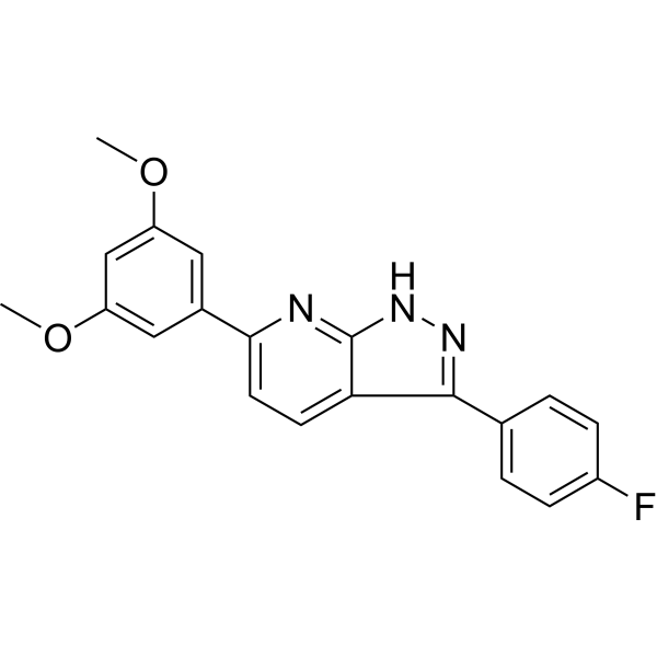
APcK110 is a potent Kit inhibitor that can be used for the research of acute myeloid leukemia (AML). APcK110 induces AML cell apoptosis.
Nur für Forschungszwecke. Wir verkaufen nicht an Patienten.
- CAS. Nr.: 1001083-74-4
- Formel: C20H16FN3O2
- Molecular Weight:349.36
-
Speicherung:
Please store the product under the recommended conditions in the Certificate of Analysis.

To place orders, for customer services and technical support, please contact: MedChemExpress USA
Tel: 609-228-6898 E-mail: [email protected] [email protected]


-
 Biologische Aktivität
-
Chemical Information
- Protokoll
- Reinheit & Dokumentation
- Verweise
-
 Help & FAQs




Biologische Aktivität
Beschreibung
In Vitro
APcK110 (0-500 nM; 0-72 h) inhibits AML cell viability and proliferation. APcK110 preferentially inhibits KIT mutated BaF3 cell lines[1].
APcK110 (0-500 nM; 30 min) inhibits the phosphorylation of Kit, Stat3, Stat5, and Akt[1].
APcK110 (500 nM; overnight) induces caspase-dependent apoptosis in OCI/AML3 cells[1].
APcK110 (50-500 nM; 7 days) inhibits AML blast colony-forming cell proliferation[1].
MedChemExpress (MCE) has not independently confirmed the accuracy of these methods. They are for reference only.
-
Cell Line:OCI/AML3, HMC1.2, BaF3 and mutant KIT-expressing BaF3 cells
-
Concentration:0-500 nM
-
Incubation Time:0, 24, 48 and 72 h
-
Result:Inhibited proliferation in a dose-dependent manner.
-
Cell Line:OCI/AML3 and HMC1.2
-
Concentration:0, 50, 100, 250 and 500 nM
-
Incubation Time:30 min
-
Result:Showed a dose-dependent inhibition of the phosphorylation of Kit, Stat3, Stat5, and Akt, a downstream effector of phosphatidylinositol 3-kinase.
-
Cell Line:OCI/AML3 cells
-
Concentration:500 nM
-
Incubation Time:Overnight
-
Result:Induced apoptosis by activation of the caspase pathway.
-
Cell Line:OCI/AML3 cells
-
Concentration:500 nM
-
Incubation Time:2 h
-
Result:Showed a shift of cells into sub-G0 following a 2 h incubation.
In Vivo
MedChemExpress (MCE) has not independently confirmed the accuracy of these methods. They are for reference only.
-
Animal Model:Eight-weeks-old female NODSCID mice, OCI/AML3 xenograft mouse model[2]
-
Dosage:500 nM
-
Administration:Intraperitoneal injection, every other day for 60 days
-
Result:Survival of compound-treated mice was significantly longer compared with mice injected with PBS.


Chemical Information
-
CAS. Nr. 1001083-74-4
-
Molecular Weight 349.36
-
Formel C20H16FN3O2
-
SMILES
FC1=CC=C(C2=NNC3=NC(C4=CC(OC)=CC(OC)=C4)=CC=C23)C=C1
-
Versand
Room temperature in continental US; may vary elsewhere.
-
Speicherung
Please store the product under the recommended conditions in the Certificate of Analysis.


Reinheit & Dokumentation
Verweise
[1]. Faderl S, et al. Kit inhibitor APcK110 induces apoptosis and inhibits proliferation of acute myeloid leukemia cells. Cancer Res. 2009 May 1;69(9):3910-7. [Content Brief]
[2]. Faderl S, et al. Kit inhibitor APcK110 extends survival in an AML xenograft mouse model. Invest New Drugs. 2011 Oct;29(5):1094-7. [Content Brief]

Calculators
Konzentration (Stammlösung) × Volumen (Stammlösung) = Konzentration (Ziellösung) × Volumen (Ziellösung)


APcK110 Related Classifications
HY-110071 Related Classifications
 Powered by Bioz