



Gracillin
Based on 2 publication(s) in Google Scholar
Gracillin is a steroidal saponin that can be extracted from the roots of the plant and has anti-tumor properties. Gracillin can induce cancer cell apoptosis and autophagy.
For research use only. We do not sell to patients.
- Purity : 99.91%
- CAS No.: 19083-00-2
- Formula: C45H72O17
- Molecular Weight:885.04
-
Storage:
4°C, protect from light
* In solvent : -80°C, 6 months; -20°C, 1 month (protect from light)

To place orders, for customer services and technical support, please contact: MedChemExpress USA
Tel: 609-228-6898 E-mail: [email protected] [email protected]


-
 Biological Activity
-
Chemical Information
-
Solvent & Solubility
- Protocols
- Purity & Documentation
- References
-
 Help & FAQs


-
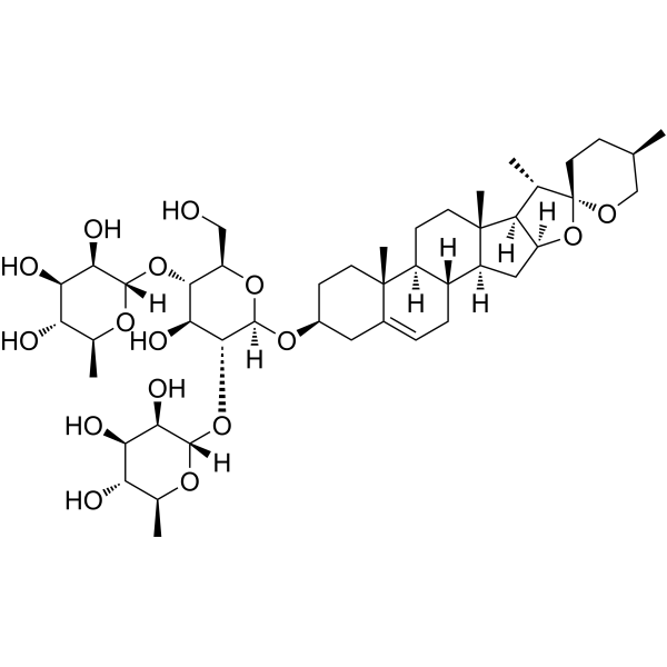
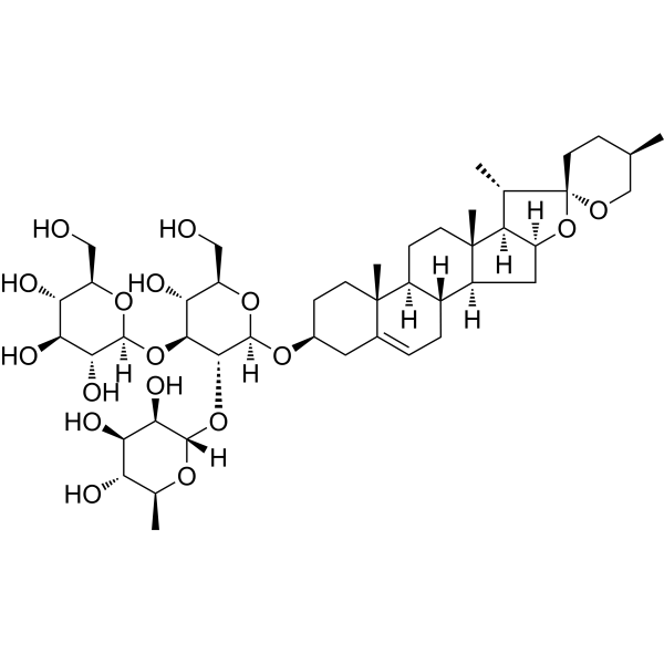
HY-N0124
DioscinAnticancer Agent -
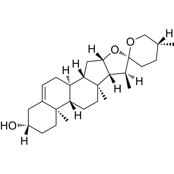
HY-N0177
DiosgeninSTAT Inhibitor -
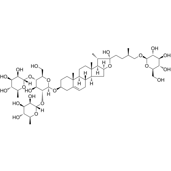
HY-N0799
ProtodioscinEndogenous Metabolite -
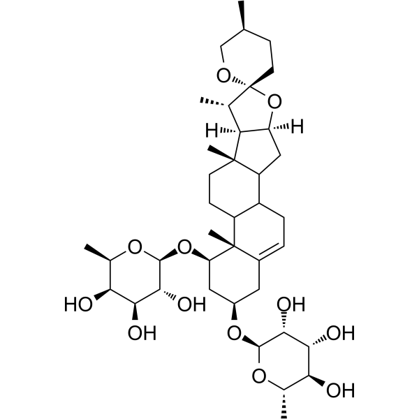
HY-N5135
Liriopesides BApoptosis Inducer -
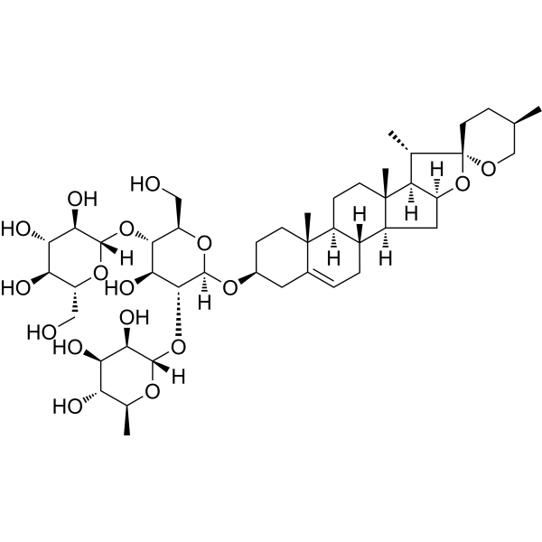
HY-N2283
DeltoninEndogenous Metabolite -
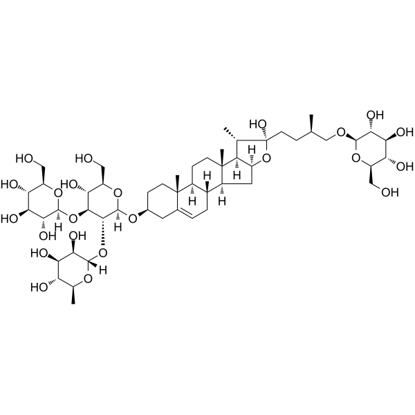
HY-N4271
ProtogracillinAntithrombotic Agent -

HY-N0706R
Gracillin (Standard)Anticancer Agent -
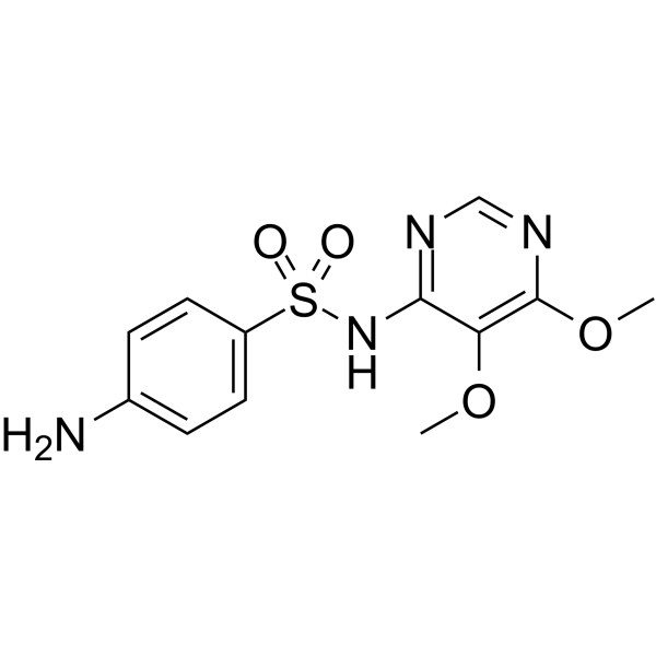
HY-B0439
SulfadoxineAntiparasitic Agent -
HY-B0439R
Sulfadoxine (Standard)Antiparasitic Agent -

HY-N0799R
Protodioscin (Standard)Endogenous Metabolite
Publications Citing Use of MedChemExpress (MCE) Gracillin
More Customer Validation & Images

-
Cell Proliferation/Viability Assay
-
WB
-
Apoptosis Analysis
-
Histological Imaging/Staining
-
In Vivo Efficacy Study


Biological Activity
Description
Cellular Effect
|
Cell Line

|
Type | Value | Description | References |
|---|---|---|---|---|
| A2780 | IC50 |
4.3 μM
Compound: Gracillin
|
Cytotoxicity against human A2780 cells after 72 hrs by MTT assay
Cytotoxicity against human A2780 cells after 72 hrs by MTT assay
|
[PMID: 19362474] |
| A549 | IC50 |
0.96 μM
Compound: Gracillin
|
Cytotoxicity against human A549 cells after 72 hrs by MTT assay
Cytotoxicity against human A549 cells after 72 hrs by MTT assay
|
[PMID: 19362474] |
| Bel-7402 | IC50 |
1.1 μM
Compound: Gracillin
|
Cytotoxicity against human Bel7402 cells after 72 hrs by MTT assay
Cytotoxicity against human Bel7402 cells after 72 hrs by MTT assay
|
[PMID: 19362474] |
| BXPC-3 | IC50 |
1.3 μM
Compound: Gracillin
|
Cytotoxicity against human BxPC3 cells after 72 hrs by MTT assay
Cytotoxicity against human BxPC3 cells after 72 hrs by MTT assay
|
[PMID: 19362474] |
| Capan-2 | IC50 |
1.3 μM
Compound: Gracillin
|
Cytotoxicity against human Capan2 cells after 72 hrs by MTT assay
Cytotoxicity against human Capan2 cells after 72 hrs by MTT assay
|
[PMID: 19362474] |
| HCT-8 | IC50 |
0.92 μM
Compound: Gracillin
|
Cytotoxicity against human HCT8 cells after 72 hrs by MTT assay
Cytotoxicity against human HCT8 cells after 72 hrs by MTT assay
|
[PMID: 19362474] |
| HeLa | IC50 |
12.74 μg/mL
Compound: 8
|
Cytotoxicity against human HeLa cells after 72 hrs by MTT assay
Cytotoxicity against human HeLa cells after 72 hrs by MTT assay
|
[PMID: 15620239] |
| HL-60 | IC50 |
3.7 μM
Compound: 9
|
Cytotoxicity against human HL60 cells after 72 hrs by MTT assay
Cytotoxicity against human HL60 cells after 72 hrs by MTT assay
|
[PMID: 12828464] |
| MCF7 | IC50 |
17.5 μM
Compound: Gracillin
|
Cytotoxicity against human MCF7 cells after 72 hrs by MTT assay
Cytotoxicity against human MCF7 cells after 72 hrs by MTT assay
|
[PMID: 19362474] |
| PANC-1 | IC50 |
0.7 μM
Compound: Gracillin
|
Cytotoxicity against human PANC1 cells after 72 hrs by MTT assay
Cytotoxicity against human PANC1 cells after 72 hrs by MTT assay
|
[PMID: 19362474] |
| SW1990 | IC50 |
1.1 μM
Compound: Gracillin
|
Cytotoxicity against human SW1990 cells after 72 hrs by MTT assay
Cytotoxicity against human SW1990 cells after 72 hrs by MTT assay
|
[PMID: 19362474] |
In Vitro
Gracillin (0-10 μM, 6-24 h) shows broad-spectrum inhibitory effects on the cancer cell viability[1].
Gracillin (5 μM) disrupts mitochondrial function, specifically, inhibits ATP production, activates AMPK, inhibits oxygen consumption rate (OCR) in H460 and A549 cells[1].
Gracillin (0-10 μM, 6 h) induces ROS generation (indicated by H2DCF-DA) in H460, H1299, H226B and A549 cells[1].
Gracillin (0-4 μM, 24 h) blocks cell cycle in G1 phase, and induces apoptosis and autophagy in A549 cells[2][3].
MedChemExpress (MCE) has not independently confirmed the accuracy of these methods. They are for reference only.
-
Cell Line:A549 cells
-
Concentration:0-4 μM
-
Incubation Time:24 h
-
Result:Induced morphological changes.
Apoptosis rate: 6.56%-67.43%.
In Vivo
MedChemExpress (MCE) has not independently confirmed the accuracy of these methods. They are for reference only.


Chemical Information
-
CAS No. 19083-00-2
-
Appearance Solid
-
Molecular Weight 885.04
-
Formula C45H72O17
-
Color White to light yellow
-
SMILES
C[C@@]12[C@]([C@@H]3C)([H])[C@](O[C@]34CC[C@@H](C)CO4)([H])C[C@@]1([H])[C@@](CC=C5[C@@]6(CC[C@H](O[C@@](O[C@H](CO)[C@@H](O)[C@@H]7O[C@]([C@@H]([C@@H](O)[C@@H]8O)O)([H])O[C@@H]8CO)([H])[C@@H]7O[C@@](O[C@@H](C)[C@H](O)[C@H]9O)([H])[C@@H]9O)C5)C)([H])[C@]6([H])CC2
-
Structure Classification
-
Initial Source
-
Shipping
Room temperature in continental US; may vary elsewhere.
-
Storage
4°C, protect from light
* In solvent : -80°C, 6 months; -20°C, 1 month (protect from light)


Publications (2)
-
Journal Impact Factor
-
Most Recent
-
Bioorg Chem
Gracillin induces mitochondria-mediated apoptosis on pancreatic ductal adenocarcinoma through disruption of redox homeostasis via inhibiting NRF2/HO-1 antioxidant axis. [Abstract]2025 Aug:163:108636. PMID: 40505322
Gracillin purchased from MedChemExpress. Usage Cited in: Bioorg Chem. 2025 Aug:163:108636. [Abstract]
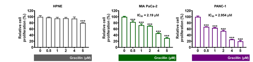
Cell viability was analyzed in human pancreatic duct (HPNE) and PDAC cells in response to Gracillin. Up to 4 μM, Gracillin did not exhibit cytotoxicity in HPNE cells, while significantly inhibiting PDAC cell viability.
Gracillin purchased from MedChemExpress. Usage Cited in: Bioorg Chem. 2025 Aug:163:108636. [Abstract]
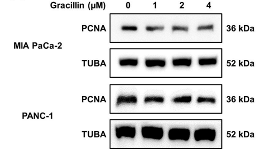
Immunoblotting analysis confirmed reduced protein expression of PCNA in PDAC cells following Gracillin (0, 1, 2, 4 μM) treatment.
Gracillin purchased from MedChemExpress. Usage Cited in: Bioorg Chem. 2025 Aug:163:108636. [Abstract]
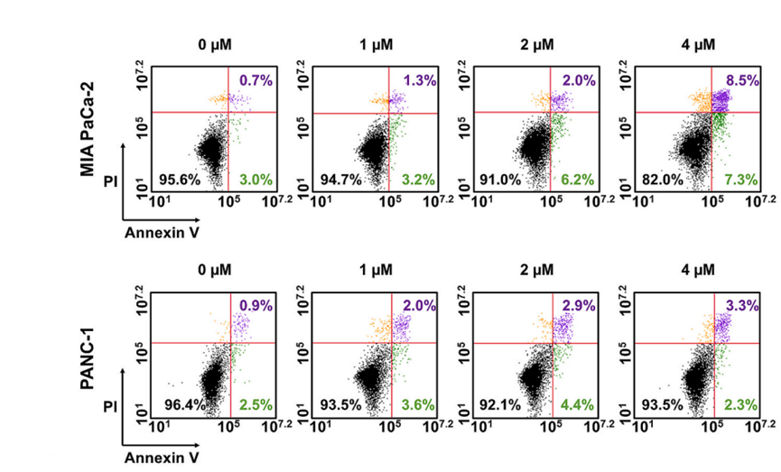
The apoptosis-inducing effects of Gracillin (0, 1, 2, 4 μM) were confirmed by Annexin V/PI staining in PDAC cells.
Gracillin purchased from MedChemExpress. Usage Cited in: Bioorg Chem. 2025 Aug:163:108636. [Abstract]
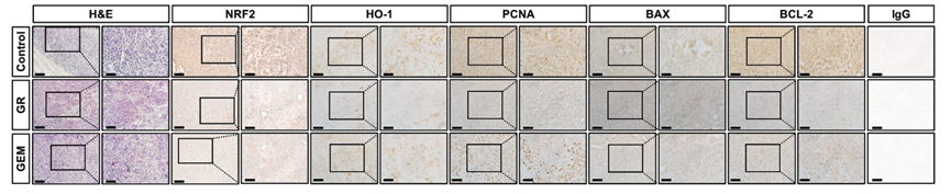
H&E and IHC performed on PANC-1 tumors demonstrating reduced NRF2, HO-1, PCNA, and BCL-2 expression and increased BAX expression following Gracillin treatment.
Gracillin purchased from MedChemExpress. Usage Cited in: Bioorg Chem. 2025 Aug:163:108636. [Abstract]
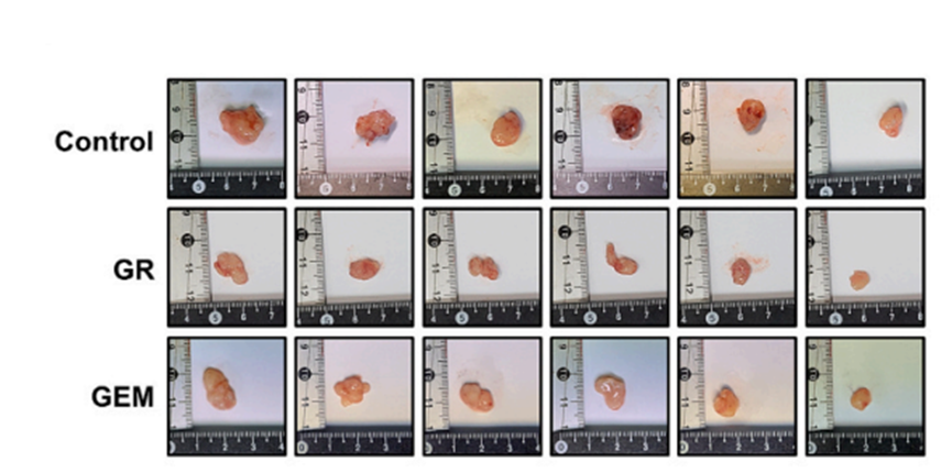
Tumor photograph showing the effects of vehicle, Gracillin (8 mg/kg, i.p.) and Gemcitabine treatments.
-
Immunopharmacol Immunotoxicol
Capillin protects against non-alcoholic steatohepatitis through suppressing NLRP3 inflammasome activation and oxidative stress. [Abstract]2021 Dec;43(6):778-789. PMID: 34618611


Solvent & Solubility
In Vitro:
DMSO : 100 mg/mL (112.99 mM; Need ultrasonic; Hygroscopic DMSO has a significant impact on the solubility of product, please use newly opened DMSO)
Please refer to the solubility information to select the appropriate solvent. Once prepared, please aliquot and store the solution to prevent product inactivation from repeated freeze-thaw cycles.
Storage method and period of stock solution: -80°C, 6 months; -20°C, 1 month (protect from light). When stored at -80°C, please use it within 6 months. When stored at -20°C, please use it within 1 month.
Please refer to the solubility information to select the appropriate solvent. Once prepared, please aliquot and store the solution to prevent product inactivation from repeated freeze-thaw cycles.
Storage method and period of stock solution: -80°C, 6 months; -20°C, 1 month (protect from light). When stored at -80°C, please use it within 6 months. When stored at -20°C, please use it within 1 month.
Concentration (start) × Volume (start) = Concentration (final) × Volume (final)
In Vivo:
Select the appropriate dissolution method based on your experimental animal and administration route.
- For the following dissolution methods, please ensure to first prepare a clear stock solution using an In Vitro approach and then sequentially add co-solvents:
- To ensure reliable experimental results, the clarified stock solution can be appropriately stored based on storage conditions. As for the working solution for In Vivo experiments, it is recommended to prepare freshly and use it on the same day.
- The percentages shown for the solvents indicate their volumetric ratio in the final prepared solution. If precipitation or phase separation occurs during preparation, heat and/or sonication can be used to aid dissolution.
In Vivo Dissolution Calculator
Please enter the basic information of animal experiments:
-
-
-
-
Recommended: Prepare an additional quantity of animals to account for potential losses during experiments.
Please enter your animal formula composition:
-
%DMSO +
Recommended: Keep the proportion of DMSO in working solution below 2% if your animal is weak.
-
%+
-
+%Tween-80 + +
-
%Saline +
The co-solvents required include: DMSO, . All of co-solvents are available by MedChemExpress (MCE). , Tween 80. All of co-solvents are available by MedChemExpress (MCE).
Working solution concentration: 0.22 mg/mL
Method for preparing stock solution: mg drug dissolved in μL DMSO. Stock solution concentration: mg/mL. * In solvent : -80°C, 6 months; -20°C, 1 month (protect from light)
1. Take μL DMSO stock solution;
2. Add μL .
μL , mix evenly;
3. Then add μL Tween 80, mix evenly;
4. Then add μL
Please ensure that the stock solution in the first step is dissolved to a clear state, and add co-solvents in sequence. You can use ultrasonic heating (ultrasonic cleaner, recommended frequency 20-40 kHz), vortexing, etc. to assist dissolution.


Purity & Documentation
-
Data Sheet (291 KB)
-
SDS (394 KB)
- English - EN (394 KB)
- Français - FR (394 KB)
- Deutsch - DE (394 KB)
- Norwegian - NO (394 KB)
- Español - ES (394 KB)
- Swedish - SV (394 KB)
- Italian - IT (394 KB)
- Korean - KR (394 KB)
- Portuguese - PT (394 KB)
-
Handling Instructions (2659 KB)
References
[1]. Min HY, et al. The natural compound gracillin exerts potent antitumor activity by targeting mitochondrial complex II. Cell Death Dis. 2019 Oct 24;10(11):810. [Content Brief]
[2]. Yang J, et al. Gracillin Isolated from Reineckia carnea Induces Apoptosis of A549 Cells via the Mitochondrial Pathway. Drug Des Devel Ther. 2021 Jan 20;15:233-243. [Content Brief]
[3]. Li Y, et al. Gracillin Shows Potential Efficacy Against Non-Small Cell Lung Cancer Through Inhibiting the mTOR Pathway. Front Oncol. 2022 Mar 22;12:851300. [Content Brief]


Complete Stock Solution Preparation Table
Please refer to the solubility information to select the appropriate solvent. Once prepared, please aliquot and store the solution to prevent product inactivation from repeated freeze-thaw cycles.
Storage method and period of stock solution: -80°C, 6 months; -20°C, 1 month (protect from light). When stored at -80°C, please use it within 6 months. When stored at -20°C, please use it within 1 month.
| Optional Solvent | Concentration Solvent Mass | 1 mg | 5 mg | 10 mg | 25 mg |
|---|---|---|---|---|---|
| DMSO | 1 mM | 1.1299 mL | 5.6495 mL | 11.2989 mL | 28.2473 mL |
| 5 mM | 0.2260 mL | 1.1299 mL | 2.2598 mL | 5.6495 mL | |
| 10 mM | 0.1130 mL | 0.5649 mL | 1.1299 mL | 2.8247 mL | |
| 15 mM | 0.0753 mL | 0.3766 mL | 0.7533 mL | 1.8832 mL | |
| 20 mM | 0.0565 mL | 0.2825 mL | 0.5649 mL | 1.4124 mL | |
| 25 mM | 0.0452 mL | 0.2260 mL | 0.4520 mL | 1.1299 mL | |
| 30 mM | 0.0377 mL | 0.1883 mL | 0.3766 mL | 0.9416 mL | |
| 40 mM | 0.0282 mL | 0.1412 mL | 0.2825 mL | 0.7062 mL | |
| 50 mM | 0.0226 mL | 0.1130 mL | 0.2260 mL | 0.5649 mL | |
| 60 mM | 0.0188 mL | 0.0942 mL | 0.1883 mL | 0.4708 mL | |
| 80 mM | 0.0141 mL | 0.0706 mL | 0.1412 mL | 0.3531 mL | |
| 100 mM | 0.0113 mL | 0.0565 mL | 0.1130 mL | 0.2825 mL |


Gracillin Related Classifications
HY-N0706 Related Classifications
 Powered by Bioz