



Tepoditamab

Based on 1 Customer Validation
Tepoditamab (MCLA-117) is a full-length human IgG1 bispecific monoclonal antibody that binds to CLEC12A of myeloid cells and CD3 of cytotoxic T cells. Among others, CLEC12A is a myeloid differentiation antigen. Tepoditamab kills AML leukaemia mother cells and AML leukaemia stem cells, induces T cell-mediated proliferative lysis of AML cells. Tepoditamab induces upto 30-fold T-cell expansion. Tepoditamab results in moderate to strong cytokine (IFNγ, IL-6, IL-8, IL-10, and TNFα) and IFNγ release in human whole blood and PBMC, respectively. Tepoditamab can be used in acute myeloid leukaemia (AML) research.
For research use only. We do not sell to patients.
- Purity : ≥99%
- CAS No.: 2044679-53-8
- Molecular Weight:144.787 kDa
-
Storage:
Please store the product under the recommended conditions in the Certificate of Analysis.


To place orders, for customer services and technical support, please contact: MedChemExpress USA
Tel: 609-228-6898 E-mail: [email protected] [email protected]


-
 Biological Activity
-
Chemical Information
- Protocols
- Purity & Documentation
- References
-
 Help & FAQs








Biological Activity
Description
Isotype
Human IgG1 kappa
Recommend Isotype Controls
Species Reactivity
Human
IC50 & Target
CLEC12A & CD3E
In Vitro
Tepoditamab binds specifically to CLEC12APOS and CD3POS cells within the normal haematopoietic compartment[2].
Tepoditamab (1,000 ng/mL, 24-48 h) induces upregulation of CD69 and CD25 on CD4 and CD8 T cells after 24 and 48 h in HL60 cells concentration-dependently[2].
Tepoditamab (0.1 ng/mL-10 μg/mL, 48 h) induces a concentration-dependent T cell-mediated lysis of HL60 cells at 100 ng/mL and above, as well as a mean EC50 of 68 ng/mL[2].
MedChemExpress (MCE) has not independently confirmed the accuracy of these methods. They are for reference only. Further protocols information, click here.
In Vivo
MedChemExpress (MCE) has not independently confirmed the accuracy of these methods. They are for reference only.
Gene ID
Accession
Conjugated
Unconjugated
Reconsititution
The product can be reconstituted/diluted with sterile PBS or saline.
Format
-
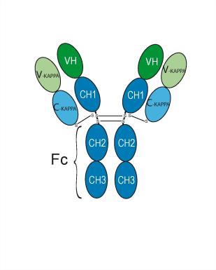
Human IgG1 kappa
Application
ELISA, FACS, Functional assay
Verified Bioactivity
-
 Loaded Tepoditamab on AHC2 biosensor, can bind CLEC12A/MICL Protein, Human (Biotinylated, HEK293, His-Avi, HY-P77633) with an affinity constant of 1.185E-08 M as determined in BLI assay.
 -
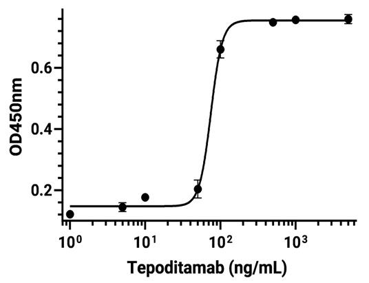 Immobilized CD3 epsilon Protein, Human (HEK293, His, HY-P70506) can bind Tepoditamab. The EC50 for this effect is 74.47 ng/mL.
 -
 Immobilized CLEC12A/MICL Protein, Human (HEK293, Fc, HY-P77631) can bind Tepoditamab. The EC50 for this effect is 67.71 ng/mL.
 -
 Flow cytometric analysis of 1×10⁶ Jurkat cells labelling CD3E (red) with Tepoditamab (HY-P99390). Cells were fixed with 4% paraformaldehyde and permeabilised with 90% methanol. Then stained with the primary antibody at 1/200 dilution for an hour at 4℃. Alexa Goat Anti-Human IgG H&L (AF488) (HY-P83776) was used as the secondary antibody at 1/1,000 dilution for 30 minutes at 4℃. Human IgG1 kappa Isotype Control (HY-P99001, blue) was used as the isotype control.


Chemical Information
-
CAS No. 2044679-53-8
-
Appearance Liquid
-
Molecular Weight 144.787 kDa
-
Color Colorless to light yellow
-
SMILES
[Tepoditamab]
-
Synonyms
MCLA 117
-
Shipping
Shipping with dry ice.
-
Formulation
Please refer to the lot-specific COA for specific buffer information.
-
Storage
Please store the product under the recommended conditions in the Certificate of Analysis.

Protocols
-
HL-60 granulocytic/neutrophil-like differentiation
HL-60 cells are a human promyelocytic leukemia cell model that can be induced toward granulocytic/neutrophil-like differentiation by DMSO, ATRA, or combined ATRA+DMSO treatment; differentiation is evaluated by morphology, reduced proliferation, CD11b gain, CD71 loss, phagocytosis, oxidative burst/NBT reduction, ROS formation, and, where relevant, NET-related assays. A literature-supported default protocol is 5 days of combined 1 µM ATRA plus 1% DMSO, because this condition produced neutrophil-like morphology, cell-cycle arrest, high CD11b positivity, low CD71 positivity, and increased phagocytic capacity compared with ATRA or DMSO alone in the cited study.
-
Research Protocol for Inflammation-related Diseases
The NLRP3 inflammasome is a cytosolic innate immune signaling platform that integrates priming signals and danger-signal activation to promote caspase-1 activation, maturation of IL-1β and IL-18, and gasdermin D-mediated pyroptotic cell death. The core experimental logic is to determine whether inflammatory disease phenotypes are driven by increased NLRP3 expression, ASC-containing inflammasome assembly, caspase-1 cleavage, GSDMD cleavage, and extracellular release of IL-1β/IL-18 rather than by nonspecific cell injury alone. The pathway is strongly linked to inflammation-related disease phenotypes because monosodium urate crystals activate NALP3/NLRP3 inflammasome signaling in gout-like crystal inflammation, cholesterol crystals activate NLRP3 inflammasomes in atherogenesis models, and DSS-induced intestinal inflammation has been reported to involve NLRP3 inflammasome activity. However, experimental colitis studies also show context-dependent protective effects of NLRP3 inflammasome co
-
Research Protocol for Cancer Immunology
Cancer immunology studies how the immune system recognizes, suppresses, edits, or fails to eliminate malignant cells through tumor antigen release, antigen presentation, T-cell priming, immune trafficking, tumor-cell killing, and feedback inhibition in the tumor microenvironment. The cancer-immunity cycle links tumor antigenicity, dendritic-cell priming, CD8+ T-cell infiltration, cytotoxic function, and immune-checkpoint regulation to tumor rejection or immune escape. Immune-checkpoint pathways such as PD-1/PD-L1 and CTLA-4 suppress antitumor T-cell activity and can be therapeutically blocked, but many tumors remain resistant because of poor antigen presentation, weak T-cell infiltration, suppressive myeloid cells, regulatory T cells, and tumor-intrinsic immune-exclusion programs. Unresolved questions include which immune-cell states predict response, how tumor-intrinsic pathways exclude immune cells, how myeloid suppression limits checkpoint blockade, and which combination strategies
-
Cell Viability Determination by MTT Colorimetric Assay
The following protocol uses the MTT colorimetric assay as a classic literature-established method for assessing cell viability/metabolic activity in cultured mammalian cells. MTT[3-(4,5-dimethylthiazol-2-yl)-2,5-diphenyltetrazolium bromide] is reduced by metabolically active cells to a colored formazan product; the amount of formazan is quantified spectrophotometrically and provides an indirect measure of metabolically active viable cells. Importantly, MTT reduction reflects cellular oxidoreductase/metabolic activity rather than an absolute direct count of living cells, so changes in cellular metabolism can alter the signal independently of cell number.


Purity & Documentation
-
Data Sheet (260 KB)
-
SDS (251 KB)
- English - EN (251 KB)
- Français - FR (251 KB)
- Deutsch - DE (251 KB)
- Norwegian - NO (251 KB)
- Español - ES (251 KB)
- Swedish - SV (251 KB)
- Italian - IT (251 KB)
- Korean - KR (251 KB)
- Portuguese - PT (251 KB)
-
Inhibitory Antibodies User Guide (603 KB)
References
[1]. Naval Daver, et al. T-cell-based immunotherapy of acute myeloid leukemia: current concepts and future developments. Leukemia. 2021 Jul;35(7):1843-1863. [Content Brief]
[2]. van Loo, P. F., et al., (2019). MCLA-117, a CLEC12AxCD3 bispecific antibody targeting a leukaemic stem cell antigen, induces T cell-mediated AML blast lysis. Expert opinion on biological therapy, 19(7), 721–733. [Content Brief]

Calculators
Concentration (start) × Volume (start) = Concentration (final) × Volume (final)


Tepoditamab Related Classifications
HY-P99390 Related Classifications
 Powered by Bioz