



Bapineuzumab

Based on 1 Customer Validation
Bapineuzumab (AAB-001) is a humanized IgG1 monoclonal antibody that binds to the N-terminal region of amyloid beta in a helical conformation. Bapineuzumab induces microglial activation by upregulating CD11B, producing pro-inflammatory cytokines (IL-1β and TNFα), clearing plaques, and reducing insoluble and aggregated Aβ. Bapineuzumab can be used for research on Alzheimer's disease.
For research use only. We do not sell to patients.
- Purity : 99.00%
- CAS No.: 648895-38-9
- Molecular Weight:144.5 kDa
-
Storage:
Please store the product under the recommended conditions in the Certificate of Analysis.


To place orders, for customer services and technical support, please contact: MedChemExpress USA
Tel: 609-228-6898 E-mail: [email protected] [email protected]


-
 Biological Activity
-
Chemical Information
- Protocols
- Purity & Documentation
- References
-
 Help & FAQs


-

HY-P99022
GantenerumabAnti-Aβ Monoclonal Antibody -

HY-P9967
AducanumabAnti-Aβ Monoclonal Antibody -

HY-P99859
DonanemabAnti-Aβ Antibody -

HY-P99317
SolanezumabAnti-Aβ Antibody -

HY-P99835
CrenezumabAnti-Aβ Antibody -

HY-P1363A
β-Amyloid (1-42), humanAmyloid β Peptide -

HY-P9999
TrontinemabAβ/TFRC Antibody -

HY-P1363B
β-Amyloid (1-42), human, HFIP-treatedβ-Amyloid (1-42) Peptide -
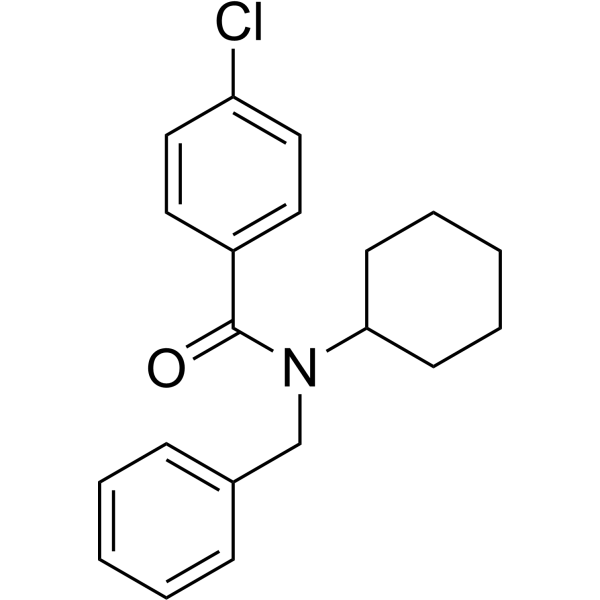
HY-19370
FPS-ZM1Amyloid-β Inhibitor -
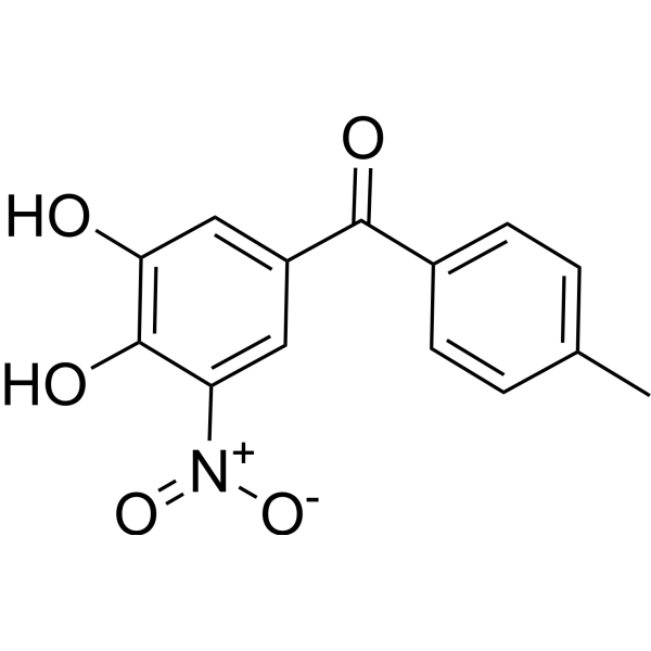
HY-17406
TolcaponeCOMT Inhibitor -
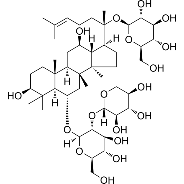
HY-N0615
Notoginsenoside R1Apoptosis Inhibitor -
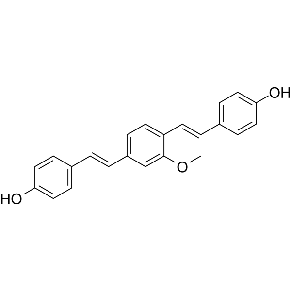
HY-103240
Methoxy-X04Fluorescent Dye


Biological Activity
Description
Isotype
Human IgG1 kappa
Recommend Isotype Controls
Species Reactivity
Human
In Vitro
The murine version of Bapineuzumab (3D6) binds recombinant Aβ 1-40 peptide with high affinity (EC50 0.17 pM)[1].
The murine version of Bapineuzumab binds Aβ plaques in human AD brain tissue without requiring formic acid antigen retrieval, with improved staining following formic acid treatment[1].
The murine version of Bapineuzumab (5 µg/mL; 24 h) activates RAW 264.7 macrophages through FcγRs, inducing TNFα secretion in vitro[1].
MedChemExpress (MCE) has not independently confirmed the accuracy of these methods. They are for reference only. Further protocols information, click here.
-
Cell Line:RAW 264.7
-
Concentration:5 µg/mL
-
Incubation Time:24 h
-
Result:Stimulated secretion of the cytokine TNFα compared to cell only controls.
In Vivo
MedChemExpress (MCE) has not independently confirmed the accuracy of these methods. They are for reference only.
-
Animal Model:APP23 mice (B6.Cg-Tg(Thy1-APP)3Somm/J, male, 27 months old)[2]
-
Dosage:12.4 mg/kg
-
Administration:i.p.; twice, 5 days apart
-
Result:Induced a mean of 4.5 new lesions at t1 and 5.3 new lesions at t2.
Induced a mean of 3.2 new lesions in the cortex at t1 and 2.8 at t2.
Induced a mean of 1.2 new lesions in the thalamus/striatum at t1 and 1.8 at t2.
Induced a mean of 0.2 new lesions in the hippocampus at t1 and 0.3 at t2.
Clinical Trial
| NCT Number | Sponsor | Condition | Start Date |
Phase

|
|---|---|---|---|---|
| NCT01329991 | Plexxikon| | 2011-05 | PHASE1 |
Gene ID
Accession
Conjugated
Unconjugated
Reconsititution
The product can be reconstituted/diluted with sterile PBS or saline.
Format
-
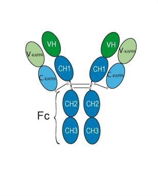
Human IgG1 kappa
Application
ELISA, FACS, Functional assay
Verified Bioactivity
-
 Flow cytometric analysis of 1.5×10⁶ HUVEC cells with Bapineuzumab (HY-P99185, red). Cells were fixed with 4% paraformaldehyde and permeabilised with 90% methanol. Then stained with the primary antibody at 1/200 dilution for an hour at 4℃. Goat Anti-Human IgG H&L (AF488) (HY-P83776) was used as the secondary antibody at 1/1,000 dilution for 30 minutes at 4℃. Human IgG1 kappa, Isotype Control (HY-P99001, blue) was used as the isotype control.


Chemical Information
-
CAS No. 648895-38-9
-
Appearance Liquid
-
Molecular Weight 144.5 kDa
-
Color Colorless to light yellow
-
SMILES
[Bapineuzumab]
-
Synonyms
AAB-001
-
Shipping
Shipping with dry ice.
-
Formulation
Please refer to the lot-specific COA for specific buffer information.
-
Storage
Please store the product under the recommended conditions in the Certificate of Analysis.

Protocols
-
LPS-Induced Endotoxemia/Systemic Inflammation
Lipopolysaccharide (LPS)-induced endotoxemia is a widely used in vivo model of acute systemic inflammation in which LPS, a Gram-negative bacterial endotoxin, activates innate immune signaling primarily through TLR4, leading to rapid and transient induction of pro-inflammatory cytokines such as TNF-α, IL-6, and IL-1β in circulation and tissues. This cytokine surge is commonly used as a measurable readout of systemic inflammatory activation and immune dysregulation, and is typically assessed within hours after intraperitoneal LPS administration in mouse models of endotoxemia. The model captures key features of systemic inflammatory response syndrome, including cytokine release, immune cell activation, and downstream tissue responses, and has been used to evaluate anti-inflammatory interventions such as cytokine modulation, lipid mediators, and immune cell-targeting therapies.
-
Research Protocol for Inflammation-related Diseases
The NLRP3 inflammasome is a cytosolic innate immune signaling platform that integrates priming signals and danger-signal activation to promote caspase-1 activation, maturation of IL-1β and IL-18, and gasdermin D-mediated pyroptotic cell death. The core experimental logic is to determine whether inflammatory disease phenotypes are driven by increased NLRP3 expression, ASC-containing inflammasome assembly, caspase-1 cleavage, GSDMD cleavage, and extracellular release of IL-1β/IL-18 rather than by nonspecific cell injury alone. The pathway is strongly linked to inflammation-related disease phenotypes because monosodium urate crystals activate NALP3/NLRP3 inflammasome signaling in gout-like crystal inflammation, cholesterol crystals activate NLRP3 inflammasomes in atherogenesis models, and DSS-induced intestinal inflammation has been reported to involve NLRP3 inflammasome activity. However, experimental colitis studies also show context-dependent protective effects of NLRP3 inflammasome co
-
Amyloid: Congo Red Amyloid Staining
Congo red amyloid staining is a histochemical method used to detect extracellular amyloid deposits in tissue sections based on the affinity of Congo red dye for β-pleated sheet-rich protein aggregates. When bound to amyloid, Congo red produces characteristic apple-green birefringence under polarized light microscopy, which is widely regarded as a diagnostic feature of amyloid deposition in histopathology. The diagnostic principle relies on the combination of dye binding (congophilia) and optical anisotropy under polarized illumination, which distinguishes amyloid from most non-amyloid eosinophilic extracellular deposits in routine histological evaluation. Amyloid identification by Congo red staining remains a cornerstone in diagnostic pathology despite the availability of adjunct methods such as immunohistochemistry and mass spectrometry, particularly because of its ability to localize deposits directly within tissue architecture. The specificity of Congo red-positive deposits is incre
-
Alzheimer’s Disease Modeling
Alzheimer’s Disease (AD) is a neurodegenerative disorder characterized by a progressive decline in cognitive functions and loss of specific types of neurons and synapses. Alzheimer's symptoms can be simulated in mice by injecting drugs (such as Aβ) or genetically modified.
-
Pyroptosis Solutions
Pyroptosis is a lytic inflammatory cell-death pathway executed by gasdermin pores, most classically through inflammasome-mediated activation of caspase-1, cleavage of gasdermin D, membrane pore formation, LDH release, and secretion of IL-1β and IL-18. The canonical pathway is commonly modeled by priming cells with an inflammatory signal such as LPS to induce pro-IL-1β and inflammasome components, followed by an activation signal such as ATP or nigericin to activate NLRP3, ASC speck formation, caspase-1 cleavage, GSDMD cleavage, cytokine release, and pyroptotic membrane rupture. The non-canonical pathway is triggered when cytosolic LPS activates mouse caspase-11 or human caspase-4/5, leading to GSDMD cleavage and pyroptosis, and this can secondarily activate NLRP3-dependent IL-1β release. Pyroptosis is linked to inflammatory injury, infection, cancer, liver disease, ocular disease, placental inflammation, and other disease phenotypes, but unresolved questions include which gasdermin fam


Purity & Documentation
-
Data Sheet (275 KB)
-
SDS (251 KB)
- English - EN (251 KB)
- Français - FR (251 KB)
- Deutsch - DE (251 KB)
- Norwegian - NO (251 KB)
- Español - ES (251 KB)
- Swedish - SV (251 KB)
- Italian - IT (251 KB)
- Korean - KR (251 KB)
- Portuguese - PT (251 KB)
-
Inhibitory Antibodies User Guide (603 KB)
References
[1]. Fuller JP, et al. Comparing the efficacy and neuroinflammatory potential of three anti-abeta antibodies. Acta neuropathologica. 2015 Nov;130(5):699-711. [Content Brief]
[3]. Salloway S, et al. Two phase 3 trials of bapineuzumab in mild-to-moderate Alzheimer's disease. The New England journal of medicine. 2014 Jan 23;370(4):322-33. [Content Brief]

Calculators
Concentration (start) × Volume (start) = Concentration (final) × Volume (final)


Bapineuzumab Related Classifications
HY-P99185 Related Classifications
 Powered by Bioz