



Biotin-β-Amyloid (1-40)
Biotin-β-Amyloid (1-40) is a N-terminal-labelled biotinylated amyloid-β-(1-40) peptide.
For research use only. We do not sell to patients.
- CAS No.: 183906-14-1
- Formula: C204H309N55O60S2
- Molecular Weight:4556.20
-
Storage:
Please store the product under the recommended conditions in the Certificate of Analysis.

To place orders, for customer services and technical support, please contact: MedChemExpress USA
Tel: 609-228-6898 E-mail: [email protected] [email protected]


-
 Biological Activity
-
Chemical Information
- Protocols
- Purity & Documentation
- References
-
 Help & FAQs


-

HY-P1363A
β-Amyloid (1-42), humanAmyloid β Peptide -

HY-P1363B
β-Amyloid (1-42), human, HFIP-treatedβ-Amyloid (1-42) Peptide -
HY-P0128
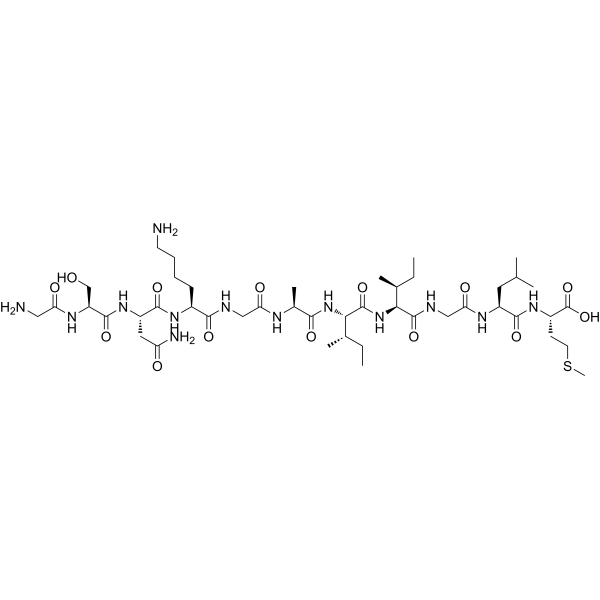
β-Amyloid (25-35)Amyloid β Peptide -

HY-P0128A
β-Amyloid (25-35), HFIP-treatedAmyloid β Peptide -

HY-P1053
β-Amyloid (10-20)β-Amyloid Peptide Fragment -

HY-P5361
β-Amyloid (40-1)Bioactive Peptide -

HY-P3688
β-Amyloid (1-38)Aβ Peptide -
HY-P4740
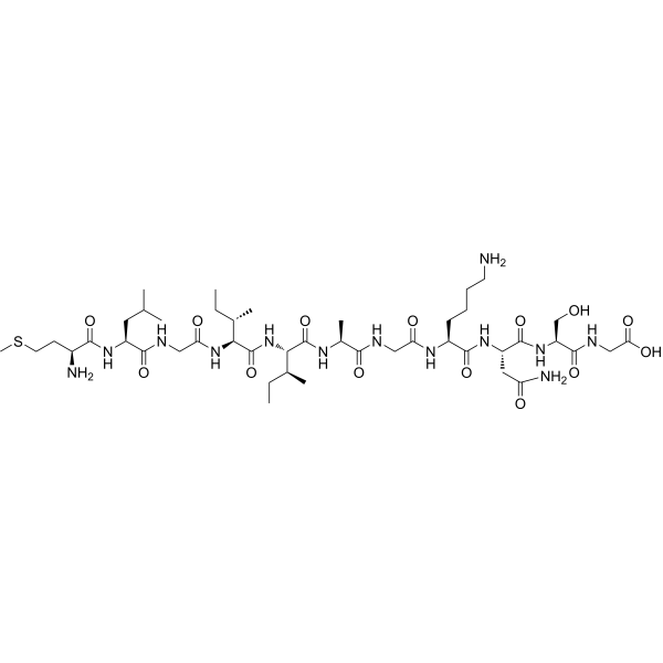
β-Amyloid (35-25)reverse version of β-Amyloid (25-35) (HY-P0128) -

HY-P4882
(Pyr3)-Amyloid β-Protein (3-42)Amyloid-β 肽 -

HY-P4888
(Pyr11)-Amyloid β-Protein (11-40)Peptide -
HY-P4889
(Gly22)-amyloid beta-protein(1-40)Peptide


Biological Activity
Description
In Vitro
β-Amyloid (1-40) is a primary protein in plaques found in the brains of patients with Alzheimer's disease. It is the major component of senile plaque amyloids, are physiological peptides present in the brain, cerebrospinal fluid (CSF) and plasma[1]. Biotin is widely used throughout the biotechnology industry to conjugate proteins for biochemical assays.?Biotin can be used for qualitative and quantitative detection, labelling or immobilisation.
MedChemExpress (MCE) has not independently confirmed the accuracy of these methods. They are for reference only. Further protocols information, click here.


Chemical Information
-
CAS No. 183906-14-1
-
Molecular Weight 4556.20
-
Formula C204H309N55O60S2
-
Sequence
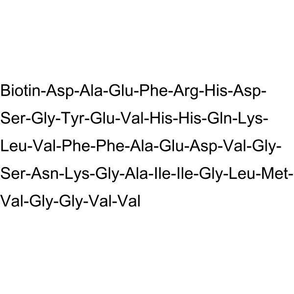
Biotin-Asp-Ala-Glu-Phe-Arg-His-Asp-Ser-Gly-Tyr-Glu-Val-His-His-Gln-Lys-Leu-Val-Phe-Phe-Ala-Glu-Asp-Val-Gly-Ser-Asn-Lys-Gly-Ala-Ile-Ile-Gly-Leu-Met-Val-Gly-Gly-Val-Val
-
Sequence Shortening
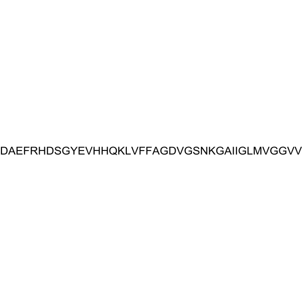
Biotin-DAEFRHDSGYEVHHQKLVFFAEDVGSNKGAIIGLMVGGVV
-
Shipping
Room temperature in continental US; may vary elsewhere.
-
Storage
Please store the product under the recommended conditions in the Certificate of Analysis.

Protocols
-
Amyloid: Congo Red Amyloid Staining
Congo red amyloid staining is a histochemical method used to detect extracellular amyloid deposits in tissue sections based on the affinity of Congo red dye for β-pleated sheet-rich protein aggregates. When bound to amyloid, Congo red produces characteristic apple-green birefringence under polarized light microscopy, which is widely regarded as a diagnostic feature of amyloid deposition in histopathology. The diagnostic principle relies on the combination of dye binding (congophilia) and optical anisotropy under polarized illumination, which distinguishes amyloid from most non-amyloid eosinophilic extracellular deposits in routine histological evaluation. Amyloid identification by Congo red staining remains a cornerstone in diagnostic pathology despite the availability of adjunct methods such as immunohistochemistry and mass spectrometry, particularly because of its ability to localize deposits directly within tissue architecture. The specificity of Congo red-positive deposits is incre
-
Alzheimer’s Disease Modeling
Alzheimer’s Disease (AD) is a neurodegenerative disorder characterized by a progressive decline in cognitive functions and loss of specific types of neurons and synapses. Alzheimer's symptoms can be simulated in mice by injecting drugs (such as Aβ) or genetically modified.


Purity & Documentation
References

Calculators
Concentration (start) × Volume (start) = Concentration (final) × Volume (final)


Biotin-β-Amyloid (1-40) Related Classifications
HY-P2549 Related Classifications
 Powered by Bioz