



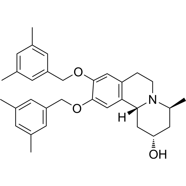
BQZ-485
BQZ-485 is a GDI2 inhibitor and a paraptosis inducer with antitumor activity. BQZ-485 binds to the Rab-binding platform domain of GDI2, disrupts the interaction between GDI2 and Rab1A, and impairs the membrane recycling of Rab1A. BQZ-485 blocks vesicle trafficking from the endoplasmic reticulum to the Golgi apparatus, and induces endoplasmic reticulum expansion, endoplasmic reticulum fusion, endoplasmic reticulum stress, unfolded protein response and cytoplasmic vacuolization. BQZ-485 is applicable to pancreatic cancer-related research.
For research use only. We do not sell to patients.
- CAS No.: 1906915-49-8
- Formula: C32H39NO3
- Molecular Weight:485.66
-
Storage:
Please store the product under the recommended conditions in the Certificate of Analysis.

To place orders, for customer services and technical support, please contact: MedChemExpress USA
Tel: 609-228-6898 E-mail: [email protected] [email protected]


-
 Biological Activity
-
Chemical Information
- Protocols
- Purity & Documentation
- References
-
 Help & FAQs




Biological Activity
Description
IC50 & Target
[1]|
eIF2-α |
Cellular Effect
|
Cell Line

|
Type | Value | Description | References |
|---|---|---|---|---|
| PC-3 | IC50 |
0.57 μM
|
Antiproliferative activity against human PC-3 prostate cancer cells assessed as reduction in cell viability incubated for 48 h by cell viability assay.
Antiproliferative activity against human PC-3 prostate cancer cells assessed as reduction in cell viability incubated for 48 h by cell viability assay.
|
PMC10598831 |
In Vitro
BQZ-485 (compound 1) (0.1-10 μM; 48 h) potently inhibits the proliferation of human prostate cancer cell line PC-3, with an IC50 value of 0.57 μM[1].
BQZ-485 stabilizes the bindable and purified wild-type GDI2 protein; it directly binds to the purified GDI2 protein with a KD value of 46 μM[1].
BQZ-485 binds to domain I within the Rab-binding platform of GDI2, with a key interaction occurring with the Tyr245 residue; it inhibits the GDI2-Rab1A interaction in a NanoLuc-based cell-free assay, with an IC50 of 6.85 μM; it can inhibit GDI2WT-mediated retrieval of Rab1A from membranes, with an EC50 of 4.96 μM[1].
BQZ-485 (2 μM; 24 h) disrupts the subcellular distribution of Rab1A in human prostate cancer cell line PC-3, reducing cytoplasmic Rab1A while increasing membrane-bound Rab1A; it can disrupt the interaction between endogenous GDI2 and Rab1A in human prostate cancer cell line PC-3 expressing wild-type GDI2[1].
BQZ-485 (1 μM; 8 h, 0-720 min) induces endoplasmic reticulum-derived cytoplasmic vacuolization and paraptotic cell death in human prostate cancer cells PC-3[1].
BQZ-485 (1 μM; 24 h) induces endoplasmic reticulum stress and activates the unfolded protein response in human prostate cancer PC-3 cells, which is characterized by increased expression levels of GRP78, CHOP and p-eIF2α[1].
MedChemExpress (MCE) has not independently confirmed the accuracy of these methods. They are for reference only. Further protocols information, click here.
-
Cell Line:PC-3 human prostate cancer cells
-
Concentration:2 μM
-
Incubation Time:24 h
-
Result:Caused depletion of Rab1A in the cytosolic fraction and accumulation of Rab1A in the membrane fraction, indicating disrupted Rab1A recycling.
-
Cell Line:PC-3 human prostate cancer cells
-
Concentration:1 μM
-
Incubation Time:24 h
-
Result:Upregulated the ER stress/UPR markers GRP78, CHOP, and phosphorylated eIF2α (p-eIF2α) compared to DMSO control.
-
Cell Line:PC-3 human prostate cancer cells
-
Concentration:0, 0.63, 1.25, 2.5, 5, 10, 20 μM
GDI2WT or GDI2Y245A 1 μM -
Incubation Time:30 min
-
Result:Reduced GDI2‑mediated extraction of membrane‑bound Rab1A with an EC50 value of 4.96 μM.


Chemical Information
-
CAS No. 1906915-49-8
-
Molecular Weight 485.66
-
Formula C32H39NO3
-
SMILES
CC1=CC(C)=CC(COC2=CC3=C([C@](C[C@@H](O)C[C@@H]4C)([H])N4CC3)C=C2OCC5=CC(C)=CC(C)=C5)=C1
-
Shipping
Room temperature in continental US; may vary elsewhere.
-
Storage
Please store the product under the recommended conditions in the Certificate of Analysis.

Protocols
-
Subcutaneous Cell-Line-Derived Xenograft
Subcutaneous cell-line-derived xenograft (CDX) models are established by implanting cultured human cancer cell lines into immunodeficient mice, where the injected cells form localized tumors that can be monitored in vivo as a measure of tumorigenic potential, growth kinetics, and treatment response. These models are widely used in oncology research because they allow reproducible tumor formation and enable comparative assessment of tumor growth between different cell lines or genetic manipulations in a controlled in vivo microenvironment. Subcutaneous implantation of cancer cells in immunodeficient mice is a standard approach for evaluating tumor growth behavior and therapeutic response across multiple cancer types, including prostate, esophageal, pancreatic, and colon cancer models.


Purity & Documentation
References

Calculators
Concentration (start) × Volume (start) = Concentration (final) × Volume (final)


BQZ-485 Related Classifications
HY-156242 Related Classifications
 Powered by Bioz