



Briquilimab

Based on 1 Customer Validation
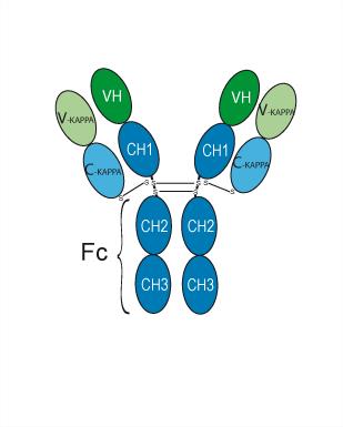
Briquilimab (JSP-191 or AMG-191) is a humanized IgG1 monoclonal antibody that binds human CD117 (c-Kit). Briquilimab blocks the interaction between CD117 receptor and stem cell factor on various CD117 expressing tissues. Briquilimab can lead to inhibition of SCF/c-Kit signaling and MC apoptosis. Briquilimab is a non-toxic approach to target and deplete HSC, enabling blood and immune reconstitution with minimal toxicity with the other agents being used for transient immune suppression to prevent immunologic rejection. Briquilimab can be used in various disease research such as severe combined immunodeficiency (SCID), myelodyplastic syndromes (MDS), acute myeloid leukemia (AML), chronic spontaneous urticarial (CSU), chronic inducible urticarial (CIndU) and asthema.
연구목적의 판매만을 진행합니다. 환자를 대상으로 한 판매는 하지 않습니다.
- Purity : 99.00%
- CAS No.: 2574591-89-0
- 분자량:145.61 kDa
-
보관:
Please store the product under the recommended conditions in the Certificate of Analysis.


To place orders, for customer services and technical support, please contact: MedChemExpress USA
Tel: 609-228-6898 E-mail: [email protected] [email protected]


-
 Biological Activity
-
Chemical Information
- Protocol
- 순도&문서
- References
-
 Help & FAQs


-

HY-P99462
Barzolvolimab (WT IgG1)Anti-KIT Antibody -
HY-15531
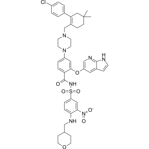
VenetoclaxBCL-2 Inhibitor -
HY-13740
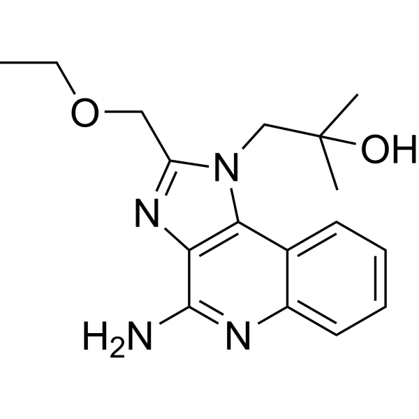
ResiquimodTLR7/8 Agonist -
HY-12726
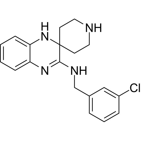
Liproxstatin-1Ferroptosis Inhibitor -

HY-P9966
EnoblituzumabB7-H3 mAb -

HY-P99001
Human IgG1 kappa, Isotype ControlIgG1κ Isotype Control -
HY-12335
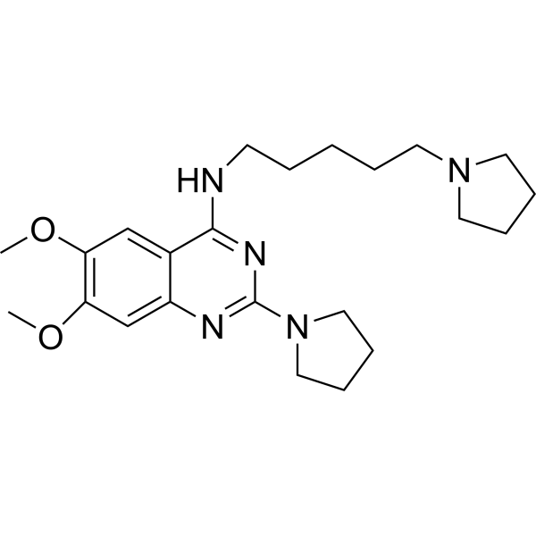
UNC0379SETD8 Inhibitor -

HY-P99029
MagrolimabAnti-CD47 Antibody -

HY-P10746
EB1002NK2R Agonist


Biological Activity
제품 설명
Isotype
Human IgG1 kappa
Recommend Isotype Controls
Species Reactivity
Human
IC50 & Target
c-Kit/CD117
In Vivo
Briquilimab (0.3-1 mg/kg, i.v., one single dose) depletes human HSPCs in all animals after 2 weeks, but the levels of depletion varies among NSG mice model[4].
Briquilimab (0.1-25 mg/kg, i.v., one single dose) has no adverse clinical observations in immunocompetent juvenile cynomolgus macaques[4].
MedChemExpress (MCE) has not independently confirmed the accuracy of these methods. They are for reference only.
-
Animal Model:Human CD34+ hematopoietic stem cell-engrafted humanized NSG mice (hu-CD34 NSG, 10-11 weeks old)[1]
-
Dosage:0.3 and 1.0 mg/kg
-
Administration:Intravenous injection (i.v.), one single dose
-
Result:Decreased the ratio of human CD45+/mouse CD45+ in most mice.
Decreased the frequency of human myeloid and T cells.
Increased the frequency of B cells.
Results in gene-marked second human donor cells among hCD45+hCD34+ and hCD45+hCD19+ cell in all mice treated with Briquilimab but not in unconditioned mice, suggesting that Briquilimab conditioning permitted second donor HSPC engraftment.
Led to higher donor chimerism in mice treated with 1.0 mg/kg dose.
-
Animal Model:Juvenile 37-39 months old male cynomolgus macaques (3.4−4.0 kg)[4]
-
Dosage:0.1, 1, 5, or 25 mg/kg
-
Administration:Intravenous injection (i.v.), one single dose
-
Result:Resulted in HSPC depletion lasted up to 21 days in most animals and >42 days in animal that received the highest dose.
Caused delayed HSPC recovery with high doses.
Clinical Trial
| NCT Number | Sponsor | Condition | Start Date |
Phase

|
|---|---|---|---|---|
| NCT01329991 | Plexxikon| | 2011-05 | PHASE1 |
Gene ID
Accession
Conjugated
Unconjugated
Reconsititution
The product can be reconstituted/diluted with sterile PBS or saline.
Format
-
Human IgG1 kappa
신청
ELISA, FACS, Functional assay
Verified Bioactivity
-
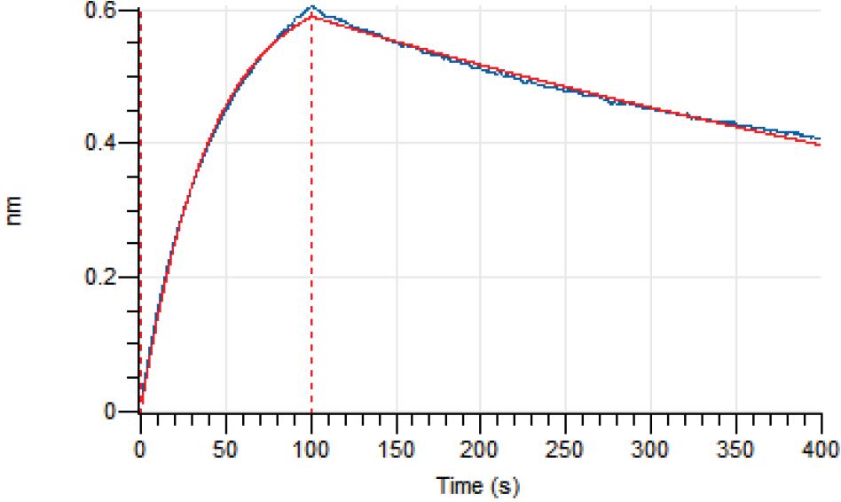 Loaded Briquilimab on AHC2 biosensor, can bind CD117/c-kit Protein, Human (HEK293, His, HY-P72752) with an affinity constant of 5.634E-09 M as determined in BLI assay.
 -
 Immobilized Human c-Kit Protein (ECD, His Tag) can bind Briquilimab. The EC50 for this effect is 1.72 ng/mL.
 -
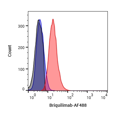 Flow Cytometry analysis of THP-1 cells labelling c-Kit/CD117 (red) with Briquilimab (HY-P99488). Cells were fixed with 4% paraformaldehyde and permeabilised with 90% methanol. Then cells were stained with the primary antibody at 1/200 dilution for an hour at 4℃. Goat Anti-Human IgG H&L (AF488) (HY-P83776) was used as the secondary antibody at 1/1,000 dilution for 30 minutes at 4℃. Human IgG1 kappa (HY-P99001, blue) was used as the isotype control, cells without incubation with primary antibody were used as the unlabeled control (black).


Chemical Information
-
CAS No. 2574591-89-0
-
Appearance Liquid
-
분자량 145.61 kDa
-
Color Colorless to light yellow
-
SMILES
[Briquilimab]
-
Synonyms
JSP-191; AMG-191
-
선적
Shipping with dry ice.
-
Formulation
Please refer to the lot-specific COA for specific buffer information.
-
보관
Please store the product under the recommended conditions in the Certificate of Analysis.

Protocol
-
Detection of Apoptosis
Apoptosis, also called programmed cell death, is generally characterized by distinct morphological characteristics.
-
TUNEL staining for apoptotic DNA fragmentation
TUNEL staining detects DNA strand breaks by using terminal deoxynucleotidyl transferase to add labeled nucleotides to exposed 3′-OH DNA termini, generating either microscopic staining in fixed cells or tissue sections, or fluorescence/cytometric signal in cell suspensions. TUNEL positivity reflects DNA fragmentation but should not be interpreted alone as definitive apoptosis, because TUNEL can also label necrotic, autolytic, mechanically damaged, or DNA-repair-associated DNA breaks.
-
Annexin V plus membrane-impermeant dye apoptosis staining
Annexin V-based apoptosis assays rely on the detection of phosphatidylserine (PS) externalization from the inner leaflet of the plasma membrane to the outer leaflet, an early biochemical hallmark of apoptosis. Fluorescently labeled Annexin V binds PS in a calcium-dependent manner, enabling identification of early apoptotic cells by flow cytometry or fluorescence microscopy. When combined with a membrane-impermeant DNA-binding dye (e. g. , propidium iodide), this approach allows discrimination between viable (Annexin V−/dye−), early apoptotic (Annexin V+/dye−), and late apoptotic or necrotic (Annexin V+/dye+) cell populations by assessing membrane integrity and PS exposure.
-
Apoptosis Solutions
Apoptosis is a regulated, generally non-lytic cell-death pathway that removes unwanted, damaged, infected, or abnormal cells through coordinated morphological changes, caspase activation, DNA fragmentation, and membrane remodeling. The intrinsic apoptosis pathway is controlled mainly by mitochondrial outer membrane permeabilization, BCL-2 family proteins, cytochrome c release, apoptosome formation, caspase-9 activation, and downstream executioner caspase-3/7 activation. The extrinsic apoptosis pathway is initiated by death receptors such as Fas, TNFR, and TRAIL receptors, which recruit adaptor proteins and activate caspase-8 before engaging executioner caspases or mitochondrial amplification through BID cleavage. Apoptosis is linked to many phenotypes, including cancer cell killing, tissue homeostasis, immune regulation, neurodegeneration, infection response, and treatment-induced cytotoxicity; unresolved questions include how apoptosis interacts with necroptosis, pyroptosis, ferroptos


순도&문서
-
Data Sheet (262 KB)
-
SDS (251 KB)
- English - EN (251 KB)
- Français - FR (251 KB)
- Deutsch - DE (251 KB)
- Norwegian - NO (251 KB)
- Español - ES (251 KB)
- Swedish - SV (251 KB)
- Italian - IT (251 KB)
- Korean - KR (251 KB)
- Portuguese - PT (251 KB)
-
Inhibitory Antibodies User Guide (603 KB)
References
[3]. Pang, W. W., et al., (2019). Anti-CD117 antibody depletes normal and myelodysplastic syndrome human hematopoietic stem cells in xenografted mice. Blood, 133(19), 2069–2078. [Content Brief]
[4]. Kwon, H. S., et al., (2019). Anti-human CD117 antibody-mediated bone marrow niche clearance in nonhuman primates and humanized NSG mice. Blood, 133(19), 2104–2108. [Content Brief]

Calculators
Concentration (start) × Volume (start) = Concentration (final) × Volume (final)


Briquilimab Related Classifications
HY-P99488 Related Classifications
 Powered by Bioz