



c-Fms-IN-3

Based on 1 Customer Validation
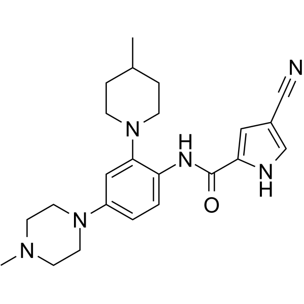
c-Fms-IN-3 is an orally active c-Fms (CSF1R) kinase inhibitor (IC50 = 0. 8 nM). c-Fms-IN-3 inhibits CSF1R kinase activity and reduces macrophage populations. c-Fms-IN-3 reduces bone erosion, pannus invasion, cartilage damage, and inflammation in collagen (HY-NP003)-induced arthritis mouse model. c-Fms-IN-3 is useful for arthritis research.
For research use only. We do not sell to patients.
- Purity : 98.83%
- CAS No.: 885704-21-2
- Formula: C23H30N6O
- Molecular Weight:406.52
-
Storage:Powder -20°C, 3 years , 4°C, 2 years ; In solvent -80°C, 2 years , -20°C, 1 year

To place orders, for customer services and technical support, please contact: MedChemExpress USA
Tel: 609-228-6898 E-mail: [email protected] [email protected]


-
 Biological Activity
-
Chemical Information
-
Solvent & Solubility
- Protocols
- Purity & Documentation
- References
-
 Help & FAQs


-
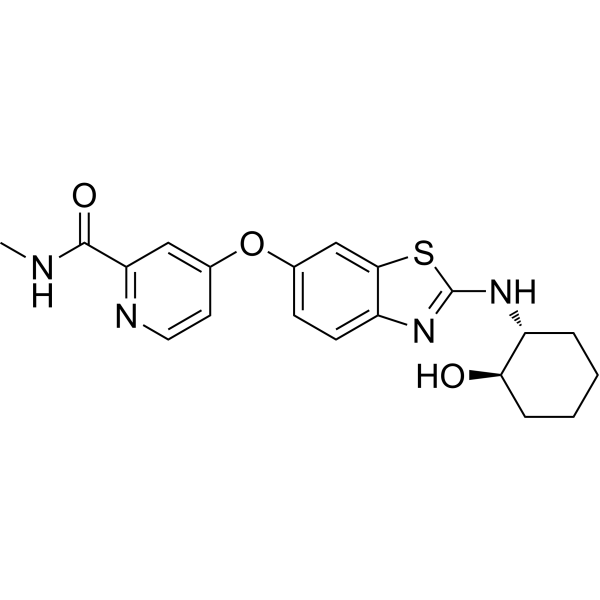
HY-12768
SotuletinibCSF-1R Inhibitor -
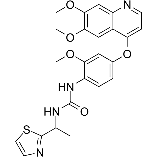
HY-10408
Ki20227CSF1R Inhibitor -
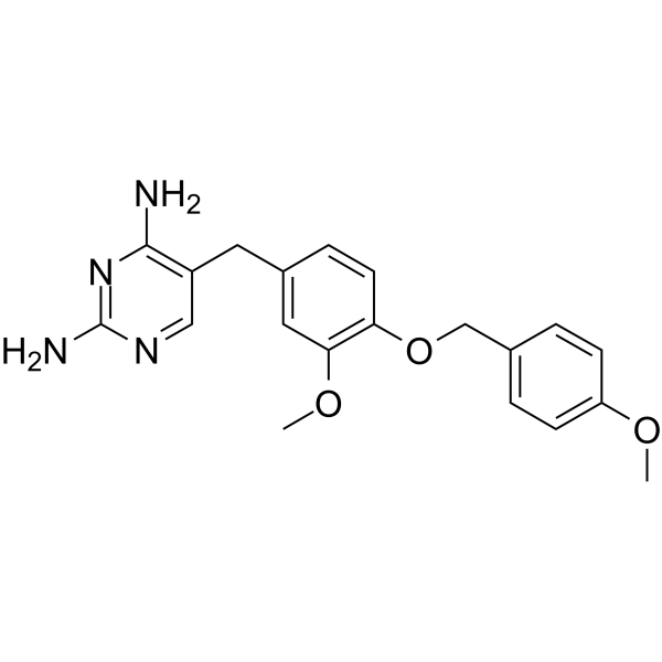
HY-10917
GW2580c-Fms Inhibitor -
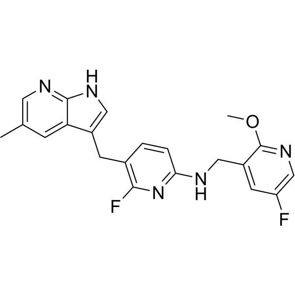
HY-114153C
PLX5622 in AIN-76A Diet (1200 ppm)PLX5622 Microglia Clearance Diet -
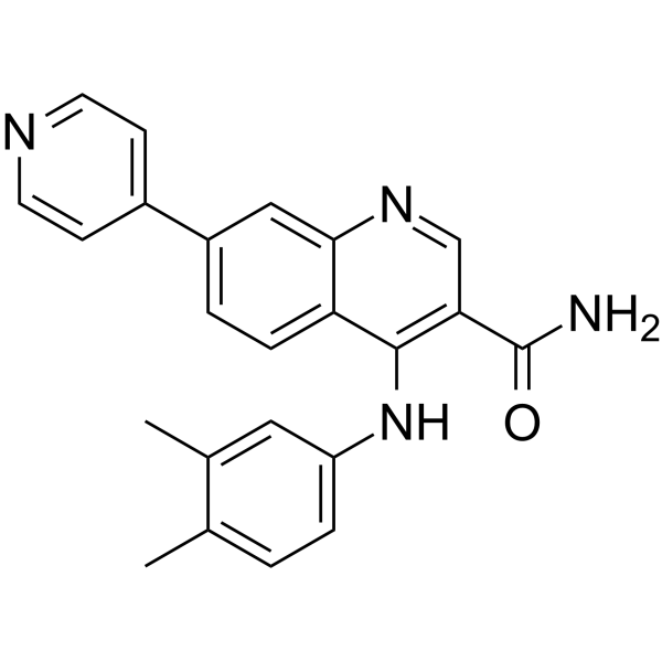
HY-112451
cFMS Receptor Inhibitor IIc-Fms Inhibitor -
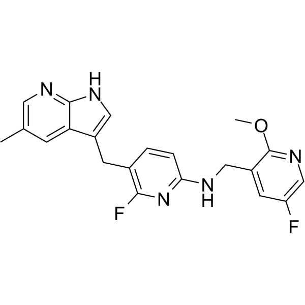
HY-114153
PLX5622CSF1R Inhibitor -
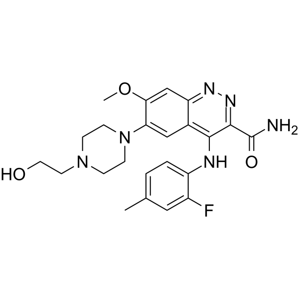
HY-117244
AZD7507c-Fms Inhibitor -
HY-13838
PLX647FMS/KIT Inhibitor -

HY-10408R
Ki20227 (Standard)Reference Standard -
HY-18787
c-Fms-IN-2c-Fms Inhibitor -
HY-109086
EdicotinibCSF-1R Inhibitor -
HY-136256
Vimseltinibc-FMS(CSF-IR)/c-Kitdual Inhibitor


Biological Activity
Description
IC50 & Target
c-Fms.
Cellular Effect
|
Cell Line

|
Type | Value | Description | References |
|---|---|---|---|---|
| Sf9 | IC50 |
0.00078 μM
Compound: 8
|
Inhibition of human cytoplasmic FMS expressed in Sf9-baculovirus system after 80 mins by fluorescence polarization
Inhibition of human cytoplasmic FMS expressed in Sf9-baculovirus system after 80 mins by fluorescence polarization
|
[PMID: 18242992] |
| Sf9 | IC50 |
0.78 nM
Compound: 8
|
Inhibition of human cytoplasmic FMS expressed in Sf9-baculovirus system after 80 mins by fluorescence polarization
Inhibition of human cytoplasmic FMS expressed in Sf9-baculovirus system after 80 mins by fluorescence polarization
|
[PMID: 18242992] |
In Vitro
c-Fms-IN-3 (Compound 8) inhibits the receptor tyrosine kinases Kit, Axl, TrkA, and Flt-3 and IRK-β, with IC50s of 0.0035, 0.0064, 0.011, 0.018, 0.083 μM[1].
MedChemExpress (MCE) has not independently confirmed the accuracy of these methods. They are for reference only. Further protocols information, click here.
Parmacokinetics
| Species | Dose | Route | T1/2 | Cmax | Vss | CL | F |
|---|---|---|---|---|---|---|---|
| Mice[1] | 10 mg/kg | p.o. | 244 min | 853 ng/mL | 5635 mL/kg | 16 | 45 % |
In Vivo
MedChemExpress (MCE) has not independently confirmed the accuracy of these methods. They are for reference only.
-
Animal Model:Murine collagen-induced model of arthritis (CIA)[1]
-
Dosage:30 and 5 mg/kg
-
Administration:p.o., twice-daily, 14 days
-
Result:Reduced pannus formation and the destruction of bone and cartilage, reduced pannus growth and bone destruction by 80%.
Depleted macrophages, demonstrated that macrophages are the target.


Chemical Information
-
CAS No. 885704-21-2
-
Appearance Solid
-
Molecular Weight 406.52
-
Formula C23H30N6O
-
Color White to gray
-
SMILES
CN(CC1)CCN1C2=CC=C(NC(C3=CC(C#N)=CN3)=O)C(N4CCC(C)CC4)=C2
-
Shipping
Room temperature in continental US; may vary elsewhere.
-
Storage
Powder -20°C 3 years 4°C 2 years In solvent -80°C 2 years -20°C 1 year


Solvent & Solubility
In Vitro:
DMSO : 25 mg/mL (61.50 mM; Need ultrasonic; Hygroscopic DMSO has a significant impact on the solubility of product, please use newly opened DMSO)
Please refer to the solubility information to select the appropriate solvent. Once prepared, please aliquot and store the solution to prevent product inactivation from repeated freeze-thaw cycles.
Storage method and period of stock solution: -80°C, 2 years; -20°C, 1 year. When stored at -80°C, please use it within 2 years. When stored at -20°C, please use it within 1 year.
Please refer to the solubility information to select the appropriate solvent. Once prepared, please aliquot and store the solution to prevent product inactivation from repeated freeze-thaw cycles.
Storage method and period of stock solution: -80°C, 2 years; -20°C, 1 year. When stored at -80°C, please use it within 2 years. When stored at -20°C, please use it within 1 year.
Concentration (start) × Volume (start) = Concentration (final) × Volume (final)
In Vivo:
Select the appropriate dissolution method based on your experimental animal and administration route.
- For the following dissolution methods, please ensure to first prepare a clear stock solution using an In Vitro approach and then sequentially add co-solvents:
- To ensure reliable experimental results, the clarified stock solution can be appropriately stored based on storage conditions. As for the working solution for In Vivo experiments, it is recommended to prepare freshly and use it on the same day.
- The percentages shown for the solvents indicate their volumetric ratio in the final prepared solution. If precipitation or phase separation occurs during preparation, heat and/or sonication can be used to aid dissolution.
In Vivo Dissolution Calculator
Please enter the basic information of animal experiments:
-
-
-
-
Recommended: Prepare an additional quantity of animals to account for potential losses during experiments.
Please enter your animal formula composition:
-
%DMSO +
Recommended: Keep the proportion of DMSO in working solution below 2% if your animal is weak.
-
%+
-
+%Tween-80 + +
-
%Saline +
The co-solvents required include: DMSO, . All of co-solvents are available by MedChemExpress (MCE). , Tween 80. All of co-solvents are available by MedChemExpress (MCE).
Working solution concentration: 0.22 mg/mL
Method for preparing stock solution: mg drug dissolved in μL DMSO. Stock solution concentration: mg/mL.
1. Take μL DMSO stock solution;
2. Add μL .
μL , mix evenly;
3. Then add μL Tween 80, mix evenly;
4. Then add μL
Please ensure that the stock solution in the first step is dissolved to a clear state, and add co-solvents in sequence. You can use ultrasonic heating (ultrasonic cleaner, recommended frequency 20-40 kHz), vortexing, etc. to assist dissolution.

Protocols
-
Kinase activity and phosphorylation assays
Kinase activity assays measure the ability of kinases to transfer phosphate groups from ATP to specific substrates, while phosphorylation assays detect the presence and levels of phosphorylated proteins. Common methods include radiolabeled ATP incorporation (e. g. ,), ADP release detection via bioluminescence (e. g. ,[3]), enzyme-linked immunosorbent assays (ELISA) for phospho-specific epitopes (e. g. ,[6]), and microtiter-based formats for high-throughput screening (e. g. ,[8]). The ADP-Glo assay quantifies kinase activity by measuring ADP produced during phosphorylation using a luciferase-based system. Radiometric assays involve autoradiography or scintillation counting after incorporation of 32P-labeled ATP into substrate proteins. ELISA-based approaches rely on phospho-specific antibodies to detect activated kinases in cell lysates or purified samples.
-
Collagen-Induced Arthritis
Collagen-induced arthritis (CIA) is an autoimmune murine model of rheumatoid arthritis in which immunization with type II collagen (CII) emulsified in an adjuvant induces a T cell- and autoantibody-driven inflammatory arthritis characterized by synovial hyperplasia, immune cell infiltration, and joint destruction. The model typically relies on genetically susceptible mouse strains (e. g. , DBA/1) and reproduces key features of human rheumatoid arthritis, including anti-collagen immune responses and progressive joint inflammation. Disease onset generally occurs within ~3-4 weeks after immunization, depending on antigen/adjuvant combinations and protocol variation. The immunopathology is driven by adaptive immune activation against CII, leading to systemic and local joint inflammation mediated by pro-inflammatory cytokines and effector immune cells, making CIA a standard preclinical platform for evaluating immunomodulatory and anti-arthritic interventions.
-
Cell Cytotoxicity Assay
Cytotoxicity assays are usually based on the assessment of cell membrane damage, which can also be indirectly detected by measuring cell viability. Detection methods include MTT assay, CKK-8 assay, LDH assay and ATP assay, etc.
-
Cell invasion
Cell invasion is the ability of cells to migrate from one area to another via the extracellular matrix. Cell invasion is the response of normal and cancer cells to chemical and mechanical stimuli. Before migrating to a new region, the extracellular matrix is degraded by proteases within the cell. Cell invasion often occurs during wound repair, vascularization and inflammation, abnormal tissue invasion, and tumor cell metastasis.
-
Protocol for Kinase activity and phosphorylation assays
Kinase activity assays measure transfer of phosphate from ATP to a protein or peptide substrate, generating phosphorylated substrate, ADP, or incorporated radiolabeled phosphate as the readout; phosphorylation assays measure site-specific phosphorylation in cells or tissues as a proxy for kinase-pathway activation, inhibition, or substrate regulation. Phosphorylation can be detected by phospho-specific Western blot, immunoprecipitation kinase assay, phospho-immunofluorescence, phospho-flow cytometry, luminescent ADP detection, radiolabeled ATP incorporation, or reporter-based pathway assays, and these readouts can be applied to cancer cells, primary neurons, mouse tumors, organoids, inflammatory macrophages, ferroptosis studies, and mitophagy studies when the kinase target is biologically relevant.
-
Research Protocol for Inflammation-related Diseases
The NLRP3 inflammasome is a cytosolic innate immune signaling platform that integrates priming signals and danger-signal activation to promote caspase-1 activation, maturation of IL-1β and IL-18, and gasdermin D-mediated pyroptotic cell death. The core experimental logic is to determine whether inflammatory disease phenotypes are driven by increased NLRP3 expression, ASC-containing inflammasome assembly, caspase-1 cleavage, GSDMD cleavage, and extracellular release of IL-1β/IL-18 rather than by nonspecific cell injury alone. The pathway is strongly linked to inflammation-related disease phenotypes because monosodium urate crystals activate NALP3/NLRP3 inflammasome signaling in gout-like crystal inflammation, cholesterol crystals activate NLRP3 inflammasomes in atherogenesis models, and DSS-induced intestinal inflammation has been reported to involve NLRP3 inflammasome activity. However, experimental colitis studies also show context-dependent protective effects of NLRP3 inflammasome co
-
How to Choose the Right Model Animal
Choosing the right model animal is a validity-driven decision in which the species, strain, sex, age, genetic background, disease-induction method, outcome measures, and welfare burden must match the scientific question rather than laboratory tradition or convenience. A model should be selected by judging face validity, construct validity, and predictive validity: whether it resembles the human phenotype, whether it reproduces relevant mechanisms, and whether results are likely to predict human biology or treatment response. Animal studies often fail to translate because of species differences, weak disease resemblance, poor experimental design, inadequate reporting, publication bias, and underuse of randomization, blinding, and sample-size justification. Unresolved questions include how to rank competing models objectively, how much human-disease complexity must be reproduced for a given objective, and when non-animal systems such as organoids, ex vivo tissue, or computational models


Purity & Documentation
-
Data Sheet (275 KB)
-
SDS (251 KB)
- English - EN (251 KB)
- Français - FR (251 KB)
- Deutsch - DE (251 KB)
- Norwegian - NO (251 KB)
- Español - ES (251 KB)
- Swedish - SV (251 KB)
- Italian - IT (251 KB)
- Korean - KR (251 KB)
- Portuguese - PT (251 KB)
-
Handling Instructions (2659 KB)
References
[1]. Illig, Carl R.; Chen, Jinsheng; Wall, Mark J. et al. Discovery of novel FMS kinase inhibitors as anti-inflammatory agents. Bioorganic & Medicinal Chemistry Letters (2008), 18(5), 1642-1648. [Content Brief]
[2]. Meegalla, Sanath K.; Wall, Mark J.; Chen, Jinsheng et al. Structure-based optimization of a potent class of arylamide FMS inhibitors. Bioorganic & Medicinal Chemistry Letters (2008), 18(12), 3632-3637. [Content Brief]


Complete Stock Solution Preparation Table
Please refer to the solubility information to select the appropriate solvent. Once prepared, please aliquot and store the solution to prevent product inactivation from repeated freeze-thaw cycles.
Storage method and period of stock solution: -80°C, 2 years; -20°C, 1 year. When stored at -80°C, please use it within 2 years. When stored at -20°C, please use it within 1 year.
| Optional Solvent | Concentration Solvent Mass | 1 mg | 5 mg | 10 mg | 25 mg |
|---|---|---|---|---|---|
| DMSO | 1 mM | 2.4599 mL | 12.2995 mL | 24.5990 mL | 61.4976 mL |
| 5 mM | 0.4920 mL | 2.4599 mL | 4.9198 mL | 12.2995 mL | |
| 10 mM | 0.2460 mL | 1.2300 mL | 2.4599 mL | 6.1498 mL | |
| 15 mM | 0.1640 mL | 0.8200 mL | 1.6399 mL | 4.0998 mL | |
| 20 mM | 0.1230 mL | 0.6150 mL | 1.2300 mL | 3.0749 mL | |
| 25 mM | 0.0984 mL | 0.4920 mL | 0.9840 mL | 2.4599 mL | |
| 30 mM | 0.0820 mL | 0.4100 mL | 0.8200 mL | 2.0499 mL | |
| 40 mM | 0.0615 mL | 0.3075 mL | 0.6150 mL | 1.5374 mL | |
| 50 mM | 0.0492 mL | 0.2460 mL | 0.4920 mL | 1.2300 mL | |
| 60 mM | 0.0410 mL | 0.2050 mL | 0.4100 mL | 1.0250 mL |


c-Fms-IN-3 Related Classifications
HY-13075 Related Classifications
 Powered by Bioz