



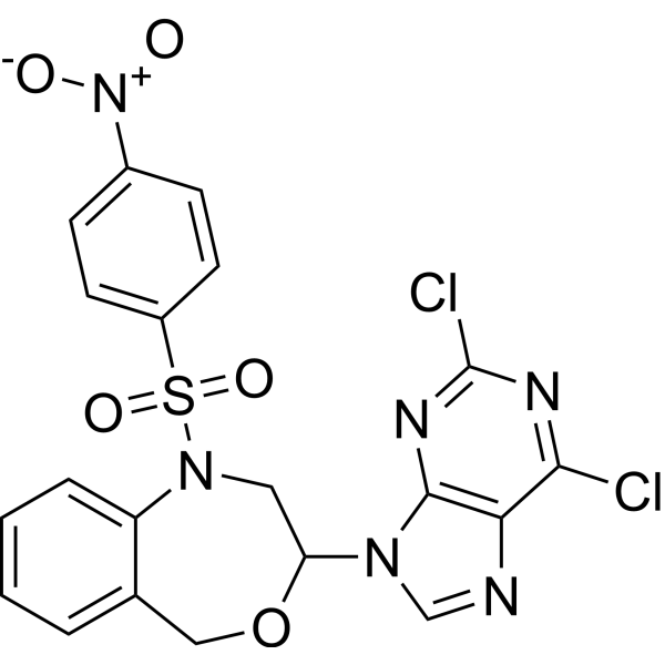
Bozepinib
Bozepinib is an anticancer agent. Bozepinib induces Apoptosis, and inhibits the HER-2 signaling pathway as well as JNK and ERK kinases. Bozepinib induces the down-regulation of c-MYC, β-catenin and SOX2 proteins, and the up-regulation of GLI-3. Bozepinib can be used in the research of breast cancer and colon cancer.
For research use only. We do not sell to patients.
- CAS No.: 1207993-83-6
- Formula: C20H14Cl2N6O5S
- Molecular Weight:521.33
-
Storage:
Please store the product under the recommended conditions in the Certificate of Analysis.

To place orders, for customer services and technical support, please contact: MedChemExpress USA
Tel: 609-228-6898 E-mail: [email protected] [email protected]


-
 Biological Activity
-
Chemical Information
- Protocols
- Purity & Documentation
- References
-
 Help & FAQs


All EGFR Isoforms
More

Biological Activity
Description
IC50 & Target
[1]|
HER2 |
Cellular Effect
|
Cell Line

|
Type | Value | Description | References |
|---|---|---|---|---|
| MCF7 | IC50 |
0.355 μM
|
Antiproliferative activity against human breast cancer MCF-7 cells.
Antiproliferative activity against human breast cancer MCF-7 cells.
|
p-Nitrobenzenesulfonamides-and-their-fluorescent |
| MDA-MB-231 | IC50 |
0.166 μM
|
Antiproliferative activity against human MDA-MB 231 breast cancer cells assessed as reduction in cell viability incubated for a total of 6 days by sulforhodamine-B assay.
Antiproliferative activity against human MDA-MB 231 breast cancer cells assessed as reduction in cell viability incubated for a total of 6 days by sulforhodamine-B assay.
|
24946763 |
| MDA-MB-468 | IC50 |
0.850 μM
|
Antiproliferative activity against human MDA-MB 468 breast cancer cells assessed as reduction in cell viability incubated for a total of 6 days by sulforhodamine-B assay.
Antiproliferative activity against human MDA-MB 468 breast cancer cells assessed as reduction in cell viability incubated for a total of 6 days by sulforhodamine-B assay.
|
24946763 |
| SK-BR-3 | IC50 |
0.330 μM
|
Antiproliferative activity against human SKBR-3 breast cancer cells assessed as reduction in cell viability incubated for a total of 6 days by sulforhodamine-B assay.
Antiproliferative activity against human SKBR-3 breast cancer cells assessed as reduction in cell viability incubated for a total of 6 days by sulforhodamine-B assay.
|
24946763 |
| Caco-2 | IC50 |
0.631 μM
|
Antiproliferative activity against human Caco-2 colon cancer cells assessed as reduction in cell viability incubated for a total of 6 days by sulforhodamine-B assay.
Antiproliferative activity against human Caco-2 colon cancer cells assessed as reduction in cell viability incubated for a total of 6 days by sulforhodamine-B assay.
|
24946763 |
| T84 | IC50 |
1.019 μM
|
Antiproliferative activity against human T-84 colon cancer cells assessed as reduction in cell viability incubated for a total of 6 days by sulforhodamine-B assay.
Antiproliferative activity against human T-84 colon cancer cells assessed as reduction in cell viability incubated for a total of 6 days by sulforhodamine-B assay.
|
24946763 |
| HT-29 | IC50 |
1.352 μM
|
Antiproliferative activity against human HT-29 colon cancer cells assessed as reduction in cell viability incubated for a total of 6 days by sulforhodamine-B assay.
Antiproliferative activity against human HT-29 colon cancer cells assessed as reduction in cell viability incubated for a total of 6 days by sulforhodamine-B assay.
|
24946763 |
| SW480 | IC50 |
0.235 μM
|
Antiproliferative activity against human SW-480 colon cancer cells assessed as reduction in cell viability incubated for a total of 6 days by sulforhodamine-B assay.
Antiproliferative activity against human SW-480 colon cancer cells assessed as reduction in cell viability incubated for a total of 6 days by sulforhodamine-B assay.
|
24946763 |
| HCT-116 | IC50 |
0.570 μM
|
Antiproliferative activity against human HCT-116 colon cancer cells assessed as reduction in cell viability incubated for a total of 6 days by sulforhodamine-B assay.
Antiproliferative activity against human HCT-116 colon cancer cells assessed as reduction in cell viability incubated for a total of 6 days by sulforhodamine-B assay.
|
24946763 |
| MCF-10A | IC50 |
1.825 μM
|
Antiproliferative activity against non-tumor human MCF-10A breast cells assessed as reduction in cell viability incubated for a total of 6 days by sulforhodamine-B assay.
Antiproliferative activity against non-tumor human MCF-10A breast cells assessed as reduction in cell viability incubated for a total of 6 days by sulforhodamine-B assay.
|
24946763 |
| CCD-18Co | IC50 |
2.012 μM
|
Antiproliferative activity against non-tumor human CCD-18Co colon cells assessed as reduction in cell viability incubated for a total of 6 days by sulforhodamine-B assay.
Antiproliferative activity against non-tumor human CCD-18Co colon cells assessed as reduction in cell viability incubated for a total of 6 days by sulforhodamine-B assay.
|
24946763 |
| RKO | IC50 |
0.13 μM
|
Antiproliferative activity against human colon cancer RKO cells assessed by Sulforhodamine-B cell survival assay with a total 6-day single-agent treatment.
Antiproliferative activity against human colon cancer RKO cells assessed by Sulforhodamine-B cell survival assay with a total 6-day single-agent treatment.
|
24194639 |
| MCF7 | IC50 |
0.78 μM
|
Antiproliferative activity against human breast cancer MCF-7 cells assessed by Sulforhodamine-B cell survival assay with a total 6-day single-agent treatment.
Antiproliferative activity against human breast cancer MCF-7 cells assessed by Sulforhodamine-B cell survival assay with a total 6-day single-agent treatment.
|
24194639 |
| HCT-116 | IC50 |
0.48 μM
|
Antiproliferative activity against human colon cancer HCT-116 cells assessed by Sulforhodamine-B cell survival assay with a total 6-day single-agent treatment.
Antiproliferative activity against human colon cancer HCT-116 cells assessed by Sulforhodamine-B cell survival assay with a total 6-day single-agent treatment.
|
24194639 |
| RKO | IC50 |
0.09 μM
|
Antiproliferative activity against human colon cancer RKO cells assessed by Sulforhodamine-B cell survival assay with a total 6-day treatment combined with 50 IU/mL interferon alpha.
Antiproliferative activity against human colon cancer RKO cells assessed by Sulforhodamine-B cell survival assay with a total 6-day treatment combined with 50 IU/mL interferon alpha.
|
24194639 |
| MCF7 | IC50 |
0.44 μM
|
Antiproliferative activity against human breast cancer MCF-7 cells assessed by Sulforhodamine-B cell survival assay with a total 6-day treatment combined with 50 IU/mL interferon alpha.
Antiproliferative activity against human breast cancer MCF-7 cells assessed by Sulforhodamine-B cell survival assay with a total 6-day treatment combined with 50 IU/mL interferon alpha.
|
24194639 |
| HCT-116 | IC50 |
0.31 μM
|
Antiproliferative activity against human colon cancer HCT-116 cells assessed by Sulforhodamine-B cell survival assay with a total 6-day treatment combined with 50 IU/mL interferon alpha.
Antiproliferative activity against human colon cancer HCT-116 cells assessed by Sulforhodamine-B cell survival assay with a total 6-day treatment combined with 50 IU/mL interferon alpha.
|
24194639 |
In Vitro
Bozepinib induces apoptosis in breast and colon cancer cells via double-stranded RNA-dependent protein kinase (PKR), and IFNα enhances this apoptotic effect while promoting autophagy and senescence[1].
Bozepinib (at low micromolar concentrations) inhibits cancer stem cell activity, suppresses the formation of mammospheres and colonospheres, eliminates the ALDH+ CSC subpopulation, and regulates key CSC pathway proteins in breast cancer and colon cancer models[1].
Bozepinib (6 days) potently inhibits the proliferation of MCF-7, MDA-MB 231, MDA-MB 468, SKBR-3, Caco-2, T-84, HT-29, SW-480 and HCT-116 cancer cells, with IC50 values ranging from 0.166 μM to 1.352 μM. Meanwhile, compared with non-tumor cells MCF-10A and CCD-18Co, this compound exhibits selective activity against cancer cells, with a maximum selectivity index of 11[2].
Bozepinib (50 μM) inhibits a variety of cancer-related kinases, including JNK, ERKs, HER2, AKT2 and VEGF receptors, in cell-free multikinase screening assays[2].
Bozepinib (5 μM; 2-16 h) inhibits the phosphorylation of HER2, AKT, JNK and ERK1/2 and reduces VEGF levels in SKBR-3, MCF-7 and MDA-MB 468 breast cancer cells, while exerting minimal effects on non-tumorigenic MCF-10A cells[2].
Bozepinib (0.01-5 μM; 4-8 h) dose-dependently inhibits capillary-like structure formation in HUVEC cells at 4 h and 8 h, while maintaining high cell viability and inducing extremely low levels of apoptosis[2].
Bozepinib (0.2-5 μM; 48 h) inhibits the migration of SKBR-3 breast cancer cells and HCT-116 colon cancer cells in a dose-dependent manner in scratch wound healing assays, with a treatment duration of 48 h[2].
Bozepinib (4-16 h) alters gene expression in MDA-MB 468 breast cancer cells. After 4 h and 16 h of exposure, it upregulates antiproliferative and antiangiogenic genes, while downregulating oncogenes and cell cycle-related genes[2].
Bozepinib potently inhibits the proliferation of breast adenocarcinoma MDA-MB-231 cells, with an IC50 of 0.166 µM[3].
Bozepinib (5 µM; 4-24 h) upregulates and activates PKR, induces eIF2α phosphorylation, but has no effect on the expression or phosphorylation of p53 in breast cancer MCF-7 cells and colon cancer HCT-116 cells[3].
MedChemExpress (MCE) has not independently confirmed the accuracy of these methods. They are for reference only. Further protocols information, click here.
-
Cell Line:human breast cancer cell lines (MCF-7, MDA-MB 231, MDA-MB 468, SKBR-3), human colon cancer cell lines (Caco-2, T-84, HT-29, SW-480, HCT-116), non-tumor cell lines (MCF-10A, CCD-18Co)
-
Concentration:dose range
-
Incubation Time:6 days
-
Result:Exhibited selective antiproliferative activity with IC50 values: MCF-7 (0.355 μM), MDA-MB 231 (0.166 μM), MDA-MB 468 (0.850 μM), SKBR-3 (0.330 μM), Caco-2 (0.631 μM), T-84 (1.019 μM), HT-29 (1.352 μM), SW-480 (0.235 μM), HCT-116 (0.570 μM), MCF-10A (1.825 μM), CCD-18Co (2.012 μM).
Showed selectivity indices ranging from 1.48 (HT-29) to 11 (MDA-MB 231).
-
Cell Line:SKBR-3, MCF-7, MDA-MB 468, MCF-10A human breast cell lines
-
Concentration:5 μM
-
Incubation Time:2 h, 4 h, 8 h, 16 h
-
Result:Completely inhibited phosphorylated HER2 after 2 h, inhibited phosphorylated AKT, and decreased total VEGF levels over time in SKBR-3 cells.
Inhibited phosphorylated JNK and ERK1/2 starting at 4 h, with greater inhibition at later time points in MCF-7 cells.
Inhibited phosphorylated JNK and ERK1/2 starting at 8 h in MDA-MB 468 cells.
Weakly up-regulated phosphorylated ERK1/2 at 8 h and returned to control levels by 16 h, while phosphorylated JNK was undetectable in non-tumor MCF-10A cells.
-
Cell Line:human breast cancer MCF-7 cells, human colon cancer HCT-116 cells
-
Concentration:5 µM
-
Incubation Time:4 h, 8 h, 16 h, 24 h
-
Result:Increased phosphorylation of PKR and its substrate eIF2α in both MCF-7 and HCT-116 cells.
Increased total PKR protein levels, more notably in HCT-116 cells.
Left levels of total p53 and phosphorylated p53 unchanged in both cell lines.
In Vivo
Bozepinib (25 mg/kg; i.p.; three times per week; for 45 consecutive days) inhibits the growth of MDA-MB 468 breast cancer xenografts in BALB/c nude mice and reduces the incidence of lung metastasis to 16.6%[2].
Bozepinib (25 mg/kg; i.p.; three times per week; for 2 consecutive weeks) inhibits the growth of HT-29 colon cancer xenografts in BALB/c nude mice[2].
Bozepinib (100 mg/kg; i.p.; twice weekly; for 29 consecutive days) causes no subacute toxicity in female BALB/c mice[2].
MedChemExpress (MCE) has not independently confirmed the accuracy of these methods. They are for reference only.
-
Animal Model:BALB/c nude (female, 6-8 weeks old, subcutaneous xenograft of MDA-MB 468 human breast cancer cells)[2]
-
Dosage:25 mg/kg
-
Administration:i.p.; three times a week; 45 days
-
Result:Reduced average tumor volume to approximately 30 mm3 by day 45, compared to 150 mm3 in control mice.
Reduced lung metastasis incidence to 16.6%, compared to 83.3% in control mice.
-
Animal Model:BALB/c nude (female, 6-8 weeks old, subcutaneous xenograft of HT-29 human colon cancer cells)[2]
-
Dosage:25 mg/kg
-
Administration:i.p.; three times a week; two weeks
-
Result:Significantly reduced tumor volume from day 3 post-injection onward compared to control mice.
-
Animal Model:BALB/c (female, 6 weeks old)[2]
-
Dosage:100 mg/kg
-
Administration:i.p.; twice a week; 29 days
-
Result:Caused no weight loss, unusual behavior, or histopathologic damage to liver or kidneys.


Chemical Information
-
CAS No. 1207993-83-6
-
Molecular Weight 521.33
-
Formula C20H14Cl2N6O5S
-
SMILES
O=S(N1CC(OCC2=CC=CC=C21)N3C=NC4=C3N=C(N=C4Cl)Cl)(C5=CC=C(C=C5)[N+]([O-])=O)=O
-
Shipping
Room temperature in continental US; may vary elsewhere.
-
Storage
Please store the product under the recommended conditions in the Certificate of Analysis.

Protocols
-
Apoptosis
Apoptosis, also called programmed cell death, is generally characterized by distinct morphological characteristics.
-
TUNEL staining for apoptotic DNA fragmentation
TUNEL staining detects DNA strand breaks by using terminal deoxynucleotidyl transferase to add labeled nucleotides to exposed 3′-OH DNA termini, generating either microscopic staining in fixed cells or tissue sections, or fluorescence/cytometric signal in cell suspensions. TUNEL positivity reflects DNA fragmentation but should not be interpreted alone as definitive apoptosis, because TUNEL can also label necrotic, autolytic, mechanically damaged, or DNA-repair-associated DNA breaks.
-
Annexin V plus membrane-impermeant dye apoptosis staining
Annexin V-based apoptosis assays rely on the detection of phosphatidylserine (PS) externalization from the inner leaflet of the plasma membrane to the outer leaflet, an early biochemical hallmark of apoptosis. Fluorescently labeled Annexin V binds PS in a calcium-dependent manner, enabling identification of early apoptotic cells by flow cytometry or fluorescence microscopy. When combined with a membrane-impermeant DNA-binding dye (e. g. , propidium iodide), this approach allows discrimination between viable (Annexin V−/dye−), early apoptotic (Annexin V+/dye−), and late apoptotic or necrotic (Annexin V+/dye+) cell populations by assessing membrane integrity and PS exposure.
-
Apoptosis Solutions
Apoptosis is a regulated, generally non-lytic cell-death pathway that removes unwanted, damaged, infected, or abnormal cells through coordinated morphological changes, caspase activation, DNA fragmentation, and membrane remodeling. The intrinsic apoptosis pathway is controlled mainly by mitochondrial outer membrane permeabilization, BCL-2 family proteins, cytochrome c release, apoptosome formation, caspase-9 activation, and downstream executioner caspase-3/7 activation. The extrinsic apoptosis pathway is initiated by death receptors such as Fas, TNFR, and TRAIL receptors, which recruit adaptor proteins and activate caspase-8 before engaging executioner caspases or mitochondrial amplification through BID cleavage. Apoptosis is linked to many phenotypes, including cancer cell killing, tissue homeostasis, immune regulation, neurodegeneration, infection response, and treatment-induced cytotoxicity; unresolved questions include how apoptosis interacts with necroptosis, pyroptosis, ferroptos
-
Breast Cancer Modeling
Breast cancer is a heterogeneous cancer, and it has been distinguished into four subtypes: luminal A, luminal B, HER2-positive and basal-like. Molecular mutations, epigenetic alterations, hormone exposure and immune microenvironment are related to the progression of breast cancer.


Purity & Documentation
References
[2]. Ramírez A, et al. HER2-signaling pathway, JNK and ERKs kinases, and cancer stem-like cells are targets of Bozepinib small compound. Oncotarget. 2014 Jun 15;5(11):3590-606. [Content Brief]
[3]. Marchal JA, et al. Bozepinib, a novel small antitumor agent, induces PKR-mediated apoptosis and synergizes with IFNα triggering apoptosis, autophagy and senescence. Drug design, development and therapy. 2013;7:1301-13. [Content Brief]

Calculators
Concentration (start) × Volume (start) = Concentration (final) × Volume (final)


Bozepinib Related Classifications
HY-118032 Related Classifications
 Powered by Bioz