



Flupirtine
Based on 1 publication(s) in Google Scholar
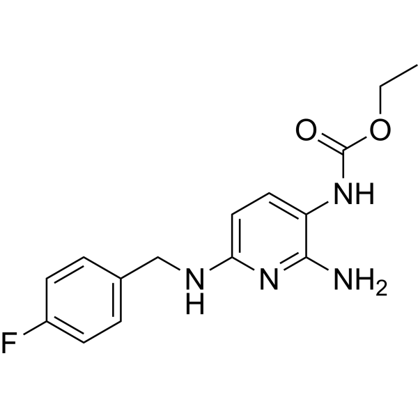
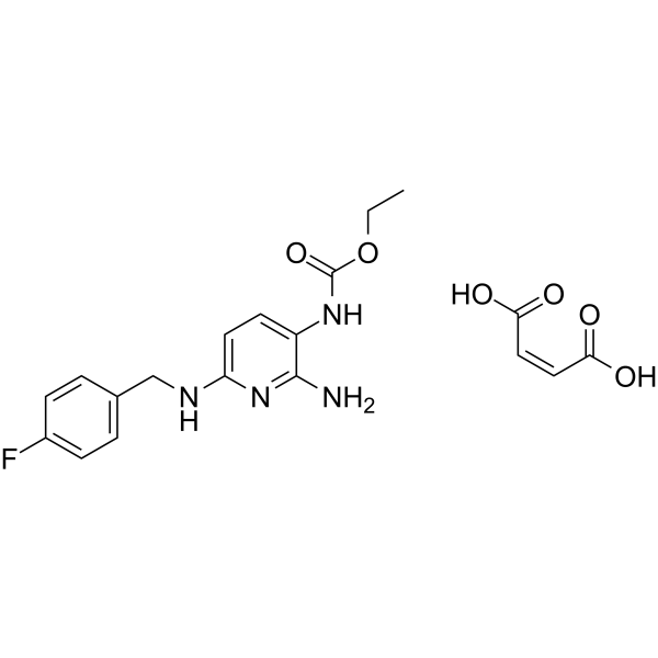
Flupirtine (D 9998) is an orally active, blood-brain barrier-crossing non-opioid analgesic and neuroprotective agent. Flupirtine is a neuronal potassium channel opener (Kv7 activator), a NMDA receptor antagonist and a GABA receptor activator. Flupirtine stabilizes blood-brain-barrier integrity, reduces oxidative stress and brain leukocyte infiltration, enhances angioneurogenesis, suppresses calcium influx, stabilizes neuronal resting membrane potential, and counteracts focal cerebral ischemia. Flupirtine exhibits analgesic, muscle relaxant properties, protects neurons from excitotoxic, ischemic, or cytokine-mediated death. Flupirtine functions as a non-opioid analgesic without antipyretic or antiphlogistic properties, shows no relevant affinity to opiate receptor. Flupirtine can be used for the research of focal cerebral ischemia, pain, Alzheimer’s disease, or multiple sclerosis.
For research use only. We do not sell to patients.
- Purity : 99.94%
- CAS No.: 56995-20-1
- Formula: C15H17FN4O2
- Molecular Weight:304.32
-
Storage:
4°C, protect from light
* In solvent : -80°C, 6 months; -20°C, 1 month (protect from light)

To place orders, for customer services and technical support, please contact: MedChemExpress USA
Tel: 609-228-6898 E-mail: [email protected] [email protected]


-
 Biological Activity
-
Chemical Information
-
Solvent & Solubility
- Protocols
- Purity & Documentation
- References
-
 Help & FAQs




Publications Citing Use of MedChemExpress (MCE) Flupirtine
MoreAll iGluR Isoforms
More

Biological Activity
Description
IC50 & Target
|
NMDA Receptor |
Kv7 |
Cellular Effect
|
Cell Line

|
Type | Value | Description | References |
|---|---|---|---|---|
| HEK293 | EC50 |
0.56 μM
Compound: Retigabine
|
Agonist activity at human KCNQ2/3 expressed in HEK293 cells by FLIPR based thallium influx assay
Agonist activity at human KCNQ2/3 expressed in HEK293 cells by FLIPR based thallium influx assay
|
[PMID: 31416667] |
In Vitro
Flupirtine (0.1-100 μM for tsA cells; 3-30 μM for SCG neurons; 3-30 μM for hippocampal, DRG, DH neurons) enhances currents through KV7 channels in tsA 201 cells expressing KV7.2/7.3 subunits, rat SCG neurons, hippocampal neurons, DRG neurons, and DH neurons with EC50 values ranging from 4.4 to 6.1 μM[2].
Flupirtine (10, 30 μM) modulates GABAA receptors (enhancing low-concentration GABA currents) and NMDA receptors (inhibiting at 30 μM) but not TRPV1, non-NMDA glutamate, or glycine receptors in rat hippocampal neurons[2].
Flupirtine (30 μM) potentiates GABAA receptors in rat DRG, DH, and SCG neurons, with greater leftward shifts of GABA concentration-response curves in DRG and DH neurons than SCG neurons[2].
Flupirtine (0.1-100 μM) is more potent at enhancing GABAA receptor currents in rat DRG neurons (EC50 22 μM) than DH (EC50 53 μM) or hippocampal (EC50 65 μM) neurons, and therapeutic concentrations (3 μM) facilitate KV7 channels and GABAA receptors similarly in DRG/DH neurons[2].
Flupirtine (10-300 μM; 1.5 minutes) antagonizes NMDA-induced currents in cultured rat superior colliculus neurones with an IC50 of 182.1 μM for steady-state responses and 228.6 μM for peak responses[3].
Flupirtine (0.001-10 mM; 24 h) inhibits the growth of U373 MG cells with a GI50 of 0.47 mM, showing significant growth reduction at 1 and 10 mM after 24 h[4].
Flupirtine (1 mM; 24, 48 h) alters the cell cycle distribution of U373 MG cells, decreasing the percentage of cells in the G0-G1 phase compared to control after 24 and 48 h, with significant variations in cell cycle phases observed after 48 h[4].
MedChemExpress (MCE) has not independently confirmed the accuracy of these methods. They are for reference only.
-
Cell Line:U373 malignant glioma (MG) cells
-
Concentration:0.001, 0.01, 0.1, 1, and 10 mM
-
Incubation Time:24 h (0.001-10 mM); 24, 48 h (1 mM)
-
Result:Inhibited U373 MG cell growth with a GI50 of 0.47. Significantly reduced cell growth at high doses (1 and 10 mM) compared to low doses (0.001 to 0.1 mM) and control.
-
Cell Line:U373 malignant glioma (MG) cells
-
Concentration:1 mM
-
Incubation Time:24 h; 48 h
-
Result:Detected G0-G0 phase percentage of 45.48, Sub G0-G0 phase percentage of 2.49, S phase percentage of 24.47, and G₂-M phase percentage of 27.56 after 24 h treatment. Detected G0-G1 phase percentage of 56.39, Sub G0-G1 phase percentage of 1.82, S phase percentage of 18.99, and G2-M phase percentage of 22.80 after 48 h treatment. Observed significant variations in cell cycle phases after 48 h but not 24 h of treatment.
In Vivo
MedChemExpress (MCE) has not independently confirmed the accuracy of these methods. They are for reference only.
-
Animal Model:C57BL6 (male, 22-25 g, transient focal cerebral ischemia via left middle cerebral artery thread occlusion for 30 min)[1]
-
Dosage:1, 5, 10 mg/kg; 10 mg/kg (majority of experiments)
-
Administration:i.p.; single dose; during reperfusion or at 3, 6, 9, 12 h post-stroke (9 h post-stroke for majority of experiments)
-
Result:Reduced infarct volumes on day 2 post-stroke at 5 and 10 mg/kg (1 mg/kg had no effect); reduced infarct volumes, TUNEL+ cell counts, rt-PA-induced acute brain toxicity, Evans blue extravasation, oxidative stress (TBARS formation), intracerebral leukocyte infiltration, calpain activity, JNK and NF-κB activation, and proteasomal activity at 10 mg/kg (given up to 9 h post-stroke); increased STAT6 abundance, neuronal density (NeuN+ cells), CD31+ endothelial cells, Dcx+ immature neurons, and BrdU+/NeuN+ mature neurons on day 84; improved performance in rota rod, tight rope, corner turn, and foot fault tests up to day 84 at 10 mg/kg (given up to 9 h post-stroke).


Chemical Information
-
CAS No. 56995-20-1
-
Appearance Solid
-
Molecular Weight 304.32
-
Formula C15H17FN4O2
-
Color White to light yellow
-
SMILES
CCOC(NC(C(N)=N1)=CC=C1NCC(C=C2)=CC=C2F)=O
-
Synonyms
D 9998
-
Shipping
Room temperature in continental US; may vary elsewhere.
-
Storage
4°C, protect from light
* In solvent : -80°C, 6 months; -20°C, 1 month (protect from light)


Publications (1)
-
Journal Impact Factor
-
Most Recent
-
Biochem Pharmacol
In vitro approach to elucidate the relevance of carboxylesterase 2 and N-acetyltransferase 2 to flupirtine-induced liver injury. [Abstract]2018 Sep:155:242-251. PMID: 30028988


Solvent & Solubility
In Vitro:
DMSO : 83.33 mg/mL (273.82 mM; Need ultrasonic; Hygroscopic DMSO has a significant impact on the solubility of product, please use newly opened DMSO)
Please refer to the solubility information to select the appropriate solvent. Once prepared, please aliquot and store the solution to prevent product inactivation from repeated freeze-thaw cycles.
Storage method and period of stock solution: -80°C, 6 months; -20°C, 1 month (protect from light). When stored at -80°C, please use it within 6 months. When stored at -20°C, please use it within 1 month.
Please refer to the solubility information to select the appropriate solvent. Once prepared, please aliquot and store the solution to prevent product inactivation from repeated freeze-thaw cycles.
Storage method and period of stock solution: -80°C, 6 months; -20°C, 1 month (protect from light). When stored at -80°C, please use it within 6 months. When stored at -20°C, please use it within 1 month.
Concentration (start) × Volume (start) = Concentration (final) × Volume (final)


Purity & Documentation
-
Data Sheet (286 KB)
-
SDS (393 KB)
- English - EN (393 KB)
- Français - FR (393 KB)
- Deutsch - DE (393 KB)
- Norwegian - NO (393 KB)
- Español - ES (393 KB)
- Swedish - SV (393 KB)
- Italian - IT (393 KB)
- Korean - KR (393 KB)
- Portuguese - PT (393 KB)
-
Handling Instructions (2659 KB)
References
[1]. Jaeger HM, et al. The indirect NMDAR inhibitor flupirtine induces sustained post-ischemic recovery, neuroprotection and angioneurogenesis. Oncotarget. 2015;6(16):14033-14044. [Content Brief]
[2]. Klinger F, et al. Concomitant facilitation of GABAA receptors and KV7 channels by the non-opioid analgesic flupirtine. Br J Pharmacol. 2012;166(5):1631-1642. [Content Brief]
[3]. Kornhuber J, et al. Flupirtine shows functional NMDA receptor antagonism by enhancing Mg2+ block via activation of voltage independent potassium channels. Rapid communication. J Neural Transm (Vienna). 1999;106(9-10):857-67. [Content Brief]
[4]. Panchanathan E, et al. Effect of flupirtine on the growth and viability of U373 malignant glioma cells. Cancer Biol Med. 2013;10(3):142-147. [Content Brief]
[5]. Dörr J, et al. Disease Modification in Multiple Sclerosis by Flupirtine-Results of a Randomized Placebo Controlled Phase II Trial. Front Neurol. 2018;9:842. Published 2018 Oct 9. [Content Brief]


Complete Stock Solution Preparation Table
Please refer to the solubility information to select the appropriate solvent. Once prepared, please aliquot and store the solution to prevent product inactivation from repeated freeze-thaw cycles.
Storage method and period of stock solution: -80°C, 6 months; -20°C, 1 month (protect from light). When stored at -80°C, please use it within 6 months. When stored at -20°C, please use it within 1 month.
| Optional Solvent | Concentration Solvent Mass | 1 mg | 5 mg | 10 mg | 25 mg |
|---|---|---|---|---|---|
| DMSO | 1 mM | 3.2860 mL | 16.4301 mL | 32.8601 mL | 82.1504 mL |
| 5 mM | 0.6572 mL | 3.2860 mL | 6.5720 mL | 16.4301 mL | |
| 10 mM | 0.3286 mL | 1.6430 mL | 3.2860 mL | 8.2150 mL | |
| 15 mM | 0.2191 mL | 1.0953 mL | 2.1907 mL | 5.4767 mL | |
| 20 mM | 0.1643 mL | 0.8215 mL | 1.6430 mL | 4.1075 mL | |
| 25 mM | 0.1314 mL | 0.6572 mL | 1.3144 mL | 3.2860 mL | |
| 30 mM | 0.1095 mL | 0.5477 mL | 1.0953 mL | 2.7383 mL | |
| 40 mM | 0.0822 mL | 0.4108 mL | 0.8215 mL | 2.0538 mL | |
| 50 mM | 0.0657 mL | 0.3286 mL | 0.6572 mL | 1.6430 mL | |
| 60 mM | 0.0548 mL | 0.2738 mL | 0.5477 mL | 1.3692 mL | |
| 80 mM | 0.0411 mL | 0.2054 mL | 0.4108 mL | 1.0269 mL | |
| 100 mM | 0.0329 mL | 0.1643 mL | 0.3286 mL | 0.8215 mL |


Flupirtine Related Classifications
HY-17001A Related Classifications
 Powered by Bioz
Keywords
- Flupirtine
- 56995-20-1
- D 9998
- D9998
- D-9998
- Potassium Channel
- iGluR
- GABA Receptor
- KV7 K+ channel
- N-methyl-D-aspartate receptors
- calpain
- c-Jun N-terminal kinase
- inwardly rectifying neuronal potassium channels
- proteasome
- G-protein-regulated inwardly rectifying potassium channels
- GABAA receptor
- nuclear factor-κB
- focal cerebral ischemia
- Inhibitor
- inhibitor
- inhibit