



LPd peroxida probe-1
Based on 2 publication(s) in Google Scholar
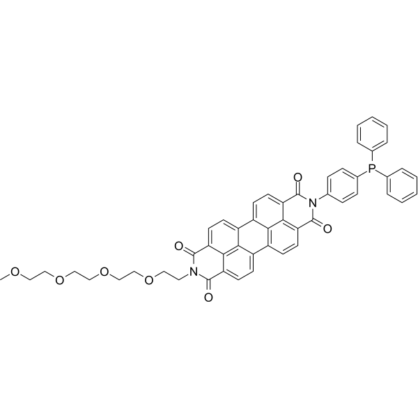
LPd peroxida probe-1, a marker of ferroptosis, is a useful fluorescent probe for investigating the roles of lipid peroxidation in a variety of cell pathophysiologies. LPd peroxida probe-1 reduces lipid hydroperoxides to lipid alcohols and is used for imaging lipid hydroperoxides in living cells.
For research use only. We do not sell to patients.
- Purity : 97.7%
- CAS No.: 1448846-35-2
- Formula: C51H41N2O8P
- Molecular Weight:840.85
-
Storage:
4°C, sealed storage, away from moisture and light
* In solvent : -80°C, 6 months; -20°C, 1 month (sealed storage, away from moisture and light)

To place orders, for customer services and technical support, please contact: MedChemExpress USA
Tel: 609-228-6898 E-mail: [email protected] [email protected]


-
 Biological Activity
-
Chemical Information
- Protocols
- Purity & Documentation
- References
-
 Help & FAQs


-

HY-100579
Ferrostatin-1Ferroptosis Inhibitor -

HY-15763
ErastinFerroptosis Inducer -
HY-D0718
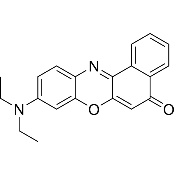
Nile RedHydrophobic Fluorescent Stain -
HY-70062
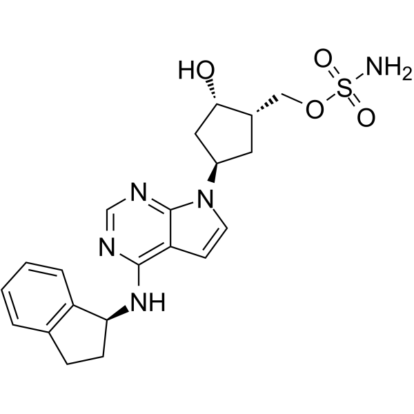
PevonedistatNAE Inhibitor -
HY-100218A
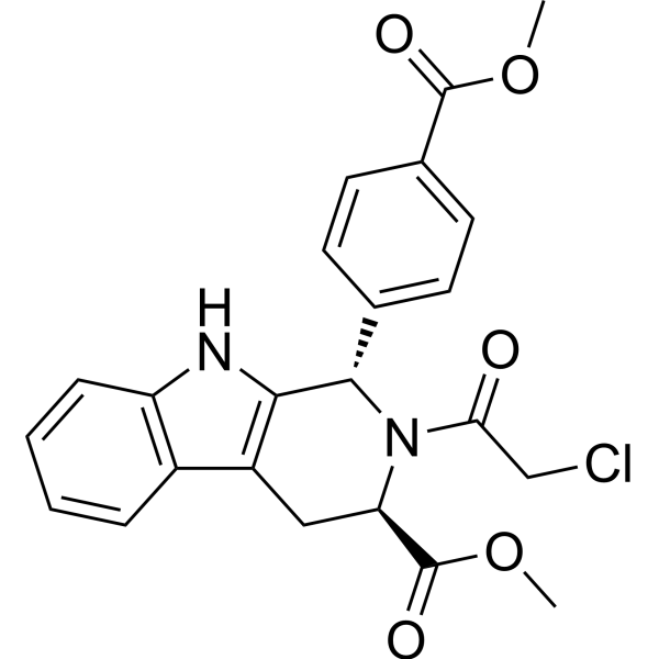
RSL3GPX4 Inhibitor -
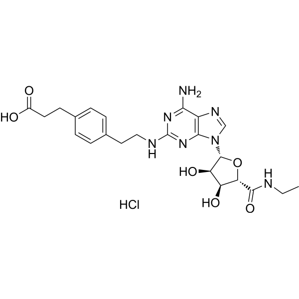
HY-13201A
CGS 21680 HydrochlorideAdenosine A2A Receptor Agonist -

HY-P7085
M-CSF Protein, Mouse -
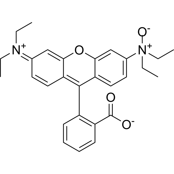
HY-D1533
RhoNox-1Fe(II) Ions Dye -

HY-D1913
Fe2OrangeBivalent Ion Fluorescent Probe -

HY-DY1008
Nile Red (solution)Hydrophobic Fluorescent Stain
Publications Citing Use of MedChemExpress (MCE) LPd peroxida probe-1
More

Biological Activity
Description
In Vitro
Guide (The following is our recommended experimental protocol. This protocol is provided as a reference only, and should be adjusted according to your specific needs)[1].
1. Cells in the culture dish are treated with 50 μM H2O2 in culture medium at 37 °C for 2 h. After removing H2O2, the cells are washed three times with Hank's Balanced Salt Solution (HBSS).
2. Cells are treated with HBSS containing 10 μM LPd peroxida probe at 37 °C for 30 min. After removing the LPd peroxida probe, the cells are washed three times with HBSS.
3. Fluorescence images are acquired using a fluorescence microscope.
MedChemExpress (MCE) has not independently confirmed the accuracy of these methods. They are for reference only.
Emission (Em)
535
Excitation (Ex)
524


Chemical Information
-
CAS No. 1448846-35-2
-
Appearance Solid
-
Molecular Weight 840.85
-
Formula C51H41N2O8P
-
Color Pink to red
-
SMILES
O=C1C2=CC=C3C4=CC=C5C(N(CCOCCOCCOCCOC)C(C6=CC=C(C4=C56)C7=C3C2=C(C(N1C8=CC=C(P(C9=CC=CC=C9)C%10=CC=CC=C%10)C=C8)=O)C=C7)=O)=O
-
Shipping
Room temperature in continental US; may vary elsewhere.
-
Storage
4°C, sealed storage, away from moisture and light
* In solvent : -80°C, 6 months; -20°C, 1 month (sealed storage, away from moisture and light)


Publications (2)
-
Journal Impact Factor
-
Most Recent
-
Free Radic Biol Med
Rab26 mitigates ferroptosis in airway epithelium induced by cigarette smoke through suppression of VDAC1 oligomerization-mediated cGAS-STING activation. [Abstract]2026 Sep:253:177-194. PMID: 42176907 -
Int J Rheum Dis
2024 Aug;27(8):e15297. PMID: 39175261


Purity & Documentation
-
Data Sheet (270 KB)
-
SDS (252 KB)
- English - EN (252 KB)
- Français - FR (252 KB)
- Deutsch - DE (252 KB)
- Norwegian - NO (252 KB)
- Español - ES (252 KB)
- Swedish - SV (252 KB)
- Italian - IT (252 KB)
- Korean - KR (252 KB)
- Portuguese - PT (252 KB)
-
Handling Instructions (2659 KB)
References
[1]. Kazuo Tomita, et al. MiR-7-5p Is Involved in Ferroptosis Signaling and Radioresistance Thru the Generation of ROS in Radioresistant HeLa and SAS Cell Lines. Int J Mol Sci. 2021 Aug 2;22(15):8300. [Content Brief]
[2]. Elizabeth M Kenny, et al. Ferroptosis Contributes to Neuronal Death and Functional Outcome After Traumatic Brain Injury. Crit Care Med. 2019 Mar;47(3):410-418. [Content Brief]

Calculators
Concentration (start) × Volume (start) = Concentration (final) × Volume (final)


LPd peroxida probe-1 Related Classifications
HY-D1412 Related Classifications
 Powered by Bioz