



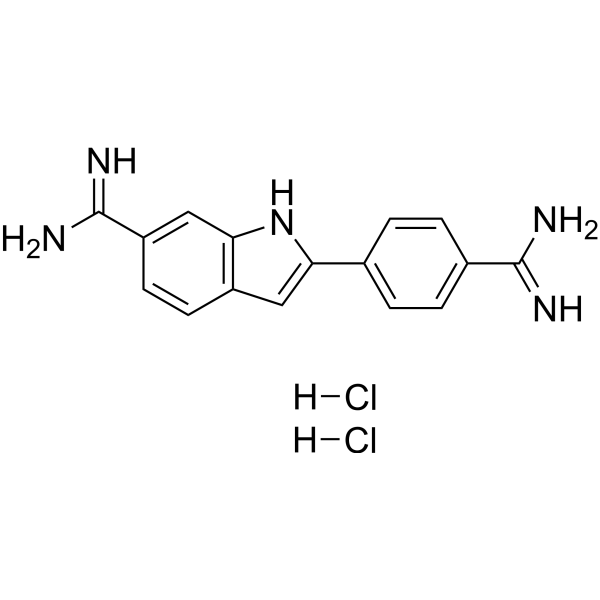
- Formule: C16H17Cl2N5
- Masse moléculaire: 350.25
DAPI (4',6-Diamidino-2-phenylindole) dihydrochloride is a DAPI dye. DAPI is a fluorescent dye that binds strongly to DNA. It binds to the AT base pair of the double-stranded DNA minor groove, and one DAPI molecule can occupy three base pair positions. The fluorescence intensity of DAPI molecules bound to double-stranded DNA is increased by about 20 times, and it is commonly observed with fluorescence microscopy, and the amount of DNA can be determined based on the intensity of fluorescence. DAPI cannot penetrate intact cell membranes and is commonly used for staining damaged and fixed cells. DAPI (Compound 3) is an acid-sensing ion channel 3 (ASIC3) inhibitor. DAPI binds to ASIC3 and blocks the channel function. DAPI can be used in the study of chronic pain treatment (Ex/Em = 356/451 nm).
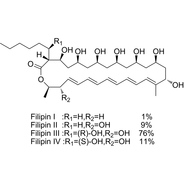
Filipin complex is a potent polyene macrolide antifungal antibiotic. Filipin complex inserts into membranes and sequester cholesterol into complexes and inhibits PRRSV entry. The Filipin complex consists of about 75.8% Filipin III (HY-N6718), 10.8% Filipin IV, 9.1% Filipin II, and 1.2% Filipin I (Ex/Em = 380/430 nm).
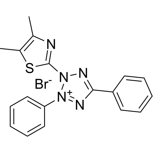
- Formule: C18H16BrN5S
- Masse moléculaire: 414.32
Thiazolyl Blue (MTT) is a cell-permeable and positively charged tetrazolium dye that is used to detect reductive metabolism in cells. Thiazolyl Blue is taken up by cells through the plasma membrane and then reduced to formazan by intracellular NAD (P) H-oxidoreductases. Thiazolyl Blue is frequently used in colorimetric assays to measure cell proliferation, cytotoxicity, and apoptosis.
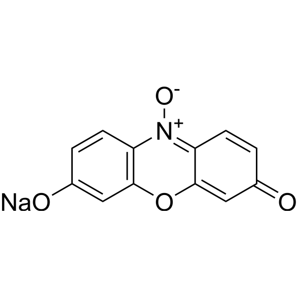
- Formule: C12H6NNaO4
- Masse moléculaire: 251.17
Resazurin sodium (Diazoresorcinol sodium) is a non-toxic, stable, membrane-permeable blue non-fluorescent dye (faintly fluorescent). Resazurin sodium is used as a redox indicator, can be reduced to pink, highly fluorescent Resorufin (Ex=530-560 nm, Em=590 nm) in living cells. Resazurin sodium can be used for the detection of cell viability, toxicity, proliferation, migration and invasion in cells (human, plant and animal, bacterial and fungal).
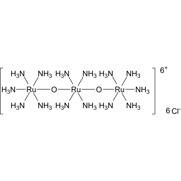
- Formule: Cl6H42N14O2Ru3
- Masse moléculaire: 786.35
Ruthenium red (Ammoniated ruthenium oxychloride) is a polycationic dye widely used for electron microscopy (EM) of cells, tissues and vegetative bacteria. Ruthenium red strongly reacts with phospholipids and fatty acids and binds to acidic mucopolysaccharides. Ruthenium red is a L-type calcium current (ICa) blocker. When using Ruthenium Red to stain plant tissue sections affected by pathogens, the breakdown of mucilaginous substances by the pathogens prevents them from taking up the stain, whereas the cell walls of healthy tissue stain red.
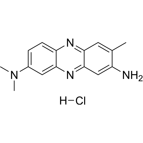
- Formule: C15H17ClN4
- Masse moléculaire: 288.78
Neutral Red is an organic dye commonly used in biology and cytology laboratories. It can be used to stain living cells, secreted proteins and other molecular structures, etc., and has a wide range of applications in cell imaging and staining. In addition, Neutral Red is widely used in industrial fields such as water treatment, food processing and paper manufacturing, for example as an indicator or colorant.

- Formule: C29H28F6N2O4
- Masse moléculaire: 582.53
CouCyCF3 is a fluorescent probe for bacterial cytoplasmic pH, with a pKa of 7.0. CouCyCF3 can be taken up by both Gram-positive and Gram-negative bacteria. Under acidic conditions, the fluorescence of CouCyCF3 in the red channel increases, while its fluorescence in the blue channel increases with elevated pH. A linear quantitative response is achieved via the I_red/I_blue ratio within the pH range of 5.0-7.5, with excitation/emission wavelengths of 445/472 nm (blue) and 640/708 nm (red). CouCyCF3 can be used for studies on bacterial acid tolerance and phagolysosome acidification.

- Formule: C35H44N4O9S2
- Masse moléculaire: 728.88
sCy5DA is a is a fluorescent D-amino acid. sCy5DA incorporates into bacterial peptidoglycan layers and cross-links via transpeptidase action, replacing peptidoglycan stem peptide D-amino acids. sCy5DA labels live bacterial cell walls, nascent peptidoglycan biosynthesis sites, and diverse bacterial species including Gram-negative, Gram-positive, and mycobacteria (Ex/Em = 646/665 nm).
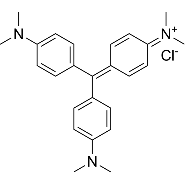
- Formule: C25H30ClN3
- Masse moléculaire: 407.98
Crystal Violet, also known as Gentian violet, methyl violet 10B, is a triphenyl-methane, an alkaline dye that binds to DNA in the nucleus of a cell, staining it a deep purple. It is often used for Gram staining to classify bacteria, or for cell or histological staining[1].
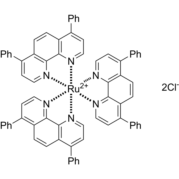
- Formule: C72H48Cl2N6Ru
- Masse moléculaire: 1169.17
Tris(4,7-diphenyl-1,10-phenanthroline)ruthenium(II) dichloride ([Ru(dpp)3]2+) is an electrochemiluminescent (ECL) probe and oxygen-sensitive sensor. Tris(4,7-diphenyl-1,10-phenanthroline)ruthenium(II) dichloride can be used to modify electrode surfaces for the detection of sulfates (S2O82-) and oxalates, based on electrochemical reactions that generate excited-state species, releasing photons through irreversible redox reactions. Tris(4,7-diphenyl-1,10-phenanthroline)ruthenium(II) dichloride utilizes the oxygen quenching of fluorescence mechanism, where fluorescence intensity reflects the metabolic rate of living microorganisms or oxygen levels within cells/tumors, allowing for real-time detection. Tris(4,7-diphenyl-1,10-phenanthroline)ruthenium(II) dichloride's main applications include microbial detection, antibacterial activity studies, and tumor microenvironment research.
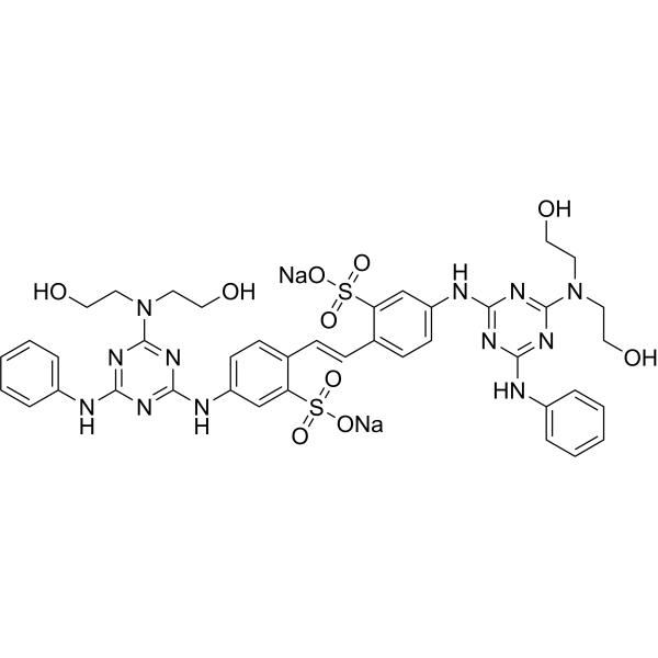
- Formule: C40H42N12Na2O10S2
- Masse moléculaire: 960.95
Fluorescent brightener 28 is a fluorescent whitening agent commonly used in the padding process of the textile industry. Fluorescent brightener 28 is capable of staining polysaccharides such as cellulose, and when the plasma membrane ruptures, it also weakly stains the cytoplasm and strongly stains the cell nucleus. Additionally, Fluorescent brightener 28 can be utilized to detect intracellular chitin in living cells. Fluorescent Brightener 28 also is a visible light emitting diode (LED)-light sensitive photoinitiator for free radical photopolymerizations.
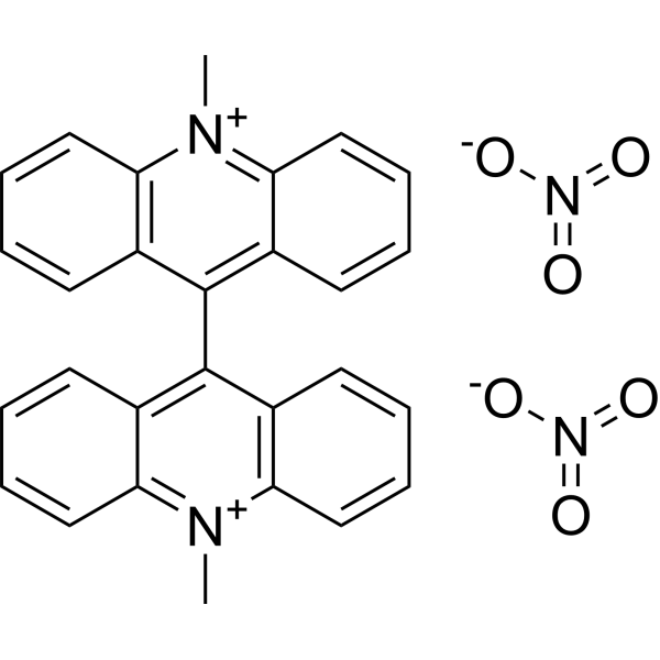
- Formule: C28H22N4O6
- Masse moléculaire: 510.50
Lucigenin is a chemiluminescence probe. Lucigenin can be used to detect the production of endogenous superoxide anion radical (O2-). Lucigenin is extremely sensitive to chloride ions, while it combined with chloride ions, the fluorescence will be quenched. Lucigenin also can be used as a chloride indicator. Ex/Em=455/505 nm.
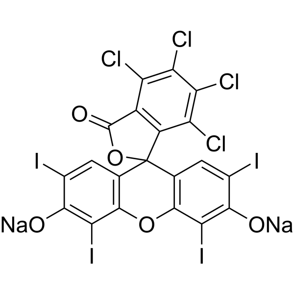
- Formule: C20H2Cl4I4Na2O5
- Masse moléculaire: 1017.64
Acid Red 94 sodium, a synthetic fluorescein derivative, is a deep red dye primarily composed of 4,5,6,7-tetrachloro-2,4,5,7-tetraiodo fluorescein. It is widely used as an ophthalmic diagnostic agent to detect dry or damaged cells on the ocular surface. Acid Red 94 sodium exhibits antitumor activity and can inhibit Friend Leukemia Virus (FLV) infection through photodynamic action. Additionally, Acid Red 94 sodium can inhibit Aβ aggregation through light irradiation. Acid Red 94 sodium holds potential for use in cancer, viral infections, and neurodegenerative disease research.
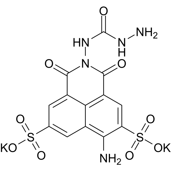
- Formule: C13H9K2N5O9S2
- Masse moléculaire: 521.57
Lucifer Yellow CH dipotassium is a high-intensity fluorescent probe containing free hydrazyl groups. Lucifer Yellow CH can react with fatty aldehydes at room temperature. Lucifer Yellow CH serves as a biological tracer to monitor neuronal branching, regeneration, gap junction detection and characterization, and selective ablation of cells after aldehyde fixation. Lucifer yellow CH displays the maximum excitation/emission of 430 nm/540 nm, respectively.
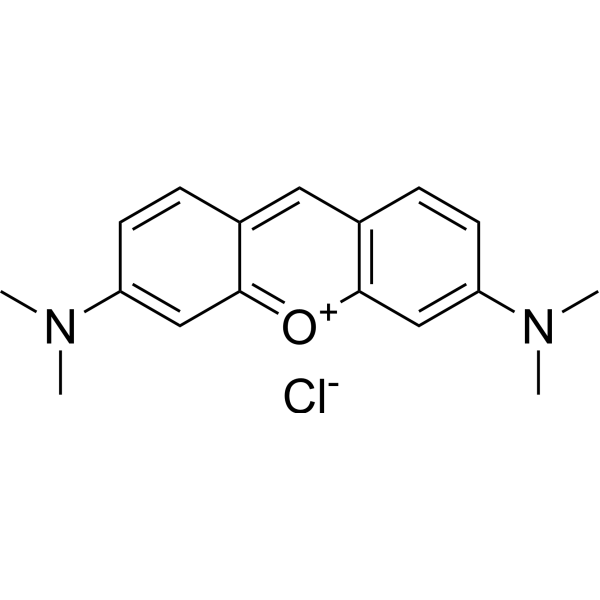
- Formule: C17H19ClN2O
- Masse moléculaire: 302.80
Pyronin Y (Pyronine G) is a cationic dye that intercalates RNA and has been used to target cell structures including RNA, DNA and organelles. Pyronin Y forms fluorescent complexes with double-stranded nucleic acids (especially RNA) enabling semi-quantitative analysis of cellular RNA. Pyronin Y can be used to identify specific RNA subspecies of ribonuclear proteins complexes in live cells.
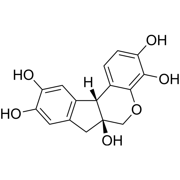
- Formule: C16H14O6
- Masse moléculaire: 302.28
Hematoxylin (Natural Black 1), a naturally occurring flavonoid compound derived from Caesalpinia sappan Linn.. Hematoxylin is a nuclear stain in histology and is also a potent Aβ42 fibrillogenesis inhibitor with an IC50 of 1.6 μM.
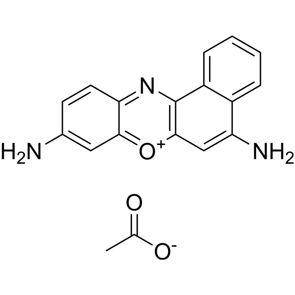
- Formule: C18H15N3O3
- Masse moléculaire: 321.33
Cresyl Violet acetate is a dye, which can be used to stain neurons.
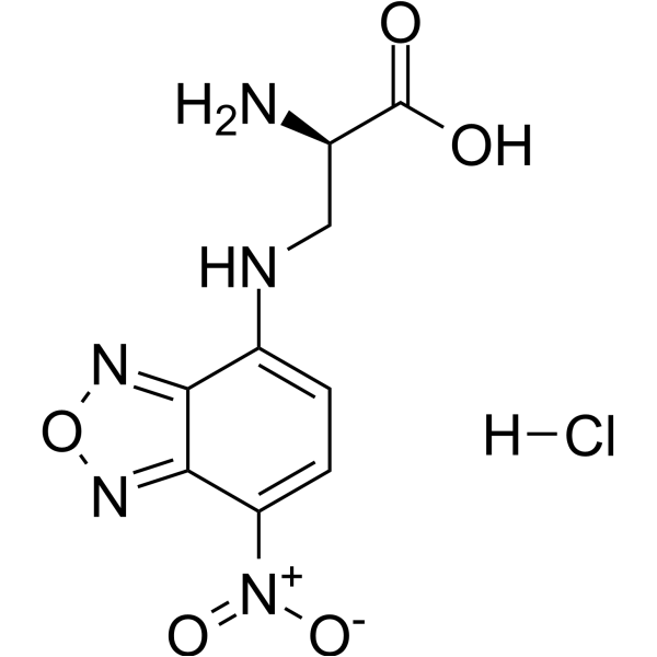
- Formule: C9H10ClN5O5
- Masse moléculaire: 303.66
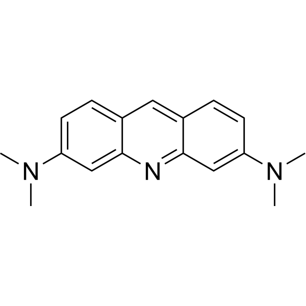
- Formule: C17H19N3
- Masse moléculaire: 265.36
Acridine Orange base is a cell-permeable fluorescent dye that stains organisms (bacteria, parasites, viruses, etc.) bright orange and, when used under appropriate conditions (pH=3.5, Ex=460 nm), distinguishes human cells in green for detection by fluorescence microscopy. Acridine Orange base fluoresces green when bound to dsDNA (Ex=488, Em=520-524) and red when bound to ssDNA (Ex=457, Em=630-644) or ssRNA (Ex=457, Em=630-644), also can be used in cell cycle assays.
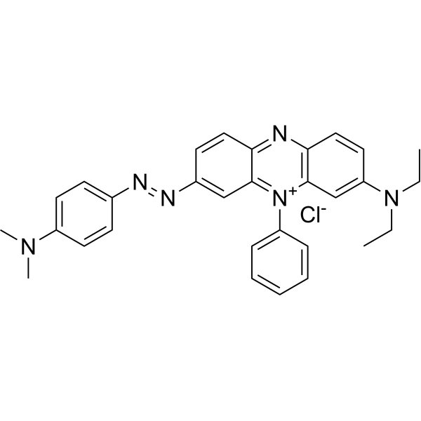
- Formule: C30H31ClN6
- Masse moléculaire: 511.06
Janus green B is a supravital stain. Janus green B staining reaction is oxygen dependent, and is reversibly inhibited by cyanide. Janus Green B has been used to stain structures such as peripheral nerves in living insects, rabbit lymphatic vessels, mitochondria, and mast cells.



