



Figitumumab

Based on 1 Customer Validation
Figitumumab (CP-751871) is a potent and fully human monoclonal anti–insulin-like growth factor 1 receptor (IGF1R) antibody. Figitumumab prevents IGF1 from binding to IGF1R with an IC50 of 1.8 nM.
Nos produits utilisent uniquement pour la recherche. Nous ne vendons pas aux patients.
- Pureté : 99.10%
- CAS No.: 943453-46-1
- Masse moléculaire:144.5 kDa
-
Stockage:
Please store the product under the recommended conditions in the Certificate of Analysis.


To place orders, for customer services and technical support, please contact: MedChemExpress USA
Tel: 609-228-6898 E-mail: [email protected] [email protected]


-
 Activité biologique
-
Chemical Information
- Protocole
- Pureté et documentation
- Références
-
 Help & FAQs


-

HY-P99294
GanitumabAnti-IGF1R Antibody -

HY-P99165
TeprotumumabIGF-1R Inhibitor -

HY-P99218
RobatumumabAnti-human IGF-1R Antibody -

HY-P99189
CixutumumabAnti-IGF-1R mAb -

HY-P99274
XentuzumabIGF1/IGF2 Antagonist -

HY-P99284
DalotuzumabHumanized Anti-IGF1R mAb -
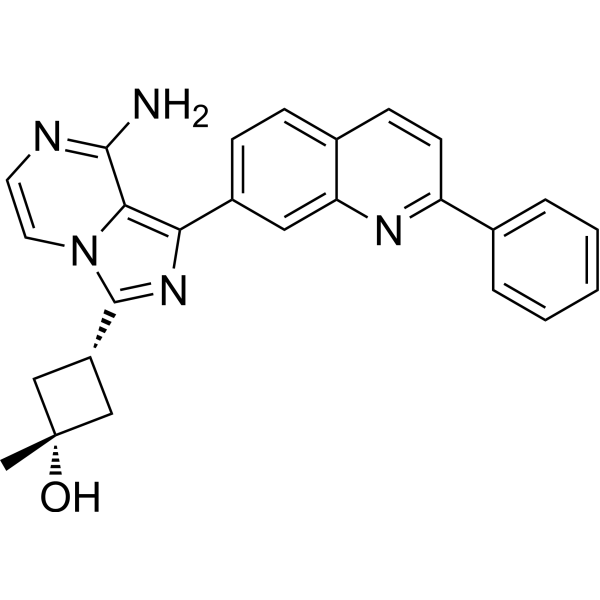
HY-10191
LinsitinibIGF-1R/Insulin Receptor Inhibitor -

HY-P990084
VeligrotugIGF-1 Receptor Antagonist -
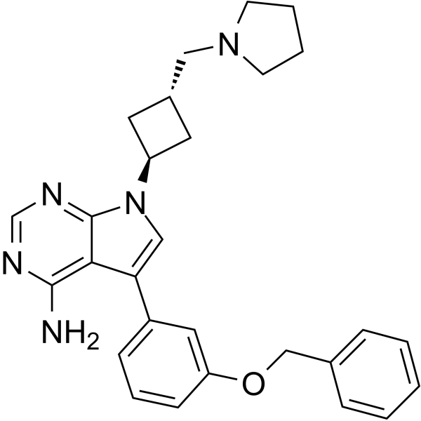
HY-10252
NVP-ADW742IGF-1R Inhibitor -
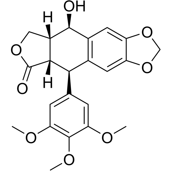
HY-15494
PicropodophyllinApoptosis Inducer -
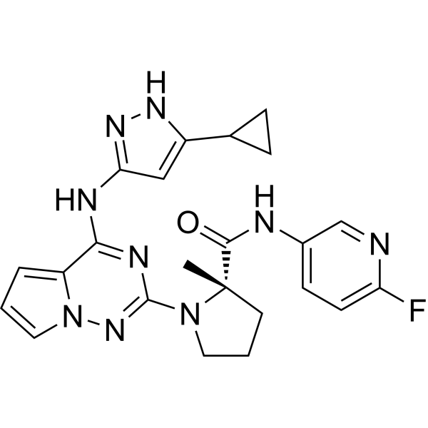
HY-10200
BMS-754807IGF-1R/IR Inhibitor -
HY-50866
NVP-AEW541IGF-1R/IR Inhibitor


Activité biologique
Description
Isotype
Human IgG2 kappa
Recommend Isotype Controls
Species Reactivity
Human
IC50 & Target
IGF1R/CD221
In Vitro
Figitumumab (CP-751871) (152 pM-10 μM; 3 days) inhibits cancer cell proliferation[1].
Figitumumab (1 μg/mL; 1 min or 24 h) induces the down-regulation of IGF-1R[2].
Figitumumab inhibits IGF1-induced autophosphorylation of IGF1R with an IC50 of 0.42 nM, and indirectly inhibits AKT activation[2].
Figitumumab recognizes the IGF-1R/IR heterodimer complex[2].
MedChemExpress (MCE) has not independently confirmed the accuracy of these methods. They are for reference only.
-
Cell Line:Breast, colon, lung small cell, and non–small cell cancer lines
-
Concentration:152 pM-10 μM
-
Incubation Time:3 days
-
Result:15 cell lines (NCIH441, NCIH526, SW403, CACO2, SW48, NCIH524, SKCO1, SNUC1, LS1034, COLO205, MDAMB361, NCIH508, LS513, MCF7, NCIH378) were highly sensitive to the drug at IC50 values ≦ 100 nM.
-
Cell Line:3T3/IGF-1R cell
-
Concentration:1 μg/mL
-
Incubation Time:1 min or 24 h
-
Result:Blocked IGF-I- or IGF-II-induced autophosphorylation of the IGF-1R.
In Vivo
Figitumumab (62.5-500 μg/mouse; i.p.; once) inhibits the growth of s.c. xenografts derived from colon (Colo-205), breast (MCF7), and lung (H460) cancer cell lines in mice[2].
MedChemExpress (MCE) has not independently confirmed the accuracy of these methods. They are for reference only.
-
Animal Model:Female athymic mice (CD-1 nu/nu) bearing NIH3T3/IGF-1R tumors[2]
-
Dosage:31 to 125 μg per mouse
-
Administration:Intraperitoneal injection, once
-
Result:Resulted in a serum Cmax between 12 and 24 hours. At 24 hours, there was a dose-dependent reduction of IGF-1R protein in tumors, with 50% reduction observed at a serum concentration of 15 μg/mL. Resulted in a down-regulation of IGF-1R from the tumor. The half-life in an athymic mouse was determined to be 4 to 6 days by longer-term studies.
-
Animal Model:Female athymic mice (CD-1 nu/nu), human Colo-205 tumor xenograft model[2]
-
Dosage:62.5 μg or 250 μg per mouse
-
Administration:Intraperitoneal injection, once
-
Result:Inhibited the tumor growth.
Essai clinique
| NCT Number | Sponsor | Condition | Start Date |
Phase

|
|---|---|---|---|---|
| NCT01329991 | Plexxikon| | 2011-05 | PHASE1 |
Gene ID
Accession
Conjugated
Unconjugated
Reconsititution
The product can be reconstituted/diluted with sterile PBS or saline.
Format
-
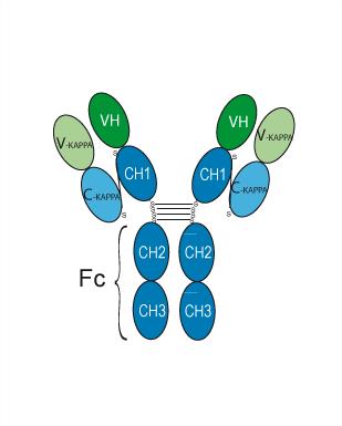
Human IgG2 kappa
Application
ELISA, FACS, Functional assay
Verified Bioactivity
-
 Immobilized IGF-I R Protein, Human (HEK293, His, HY-P72606) can bind Figitumumab. The EC50 for this effect is 17.54 ng/mL.
 -
 Flow cytometric analysis of 1X10⁶ MCF-7 cells with Figitumumab (HY-P99197, red). Cells were fixed with 4% paraformaldehyde. Then stained with the primary antibody at 1/200 dilution for an hour at 4℃. Alexa Fluor 488-conjugated AffiniPure Goat Anti-Human IgG H&L (AF488) (HY-P83776) was used as the secondary antibody at 1/1,000 dilution for 30 minutes at 4℃. Human IgG2 kappa (HY-P99002, blue) was used as the isotype control, cells without incubation with primary antibody were used as the unlabeled control (black).


Chemical Information
-
CAS No. 943453-46-1
-
Appearance Liquid
-
Masse moléculaire 144.5 kDa
-
Color Colorless to light yellow
-
SMILES
[Figitumumab]
-
Synonyms
CP-751871
-
Livraison
Shipping with dry ice.
-
Formulation
Please refer to the lot-specific COA for specific buffer information.
-
Stockage
Please store the product under the recommended conditions in the Certificate of Analysis.


Pureté et documentation
-
Fiche technique (262 KB)
-
SDS (251 KB)
- English - EN (251 KB)
- Français - FR (251 KB)
- Deutsch - DE (251 KB)
- Norwegian - NO (251 KB)
- Español - ES (251 KB)
- Swedish - SV (251 KB)
- Italian - IT (251 KB)
- Korean - KR (251 KB)
- Portuguese - PT (251 KB)
-
Inhibitory Antibodies User Guide (603 KB)
Références
[1]. Pavlicek A, et al. Molecular predictors of sensitivity to the insulin-like growth factor 1 receptor inhibitor Figitumumab (CP-751,871). Mol Cancer Ther. 2013 Dec;12(12):2929-39. [Content Brief]
[2]. Cohen BD, et al. Combination therapy enhances the inhibition of tumor growth with the fully human anti-type 1 insulin-like growth factor receptor monoclonal antibody CP-751,871. Clin Cancer Res. 2005 Mar 1;11(5):2063-73. [Content Brief]

Calculators
Concentration (start) × Volume (start) = Concentration (final) × Volume (final)


Figitumumab Related Classifications
HY-P99197 Related Classifications
 Powered by Bioz