



JS25

Based on 1 Customer Validation
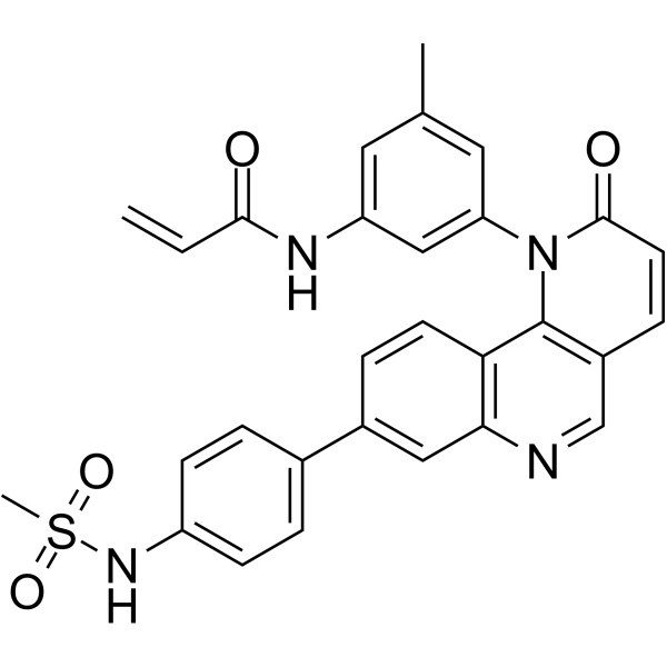
JS25 is a selective and covalent inhibitor of BTK that inactivates BTK with an IC50 value of 5.8 nM by chelating Tyr551. JS25 inhibits cancer cells proliferation, pronounces cell death, and promotes murine xenograft model of Burkitt’s lymphoma. JS25 effectively crosses the blood-brain barrier.
Nos produits utilisent uniquement pour la recherche. Nous ne vendons pas aux patients.
- Pureté : 98.06%
- CAS No.: 2411771-95-2
- Formule: C29H24N4O4S
- Masse moléculaire:524.59
-
Stockage:
4°C, sealed storage, away from moisture
* In solvent : -80°C, 6 months; -20°C, 1 month (sealed storage, away from moisture)

To place orders, for customer services and technical support, please contact: MedChemExpress USA
Tel: 609-228-6898 E-mail: [email protected] [email protected]


-
 Activité biologique
-
Chemical Information
-
Solvant et solubilité
- Protocole
- Pureté et documentation
- Références
-
 Help & FAQs


-
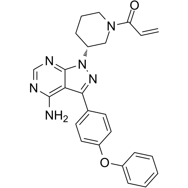
HY-10997
IbrutinibBtk Inhibitor -
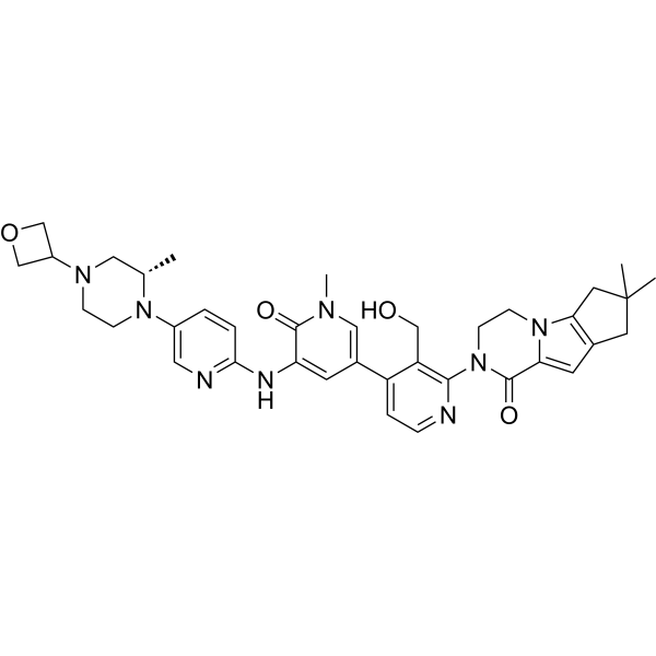
HY-19834
FenebrutinibBtk Inhibitor -
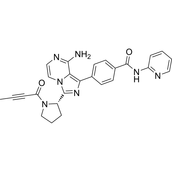
HY-17600
AcalabrutinibBTK Inhibitor -
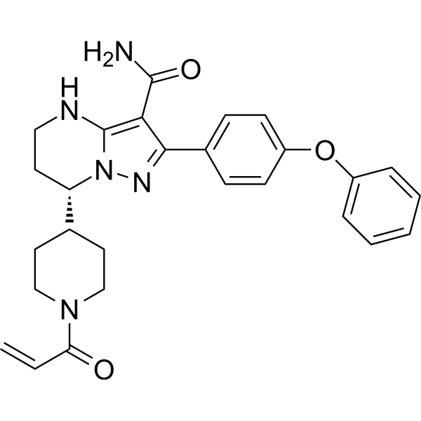
HY-101474A
ZanubrutinibBtk Inhibitor -
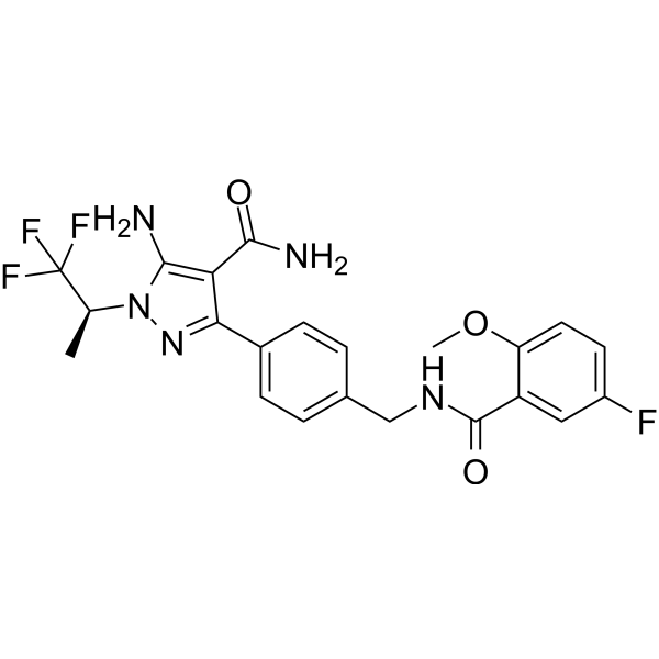
HY-131328
PirtobrutinibBTK Inhibitor -
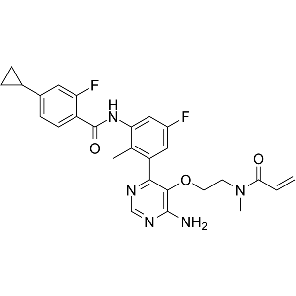
HY-128757
RemibrutinibBTK Inhibitor -
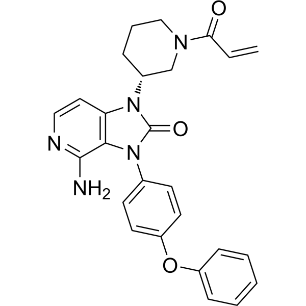
HY-109192
TolebrutinibBTK Inhibitor -
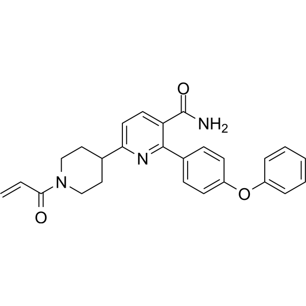
HY-129390
OrelabrutinibBTK Inhibitor -
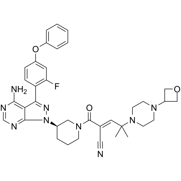
HY-112166
RilzabrutinibBTK Inhibitor -
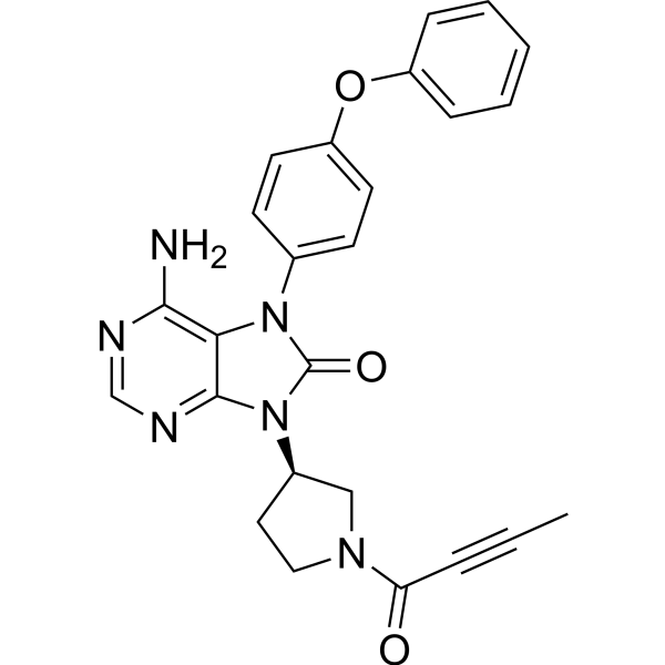
HY-15771
TirabrutinibBTK Inhibitor -
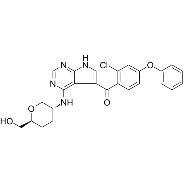
HY-112215
NemtabrutinibBTK Inhibitor -
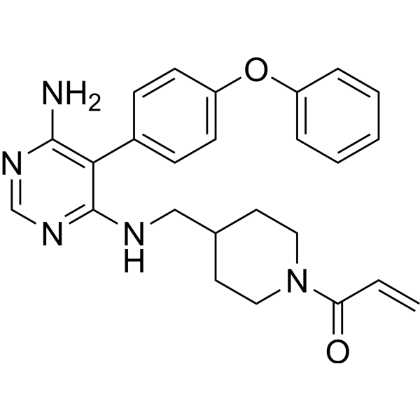
HY-101215
Evobrutinib


Activité biologique
Description
IC50 & Target
IC50: 5.8 nM (BTK), 49.0 nM (BMX), 440 nM (ITK), 190 nM (TXK), 220 nM (TEC), 2.60 μM (BLK)[1]
In Vitro
JS25 (0-50 μM; 72 h) inhibits the proliferation of myeloid and lymphoid B-cell cancer cell lines[1].
JS25 shows inhibitory capability against BTK, BMX, ITK, TXK, TEC, and BLK with IC50s of 28.5 nM, 49.0 nM, 0.44 μM, 0.19 μM, 0.22 μM, and 2.60 μM, respectively; shows little inhibition against other BTK pathway-related proteins (EGFR, ERBB2, and JAK3), with IC50>3 μM. JS25 presents a more favorable selectivity profile than Ibrutinib (HY-10997) and Acalabrutinib (HY-17600)[1].
JS25 (10 μM; 0, 4, 15 h) degrades BTK and inhibits both the catalytic activity and the expression of BTK in tumor cells[1].
JS25 (10 μM; 72 h) inhibits the tumor growth of Burkitt’s lymphoma and induces selective ex vivo cytotoxicity in primary diffuse large B-cell lymphoma (DLBCL) samples[1].
JS25 (15 μM) shows 28.5% perme-ability to the BBB at 24 h in HBEC-5i cell model[1].
MedChemExpress (MCE) has not independently confirmed the accuracy of these methods. They are for reference only.
In Vivo
JS25 (1, 2.5, and 5 μM; injection; every day for 2 days) decreases tumor burden in zebrafish-derived xenografts of chronic lymphocytic leukemia, wich efficacy is better than Ibrutinib (HY-10997)[1].
MedChemExpress (MCE) has not independently confirmed the accuracy of these methods. They are for reference only.
-
Animal Model:Female adult BALB/c/NSG mice with Raji cells (s.c.)[1]
-
Dosage:10 mg/kg and 20 mg/kg
-
Administration:Intraperitoneal injection; every 2 days for 14 days
-
Result:Caused a 30-40% reduction of the subcutaneous tumor and an overall reduction in the percentage of metastasis and secondary tumor formation.
- Cell Cytotoxicity Assay
- Mammalian live/dead viability and cytotoxicity staining
- Patient-Derived Xenograft (PDX)
- Apoptosis Solutions
- Splenic/Portal-Vein Liver Metastasis Xenograft
- Subcutaneous Cell-Line-Derived Xenograft
- Orthotopic Cell-Line Xenograft
- Cell Viability Determination by MTT Colorimetric Assay
- Intraperitoneal/Peritoneal Dissemination Xenograft


Chemical Information
-
CAS No. 2411771-95-2
-
Appearance Solid
-
Masse moléculaire 524.59
-
Formule C29H24N4O4S
-
Color Light yellow to light brown
-
SMILES
C=CC(NC1=CC(C)=CC(N2C(C=CC3=C2C4=C(N=C3)C=C(C=C4)C5=CC=C(C=C5)NS(C)(=O)=O)=O)=C1)=O
-
Livraison
Room temperature in continental US; may vary elsewhere.
-
Stockage
4°C, sealed storage, away from moisture
* In solvent : -80°C, 6 months; -20°C, 1 month (sealed storage, away from moisture)


Solvant et solubilité
In Vitro:
DMSO : 20 mg/mL (38.13 mM; ultrasonic and warming and heat to 60°C; Hygroscopic DMSO has a significant impact on the solubility of product, please use newly opened DMSO)
Please refer to the solubility information to select the appropriate solvent. Once prepared, please aliquot and store the solution to prevent product inactivation from repeated freeze-thaw cycles.
Storage method and period of stock solution: -80°C, 6 months; -20°C, 1 month (sealed storage, away from moisture). When stored at -80°C, please use it within 6 months. When stored at -20°C, please use it within 1 month.
Please refer to the solubility information to select the appropriate solvent. Once prepared, please aliquot and store the solution to prevent product inactivation from repeated freeze-thaw cycles.
Storage method and period of stock solution: -80°C, 6 months; -20°C, 1 month (sealed storage, away from moisture). When stored at -80°C, please use it within 6 months. When stored at -20°C, please use it within 1 month.
Concentration (start) × Volume (start) = Concentration (final) × Volume (final)


Pureté et documentation
-
Fiche technique (279 KB)
-
SDS (252 KB)
- English - EN (252 KB)
- Français - FR (252 KB)
- Deutsch - DE (252 KB)
- Norwegian - NO (252 KB)
- Español - ES (252 KB)
- Swedish - SV (252 KB)
- Italian - IT (252 KB)
- Korean - KR (252 KB)
- Portuguese - PT (252 KB)
-
Instruction de manipulation (2659 KB)
Références


Complete Stock Solution Preparation Table
Please refer to the solubility information to select the appropriate solvent. Once prepared, please aliquot and store the solution to prevent product inactivation from repeated freeze-thaw cycles.
Storage method and period of stock solution: -80°C, 6 months; -20°C, 1 month (sealed storage, away from moisture). When stored at -80°C, please use it within 6 months. When stored at -20°C, please use it within 1 month.
| Optional Solvent | Concentration Solvent Mass | 1 mg | 5 mg | 10 mg | 25 mg |
|---|---|---|---|---|---|
| DMSO | 1 mM | 1.9063 mL | 9.5313 mL | 19.0625 mL | 47.6563 mL |
| 5 mM | 0.3813 mL | 1.9063 mL | 3.8125 mL | 9.5313 mL | |
| 10 mM | 0.1906 mL | 0.9531 mL | 1.9063 mL | 4.7656 mL | |
| 15 mM | 0.1271 mL | 0.6354 mL | 1.2708 mL | 3.1771 mL | |
| 20 mM | 0.0953 mL | 0.4766 mL | 0.9531 mL | 2.3828 mL | |
| 25 mM | 0.0763 mL | 0.3813 mL | 0.7625 mL | 1.9063 mL | |
| 30 mM | 0.0635 mL | 0.3177 mL | 0.6354 mL | 1.5885 mL |


JS25 Related Classifications
HY-151808 Related Classifications
 Powered by Bioz