



Calcium gluconate

Based on 1 Customer Validation
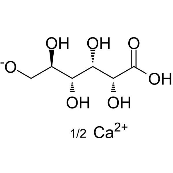
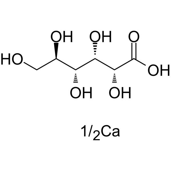
Calcium gluconate is an orally effective calcium salt supplement. Calcium gluconate reduces elevated serum potassium, decreased serum calcium, and postoperative myalgia associated with succinylcholine administration. Calcium gluconate restores calcium homeostasis, skeletal integrity, bone mineralization and bone density, and maintains levels of parathyroid hormone, bone resorption markers and osteoclasts. Calcium gluconate reverses LPS (HY-D1056)-induced ERK phosphorylation, inflammatory cytokine release and acute lung injury, alleviates airway inflammatory damage and suppresses immune responses. Calcium gluconate can be used in research related to postoperative myalgia, osteoporosis/osteomalacia and acute lung injury.
For research use only. We do not sell to patients.
- Purity : 99.86%
- CAS No.: 299-28-5
- Formula: C6H12O7.1/2Ca
- Molecular Weight:216.20
-
Storage:
4°C, sealed storage, away from moisture
* In solvent : -80°C, 6 months; -20°C, 1 month (sealed storage, away from moisture)

To place orders, for customer services and technical support, please contact: MedChemExpress USA
Tel: 609-228-6898 E-mail: [email protected] [email protected]


-
 Biological Activity
-
Chemical Information
-
Solvent & Solubility
- Protocols
- Purity & Documentation
- References
-
 Help & FAQs




Biological Activity
Description
In Vivo
Calcium gluconate (12.5-50 mg/kg; i.p.; single dose) dose-dependently inhibits LPS-induced acute lung injury in male C57BL/6J mice, with the 50 mg/kg dose producing the most significant reductions in histopathological injury, neutrophil influx, pro-inflammatory cytokine release, and ERK phosphorylation[3].
Calcium gluconate (50 mg/kg; i.p.; single dose) does not induce measurable inflammatory changes in healthy male C57BL/6J mice[3].
MedChemExpress (MCE) has not independently confirmed the accuracy of these methods. They are for reference only.
-
Animal Model:Atp4b-deficient (Atp4b−/−)[2]
-
Dosage:1%
-
Administration:p.o.; daily; 4 weeks
-
Result:Maintained serum parathyroid hormone (PTH) levels within normal ranges, matching control mice levels.
Maintained C-terminal telopeptide (CTX) levels within normal ranges, matching control mice levels.
Kept osteoclast numbers per bone perimeter (OcN/BPm) within normal ranges, equivalent to control mice levels.
-
Animal Model:C57BL/6J (male, 4-6 weeks old, LPS-induced ALI)[3]
-
Dosage:12.5 mg/kg; 25 mg/kg; 50 mg/kg
-
Administration:i.p.; single dose
-
Result:Significantly reduced LPS-induced ALI histopathological scores compared to the LPS-only group, with the highest reduction observed at the 50 mg/kg dose.
Significantly reduced LPS-induced neutrophil influx into BALF at 25 mg/kg and 50 mg/kg; the 12.5 mg/kg dose did not produce a significant reduction.
Did not significantly reduce BALF IL-1β levels at 12.5 mg/kg; significantly reduced BALF IL-1β levels at 25 mg/kg and 50 mg/kg.
Did not significantly reduce BALF IL-6 levels at 12.5 mg/kg; significantly reduced BALF IL-6 levels at 25 mg/kg and 50 mg/kg.
Significantly reduced BALF TNF-α levels at 12.5 mg/kg and 25 mg/kg; produced a more significant reduction at 50 mg/kg.
Significantly reduced LPS-induced increases in lung tissue p-ERK expression at 12.5 mg/kg; produced greater, more significant reductions at 25 mg/kg and 50 mg/kg.


Chemical Information
-
CAS No. 299-28-5
-
Appearance Solid
-
Molecular Weight 216.20
-
Formula C6H12O7.1/2Ca
-
Color White to off-white
-
SMILES
OC[C@@H](O)[C@@H](O)[C@H](O)[C@@H](O)C(O)=O.[Ca].[1/2]
-
Shipping
Room temperature in continental US; may vary elsewhere.
-
Storage
4°C, sealed storage, away from moisture
* In solvent : -80°C, 6 months; -20°C, 1 month (sealed storage, away from moisture)


Solvent & Solubility
In Vitro:
H2O : 25 mg/mL (115.63 mM; ultrasonic and warming and heat to 60°C)
Please refer to the solubility information to select the appropriate solvent. Once prepared, please aliquot and store the solution to prevent product inactivation from repeated freeze-thaw cycles.
Storage method and period of stock solution: -80°C, 6 months; -20°C, 1 month (sealed storage, away from moisture). When stored at -80°C, please use it within 6 months. When stored at -20°C, please use it within 1 month.
* Note: If you choose water as the stock solution, please dilute it to the working solution, then filter and sterilize it with a 0.22 μm filter before use.
Please refer to the solubility information to select the appropriate solvent. Once prepared, please aliquot and store the solution to prevent product inactivation from repeated freeze-thaw cycles.
Storage method and period of stock solution: -80°C, 6 months; -20°C, 1 month (sealed storage, away from moisture). When stored at -80°C, please use it within 6 months. When stored at -20°C, please use it within 1 month.
* Note: If you choose water as the stock solution, please dilute it to the working solution, then filter and sterilize it with a 0.22 μm filter before use.
Concentration (start) × Volume (start) = Concentration (final) × Volume (final)

Protocols
-
Kinase activity and phosphorylation assays
Kinase activity assays measure the ability of kinases to transfer phosphate groups from ATP to specific substrates, while phosphorylation assays detect the presence and levels of phosphorylated proteins. Common methods include radiolabeled ATP incorporation (e. g. ,), ADP release detection via bioluminescence (e. g. ,[3]), enzyme-linked immunosorbent assays (ELISA) for phospho-specific epitopes (e. g. ,[6]), and microtiter-based formats for high-throughput screening (e. g. ,[8]). The ADP-Glo assay quantifies kinase activity by measuring ADP produced during phosphorylation using a luciferase-based system. Radiometric assays involve autoradiography or scintillation counting after incorporation of 32P-labeled ATP into substrate proteins. ELISA-based approaches rely on phospho-specific antibodies to detect activated kinases in cell lysates or purified samples.
-
Western Blot
Western blotting (WB) is a commonly used experimental method in molecular biology, biochemistry, and immunogenetics for identifying and quantifying target proteins. It combines gel electrophoresis with immunoassay, enabling researchers to analyze protein expression, post-translational modifications, and molecular weight.
-
Mesenchymal stromal/stem cell osteogenic differentiation
Mesenchymal stromal/stem cells can be induced toward an osteoblast-like lineage in vitro by culture in osteogenic medium containing dexamethasone, ascorbic acid or ascorbate-2-phosphate, and β-glycerophosphate; the differentiation process is commonly evaluated by alkaline phosphatase activity, osteogenic marker expression, collagenous matrix formation, and calcium-rich matrix mineralization. The main readouts are alkaline phosphatase activity as an early osteogenic marker and Alizarin Red S staining as a calcium-deposit readout for mineralized extracellular matrix; Alizarin Red S can be inspected microscopically or extracted and measured colorimetrically at 405 nm.
-
Research Protocol for Endocrine Diseases
Endocrine diseases often arise from disrupted hormone production, hormone signaling, or target-tissue responsiveness; for diabetes-focused endocrine disease models, insulin signaling regulates glucose uptake, hepatic glucose output, lipid metabolism, and β-cell compensation. Type 2 diabetes develops through interacting defects in insulin resistance, β-cell dysfunction, adipose inflammation, hepatic glucose overproduction, altered incretin signaling, and ectopic lipid metabolism. A major unresolved question is whether endocrine dysfunction is driven primarily by target-tissue insulin resistance, intrinsic β-cell failure, immune/inflammatory stress, or combined multi-organ failure that differs by disease stage.
-
Protocol for Kinase activity and phosphorylation assays
Kinase activity assays measure transfer of phosphate from ATP to a protein or peptide substrate, generating phosphorylated substrate, ADP, or incorporated radiolabeled phosphate as the readout; phosphorylation assays measure site-specific phosphorylation in cells or tissues as a proxy for kinase-pathway activation, inhibition, or substrate regulation. Phosphorylation can be detected by phospho-specific Western blot, immunoprecipitation kinase assay, phospho-immunofluorescence, phospho-flow cytometry, luminescent ADP detection, radiolabeled ATP incorporation, or reporter-based pathway assays, and these readouts can be applied to cancer cells, primary neurons, mouse tumors, organoids, inflammatory macrophages, ferroptosis studies, and mitophagy studies when the kinase target is biologically relevant.
-
Research Protocol for Inflammation-related Diseases
The NLRP3 inflammasome is a cytosolic innate immune signaling platform that integrates priming signals and danger-signal activation to promote caspase-1 activation, maturation of IL-1β and IL-18, and gasdermin D-mediated pyroptotic cell death. The core experimental logic is to determine whether inflammatory disease phenotypes are driven by increased NLRP3 expression, ASC-containing inflammasome assembly, caspase-1 cleavage, GSDMD cleavage, and extracellular release of IL-1β/IL-18 rather than by nonspecific cell injury alone. The pathway is strongly linked to inflammation-related disease phenotypes because monosodium urate crystals activate NALP3/NLRP3 inflammasome signaling in gout-like crystal inflammation, cholesterol crystals activate NLRP3 inflammasomes in atherogenesis models, and DSS-induced intestinal inflammation has been reported to involve NLRP3 inflammasome activity. However, experimental colitis studies also show context-dependent protective effects of NLRP3 inflammasome co
-
Inhalation Toxicity Study
Inhalation toxicity studies expose rodents to a controlled aerosol, vapor, gas, or smoke atmosphere and assess respiratory and systemic toxicity using exposure-atmosphere characterization, clinical observations, body and organ weights, bronchoalveolar lavage fluid, histopathology, blood chemistry, hematology, and, when included, molecular endpoints such as transcriptomics, proteomics, lipidomics, or tissue burden analysis. The primary biological readouts are airway irritation, pulmonary inflammation, cytotoxicity, altered surfactant or lipid homeostasis, impaired particle clearance, and tissue remodeling, reflected by BALF cell differentials, BALF protein, LDH, phosphatase activities, cytokines, lung weight, microscopic respiratory-tract lesions, and retained lung burden.
-
Osteoclast differentiation from monocyte/macrophage precursors
Osteoclast differentiation is an in vitro induction assay in which monocyte/macrophage-lineage precursors are exposed to macrophage colony-stimulating factor (M-CSF) and receptor activator of NF-κB ligand (RANKL), generating multinucleated osteoclasts that are commonly identified by tartrate-resistant acid phosphatase (TRAP) staining and functionally confirmed by resorption pits on dentin, bone, or mineralized substrates. M-CSF supports survival and expansion of osteoclast precursors, while RANKL binding to RANK drives osteoclast commitment, fusion, maturation, and resorptive function; osteoprotegerin inhibits this pathway by binding RANKL and preventing RANK activation. The main readouts are the number of TRAP-positive multinucleated cells, formation of F-actin rings, and resorbed surface area; TRAP-positive multinucleated cells indicate osteoclast differentiation, whereas pit formation on dentin, bone, or mineralized coating indicates functional bone-resorbing activity.


Purity & Documentation
-
Data Sheet (276 KB)
-
SDS (393 KB)
- English - EN (393 KB)
- Français - FR (393 KB)
- Deutsch - DE (393 KB)
- Norwegian - NO (393 KB)
- Español - ES (393 KB)
- Swedish - SV (393 KB)
- Italian - IT (393 KB)
- Korean - KR (393 KB)
- Portuguese - PT (393 KB)
-
Handling Instructions (2659 KB)
References
[1]. Shrivastava OP, et al. Calcium gluconate pretreatment for prevention of succinylcholine-induced myalgia. Anesth Analg. 1983;62(1):59-62. [Content Brief]
[2]. Krause M, et al. Calcium gluconate supplementation is effective to balance calcium homeostasis in patients with gastrectomy. Osteoporos Int. 2015;26(3):987-995. [Content Brief]
[3]. Liu L, et al. Effects of calcium gluconate on lipopolysaccharide-induced acute lung injury in mice. Biochem Biophys Res Commun. 2018;503(4):2931-2935. [Content Brief]


Complete Stock Solution Preparation Table
Please refer to the solubility information to select the appropriate solvent. Once prepared, please aliquot and store the solution to prevent product inactivation from repeated freeze-thaw cycles.
Storage method and period of stock solution: -80°C, 6 months; -20°C, 1 month (sealed storage, away from moisture). When stored at -80°C, please use it within 6 months. When stored at -20°C, please use it within 1 month.
| Optional Solvent | Concentration Solvent Mass | 1 mg | 5 mg | 10 mg | 25 mg |
|---|---|---|---|---|---|
| H2O | 1 mM | 4.6253 mL | 23.1267 mL | 46.2535 mL | 115.6337 mL |
| 5 mM | 0.9251 mL | 4.6253 mL | 9.2507 mL | 23.1267 mL | |
| 10 mM | 0.4625 mL | 2.3127 mL | 4.6253 mL | 11.5634 mL | |
| 15 mM | 0.3084 mL | 1.5418 mL | 3.0836 mL | 7.7089 mL | |
| 20 mM | 0.2313 mL | 1.1563 mL | 2.3127 mL | 5.7817 mL | |
| 25 mM | 0.1850 mL | 0.9251 mL | 1.8501 mL | 4.6253 mL | |
| 30 mM | 0.1542 mL | 0.7709 mL | 1.5418 mL | 3.8545 mL | |
| 40 mM | 0.1156 mL | 0.5782 mL | 1.1563 mL | 2.8908 mL | |
| 50 mM | 0.0925 mL | 0.4625 mL | 0.9251 mL | 2.3127 mL | |
| 60 mM | 0.0771 mL | 0.3854 mL | 0.7709 mL | 1.9272 mL | |
| 80 mM | 0.0578 mL | 0.2891 mL | 0.5782 mL | 1.4454 mL | |
| 100 mM | 0.0463 mL | 0.2313 mL | 0.4625 mL | 1.1563 mL |
* Note: If you choose water as the stock solution, please dilute it to the working solution, then filter and sterilize it with a 0.22 μm filter before use.


Calcium gluconate Related Classifications
HY-W145486 Related Classifications
 Powered by Bioz
Keywords