



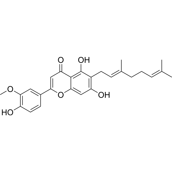
Cannflavin A

Based on 1 Customer Validation
Cannflavin A can be isolated from Cannabis sativa L.. Cannflavin A has anti-cancer, neuroprotective and anti-inflammatory activity. Cannflavin A inhibits Aβ1-42 aggregation. Cannflavin A also inhibits kynurenine-3-monooxygenase (KMO) (IC50 = 29.4 μM). Cannflavin A activates apoptosis via caspase-3 cleavage. Cannflavin A exerts anti-inflammatory effects by inhibiting pro-inflammatory enzymes, including prostaglandin E2 and cytochrome c oxidases I and II in PC12 cell line.
Nos produits utilisent uniquement pour la recherche. Nous ne vendons pas aux patients.
- Pureté : 99.20%
- CAS No.: 76735-57-4
- Formule: C26H28O6
- Masse moléculaire:436.50
-
Stockage:
-20°C, sealed storage, away from moisture
* In solvent : -80°C, 6 months; -20°C, 1 month (sealed storage, away from moisture)

To place orders, for customer services and technical support, please contact: MedChemExpress USA
Tel: 609-228-6898 E-mail: [email protected] [email protected]


-
 Activité biologique
-
Chemical Information
-
Solvant et solubilité
- Protocole
- Pureté et documentation
- Références
-
 Help & FAQs




Activité biologique
Description
Cellular Effect
|
Cell Line

|
Type | Value | Description | References |
|---|---|---|---|---|
| A549 | IC50 |
1.8 μM
Compound: 4
|
Inhibition of mPGES-1 in interleukin-1 beta-stimulated human A549 cells microsomal membranes assessed as reduction in conversion of PGH2 to PGE2 pre-incubated for 15 mins followed by PGH2 substrate addition measured after 1 min by RP-HPLC method
Inhibition of mPGES-1 in interleukin-1 beta-stimulated human A549 cells microsomal membranes assessed as reduction in conversion of PGH2 to PGE2 pre-incubated for 15 mins followed by PGH2 substrate addition measured after 1 min by RP-HPLC method
|
[PMID: 28165233] |
In Vitro
Cannflavin A (10 μM, 48 h) significantly inhibits the neurotoxicity across all concentrations of Aβ1-42 in PC12 neuronal cells[2].
Cannflavin A (2.5-40 μM) displays an IC50 value of 29.4 μM against human KMO protein[3].
Cannflavin A (75 μM) demonstrates competitive binding to KMO protein with Ro 61-8048[3].
MedChemExpress (MCE) has not independently confirmed the accuracy of these methods. They are for reference only.


Chemical Information
-
CAS No. 76735-57-4
-
Appearance Solid
-
Masse moléculaire 436.50
-
Formule C26H28O6
-
Color Light yellow to yellow
-
SMILES
OC1=C2C(OC(C3=CC(OC)=C(C=C3)O)=CC2=O)=CC(O)=C1C/C=C(C)/CC/C=C(C)\C
-
Structure Classification
-
Initial Source
-
Livraison
Room temperature in continental US; may vary elsewhere.
-
Stockage
-20°C, sealed storage, away from moisture
* In solvent : -80°C, 6 months; -20°C, 1 month (sealed storage, away from moisture)


Solvant et solubilité
In Vitro:
DMSO : ≥ 30 mg/mL (68.73 mM; Hygroscopic DMSO has a significant impact on the solubility of product, please use newly opened DMSO)
* "≥" means soluble, but saturation unknown.
Please refer to the solubility information to select the appropriate solvent. Once prepared, please aliquot and store the solution to prevent product inactivation from repeated freeze-thaw cycles.
Storage method and period of stock solution: -80°C, 6 months; -20°C, 1 month (sealed storage, away from moisture). When stored at -80°C, please use it within 6 months. When stored at -20°C, please use it within 1 month.
Please refer to the solubility information to select the appropriate solvent. Once prepared, please aliquot and store the solution to prevent product inactivation from repeated freeze-thaw cycles.
Storage method and period of stock solution: -80°C, 6 months; -20°C, 1 month (sealed storage, away from moisture). When stored at -80°C, please use it within 6 months. When stored at -20°C, please use it within 1 month.
Concentration (start) × Volume (start) = Concentration (final) × Volume (final)


Pureté et documentation
-
Fiche technique (271 KB)
-
SDS (393 KB)
- English - EN (393 KB)
- Français - FR (393 KB)
- Deutsch - DE (393 KB)
- Norwegian - NO (393 KB)
- Español - ES (393 KB)
- Swedish - SV (393 KB)
- Italian - IT (393 KB)
- Korean - KR (393 KB)
- Portuguese - PT (393 KB)
-
Instruction de manipulation (2659 KB)
Références
[1]. Rea KA, et al. Biosynthesis of cannflavins A and B from Cannabis sativa L. Phytochemistry. 2019 Aug;164:162-171. [Content Brief]
[2]. Eggers C, et al. Novel cannabis flavonoid, cannflavin A displays both a hormetic and neuroprotective profile against amyloid β-mediated neurotoxicity in PC12 cells: Comparison with geranylated flavonoids, mimulone and diplacone. Biochem Pharmacol. 2019 Nov;169:113609. [Content Brief]
[3]. Puopolo T, et al. Gram-Scale Preparation of Cannflavin A from Hemp (Cannabis sativa L.) and Its Inhibitory Effect on Tryptophan Catabolism Enzyme Kynurenine-3-Monooxygenase. Biology (Basel). 2022 Sep 28;11(10):1416. [Content Brief]
[4]. Tomko AM, et al. Anti-cancer properties of cannflavin A and potential synergistic effects with gemcitabine, cisplatin, and cannabinoids in bladder cancer. J Cannabis Res. 2022 Jul 22;4(1):41. [Content Brief]


Complete Stock Solution Preparation Table
Please refer to the solubility information to select the appropriate solvent. Once prepared, please aliquot and store the solution to prevent product inactivation from repeated freeze-thaw cycles.
Storage method and period of stock solution: -80°C, 6 months; -20°C, 1 month (sealed storage, away from moisture). When stored at -80°C, please use it within 6 months. When stored at -20°C, please use it within 1 month.
| Optional Solvent | Concentration Solvent Mass | 1 mg | 5 mg | 10 mg | 25 mg |
|---|---|---|---|---|---|
| DMSO | 1 mM | 2.2910 mL | 11.4548 mL | 22.9095 mL | 57.2738 mL |
| 5 mM | 0.4582 mL | 2.2910 mL | 4.5819 mL | 11.4548 mL | |
| 10 mM | 0.2291 mL | 1.1455 mL | 2.2910 mL | 5.7274 mL | |
| 15 mM | 0.1527 mL | 0.7637 mL | 1.5273 mL | 3.8183 mL | |
| 20 mM | 0.1145 mL | 0.5727 mL | 1.1455 mL | 2.8637 mL | |
| 25 mM | 0.0916 mL | 0.4582 mL | 0.9164 mL | 2.2910 mL | |
| 30 mM | 0.0764 mL | 0.3818 mL | 0.7637 mL | 1.9091 mL | |
| 40 mM | 0.0573 mL | 0.2864 mL | 0.5727 mL | 1.4318 mL | |
| 50 mM | 0.0458 mL | 0.2291 mL | 0.4582 mL | 1.1455 mL | |
| 60 mM | 0.0382 mL | 0.1909 mL | 0.3818 mL | 0.9546 mL |


Cannflavin A Related Classifications
HY-W748591 Related Classifications
 Powered by Bioz