



Carbamyl-PAF

Based on 1 Customer Validation
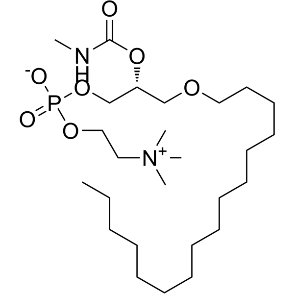
Carbamyl-PAF (C-PAF) is a metabolically stable platelet-activating factor analog and PAF receptor agonist that activates G protein-coupled PAF receptors, triggering calcium mobilization, NF-κB activation, and downstream signaling, thereby inducing neutrophil recruitment, inflammatory responses, and ROS generation. Carbamyl-PAF is used in research on obesity, pulmonary inflammation, Zellweger syndrome, and non-melanoma skin cancer.
For research use only. We do not sell to patients.
- Purity : 99.6%
- CAS No.: 91575-58-5
- Formula: C26H55N2O7P
- Molecular Weight:538.70
-
Storage:
Solution, -20°C, 2 years

To place orders, for customer services and technical support, please contact: MedChemExpress USA
Tel: 609-228-6898 E-mail: [email protected] [email protected]


-
 Biological Activity
-
Chemical Information
- Protocols
- Purity & Documentation
- References
-
 Help & FAQs


-
HY-128694
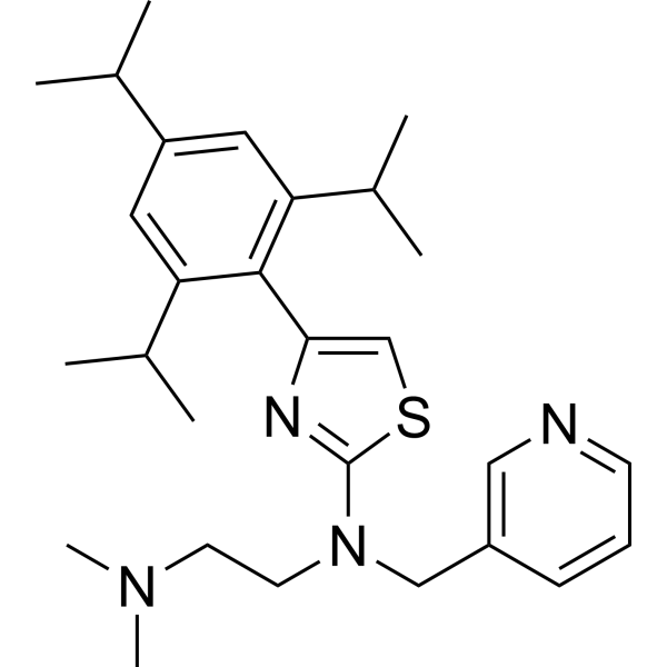
ForopafantPAF Receptor Antagonist -

HY-137798
Chromozym PLLiposome -
HY-130345
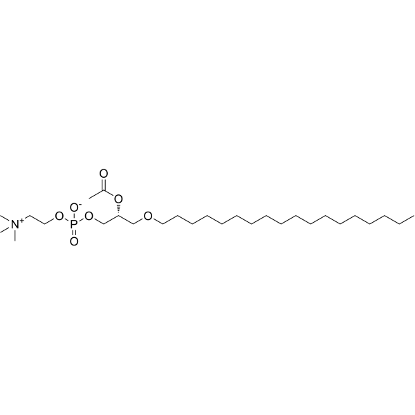
PAF (C18)Platelet Activating Factor -
HY-12357
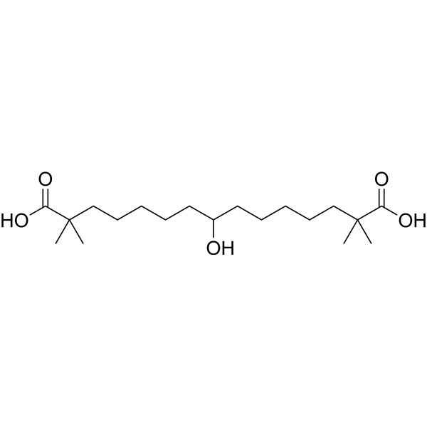
Bempedoic acidACL Inhibitor/AMPK Activator -
HY-A0183
PhosphatidylserineAnti-Inflammatory/Immunosuppressive Signal -
HY-108634
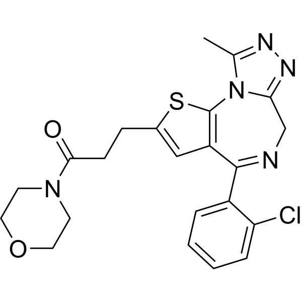
ApafantPAF Receptor Antagonist -
HY-109897
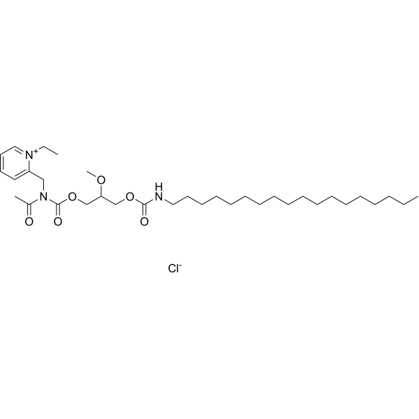
CV-6209PAF Antagonist -
HY-116084S
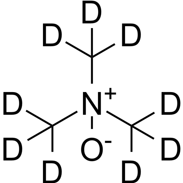
Trimethylamine N-oxide-d9Stable Isotope -

HY-W040255
1-Palmitoyl-2-glutaryl-sn-glycero-3-phosphocholineOxidized Phospholipid -
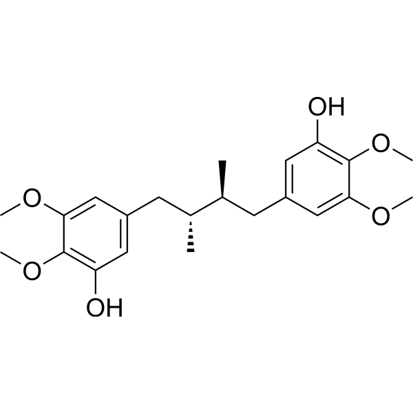
HY-N7343
PregomisinNatural Compound -

HY-148971A
PtdIns-(345)-P3 (12-dipalmitoyl) sodiumPIP3 Analog -
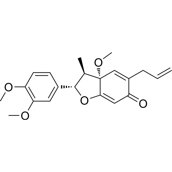
HY-N10663
KadsurenonePlatelet-activating Factor Antagonist


Biological Activity
Description
In Vitro
Carbamyl-PAF (C-PAF) (3 h) induces a classically activated macrophage phenotype in BMMs by upregulating TNF-α and IL-6 and downregulating arginase 1[2].
Carbamyl-PAF (100 nM; 4 h) reduces PAR2 protein expression and induces NF-κB p65 nuclear translocation in RAW 264.7 mouse macrophages[3].
Carbamyl-PAF (10 nM; 3 h) upregulates Ucp1 mRNA expression in 3T3-L1 adipocytes via the PAF receptor, suggesting its role in cellular thermogenesis[2].
Carbamyl-PAF (100 nM; 7 days) decreases neurosphere formation and promotes a differentiated epithelial phenotype in PE-derived neurospheres from adult BALB/cJ mouse ciliary epithelium, while inhibiting the expression of retinal progenitor and pluripotency markers[5].
Carbamyl-PAF (100 nM; 3 days) inhibits the proliferation of cultured normal human epidermal keratinocytes[6].
Carbamyl-PAF (100 nM; 7 days) alone does not affect cornified envelope formation, but reverses Apafant (WEB 2086) (HY-108634)-induced stimulation of cornified envelope formation in cultured normal human epidermal keratinocytes[6].
Carbamyl-PAF (0.5 mM; 80 seconds) pretreatment restores NMDA (HY-17551) (50 μM)-induced cytosolic calcium responses to wild-type levels in Pxr1-/- mouse embryonic neuronal cultures[8].
Carbamyl-PAF (100 nM) induces Ca2+ mobilization in RAW 264.7 mouse macrophages, whereas blockade of PAR2 with ENMD1068 (HY-124748) impairs the amplitude and responsiveness of this Carbamyl-PAF-induced signal[3].
Carbamyl-PAF (100 nM; 1-3 h) increases PAR2 mRNA expression in RAW 264.7 mouse macrophages[3].
Carbamyl-PAF (100 nM) induces Ca2+ mobilization in RAW 264.7 mouse macrophages[4].
Carbamyl-PAF (10 μM) induces reactive oxygen species production in Pam 212 keratinocytes[9].
MedChemExpress (MCE) has not independently confirmed the accuracy of these methods. They are for reference only. Further protocols information, click here.
-
Cell Line:3T3-L1 adipocytes
-
Concentration:10 nM
-
Incubation Time:3 h
-
Result:Induced the expression of Ucp1 mRNA.
These effects were absent in WEB2086-treated cells.
-
Cell Line:RAW 264.7 murine macrophages
-
Concentration:100 nM
-
Incubation Time:20 min
-
Result:Confirmed the presence of PAR2 in the PAFR immunoprecipitates.
Showed increased signal after co-stimulation of PAFR and PAR2 with their respective agonists compared to activation with C-PAF alone.
-
Cell Line:RAW 264.7 murine macrophages
-
Concentration:100 nM
-
Incubation Time:4 h
-
Result:Showed constitutive PAR2 expression in RAW 264.7 murine macrophages.
Impaired PAR2 expression in C-PAF-stimulated cells compared to non-stimulated cells.
Increased NF-κB (p65) transcription factor nuclear fluorescence compared to control cells.
PAR2 blockade with ENMD1068 reduced NF-κB (p65) nuclear fluorescence.
-
Cell Line:RAW 264.7 murine macrophages
-
Concentration:100 nM
-
Incubation Time:1, 2, 3 h
-
Result:Increased PAR2 mRNA at all analyzed time points (1, 2, and 3 h) compared to non-stimulated cells.
-
Cell Line:RAW 264.7 murine macrophages
-
Concentration:100 nM
-
Incubation Time:4 h
-
Result:Decreased PAR2 protein expression at 4 h.
Increased NF-κB p65 nuclear translocation.
-
Cell Line:RAW 264.7 murine macrophages
-
Concentration:100 nM
-
Incubation Time:1, 2, 3 h
-
Result:Increased PAR2 mRNA expression at 1, 2, and 3 h compared to non-stimulated cells.
-
Cell Line:Cultured human normal epidermal keratinocytes
-
Concentration:100 nM
-
Incubation Time:3 days
-
Result:Inhibited keratinocyte proliferation at 100 nM, an effect blocked by WEB 2086 (100 nM).
-
Cell Line:Cultured human normal epidermal keratinocytes
-
Concentration:100 nM
-
Incubation Time:7 days
-
Result:Had no effect on cornified envelope formation alone but reversed the WEB 2086 (100 nM)-induced stimulation of cornified envelope formation.
In Vivo
Carbamyl-PAF (100 nM/20 µL; i.n.) induces perivascular and peribronchial lung inflammation in BALB/c mice[3].
Carbamyl-PAF (100 nM; i.n.) increases leukocyte presence in the mouse mesenteric microcirculation[3].
Carbamyl-PAF (100 nM; i.n.; 20 µL) induces pulmonary inflammation characterized by leukocyte infiltration and elevated MPO and NAG levels in BALB/c mice, whereas blockade of PAR2 with ENMD1068 attenuates this inflammation[4].
Carbamyl-PAF (100 nM; i.n.; 20 µL) induces the production of neutrophil chemoattractants CXCL1 and CXCL2 in the lungs of BALB/c mice, whereas blocking PAR2 with ENMD1068 reduces this production[4].
Carbamyl-PAF (100 nM; i.n.) promotes leukocyte rolling and adhesion in the mesenteric microcirculation of BALB/c mice, a process that is inhibited by the PAR2 antagonist ENMD1068[4].
Carbamyl-PAF (0.25-2.5 mg/kg; i.p.; twice daily; E8 to E18) causes death of pregnant mice within 48 h, thus precluding assessment of its effects on neuronal migration[8].
Carbamyl-PAF (500 pM; i.p.) induces 8-oxo-dG formation and apoptosis in C57BL/6 mouse skin, acting as a mediator that promotes in vivo oxidative DNA damage [9].
Carbamyl-PAF (500 pM; i.p.) induces immunosuppression in vivo and acts as a mediator in suppressing delayed-type hypersensitivity responses in UV-irradiated mice[9].
MedChemExpress (MCE) has not independently confirmed the accuracy of these methods. They are for reference only.
-
Animal Model:BALB/c (male, 8-10 weeks old, 25-30 g)[3]
-
Dosage:10-8, 10-7, 10-6 M/20 µL
-
Administration:i.n.; single dose
-
Result:Increased the number of neutrophils in BALF at all doses utilized (10-8-10-6 M) compared to PBS-instilled mice, peaking 24 h after 10-7 M instillation.
-
Animal Model:BALB/c (male, 8-10 weeks old, 25-30 g)[3]
-
Dosage:10-7 M/20 µL
-
Administration:i.n.; single dose
-
Result:Lungs displayed perivascular and peribronchiolar inflammation with leukocyte infiltration after 24 h.
-
Animal Model:BALB/c (male, 8-10 weeks old, 25-30 g)[3]
-
Dosage:10-7 M
-
Administration:i.n.; single dose
-
Result:Increased the number of leukocytes surrounding the vessels in the mesentery microcirculation compared to PBS-injected mice.
-
Animal Model:BALB/c (male, 25-30 g, 8-10 weeks old)[4]
-
Dosage:10-8, 10-7, 10-6 M
-
Administration:i.n.; single dose
-
Result:Increased the number of neutrophils in BALF at all doses utilized (10-8-10-6 M) compared to PBS-instilled mice.
Peaked 24 h after 10-7 M instillation.
-
Animal Model:BALB/c (male, 25-30 g, 8-10 weeks old)[4]
-
Dosage:10-7 M
-
Administration:i.n.; single dose
-
Result:Induced perivascular and peribronchiolar inflammation with leukocyte infiltration after 24 h, with a histopathology score of approximately 7.
Increased MPO and NAG levels in lung tissue.
Reduced cellular infiltration in the perivascular region, impaired lung inflammation as assessed by the histopathology score, and reduced levels of both MPO and NAG when pretreated with ENMD1068 compared to C-PAF-instilled mice.\nIncreased the levels of neutrophil chemokines CXCL1 and CXCL2 in BALF compared to control mice (CXCL1: approximately 390 pg/mL at 1 h, 480 pg/mL at 4 h; CXCL2: approximately 280 pg/mL at 1 h, 300 pg/mL at 4 h).
Decreased the levels of CXCL1 and CXCL2 measured in BALF at 1 h and 4 h after instillation when treated with ENMD1068 compared to C-PAF-treated mice.
Low levels of CXCL1 persisted in ENMD1068-treated mice 12 h after instillation, whereas CXCL2 returned to basal levels 12 h after instillation.
-
Animal Model:BALB/c (male, 25-30 g, 8-10 weeks old)[4]
-
Dosage:10-7 M
-
Administration:i.n.; single dose
-
Result:Increased the number of leukocytes surrounding the vessels compared to PBS-injected mice.
Reduced the number of adherent, rolling, and velocity of leukocytes in the mesentery microcirculation following PAR2 antagonist ENMD1068 treatment in C-PAF-treated mice.
-
Animal Model:Pxr1 heterozygous mice (pregnant females mated with Pxr1 heterozygous males)[8]
-
Dosage:0.25, 2.5 mg/kg
-
Administration:i.p.; twice daily; E8 to E18
-
Result:All pregnant dams administered 0.25 mg/kg (n = 3) or 2.5 mg/kg (n = 3) died of shock within the first 48 hours after initiation of administration (E8).
-
Animal Model:C57BL/6 mice[9]
-
Dosage:500 pM
-
Administration:i.p.; single dose
-
Result:Increased 8-oxo-dG formation in the skin with statistically significant induction compared to normal skin.
Induced a high number of TUNEL-positive apoptotic cells in the skin.
-
Animal Model:C57BL/6 mice[9]
-
Dosage:500 pM
-
Administration:i.p.; single dose
-
Result:Induced immune suppression as measured by suppression of the delayed-type hypersensitivity response to Candida albicans.


Chemical Information
-
CAS No. 91575-58-5
-
Appearance Liquid
-
Molecular Weight 538.70
-
Formula C26H55N2O7P
-
Color Colorless to light yellow
-
SMILES
CNC(O[C@H](COCCCCCCCCCCCCCCCC)COP(OCC[N+](C)(C)C)([O-])=O)=O
-
Synonyms
C-PAF
-
Shipping
Room temperature in continental US; may vary elsewhere.
-
Storage
Solution, -20°C, 2 years

Protocols
-
ROS/oxidative-stress fluorescent staining
ROS/oxidative-stress fluorescent staining uses cell-permeant fluorogenic probes that become fluorescent after oxidation inside cells or tissues; commonly used examples include DCFH-DA/DCFDA for broad cellular oxidant detection, DHE for superoxide-related signal detection, MitoSOX for mitochondrial superoxide-related signal detection, and CellROX probes for oxidative-stress-associated fluorescence readouts. The assay detects probe oxidation rather than a single ROS species unless the probe and analysis method have been chemically validated for that species. DCFH-DA enters cells, is deacetylated by intracellular esterases to DCFH, and produces fluorescent DCF after oxidation, so the readout is used as an operational measure of total cellular oxidative stress rather than a species-specific ROS measurement. DHE and MitoSOX can report superoxide-related oxidation, but red fluorescence alone can include non-specific ethidium-like oxidation products; HPLC or optimized spectral approaches are
-
Zymosan-Induced Peritonitis
Zymosan-induced peritonitis is a sterile acute-inflammation model produced by intraperitoneal injection of zymosan, a yeast cell-wall particle preparation, followed by quantification of leukocyte recruitment and soluble inflammatory mediators in peritoneal lavage fluid. Low-dose zymosan peritonitis is commonly used as a self-resolving acute inflammation model in which neutrophil recruitment occurs early and monocyte/macrophage accumulation follows later. The assay readouts include total peritoneal leukocyte number, differential neutrophil and monocyte/macrophage counts, peritoneal cytokines and chemokines, plasma or peritoneal exudation, and optional lipidomic or metabolomic changes during inflammation and resolution. Early neutrophil recruitment after zymosan depends strongly on complement and mast-cell C5a receptor signaling, whereas later monocyte recruitment is linked to MCP-1/CCL2 production.
-
Research Protocol for Inflammation-related Diseases
The NLRP3 inflammasome is a cytosolic innate immune signaling platform that integrates priming signals and danger-signal activation to promote caspase-1 activation, maturation of IL-1β and IL-18, and gasdermin D-mediated pyroptotic cell death. The core experimental logic is to determine whether inflammatory disease phenotypes are driven by increased NLRP3 expression, ASC-containing inflammasome assembly, caspase-1 cleavage, GSDMD cleavage, and extracellular release of IL-1β/IL-18 rather than by nonspecific cell injury alone. The pathway is strongly linked to inflammation-related disease phenotypes because monosodium urate crystals activate NALP3/NLRP3 inflammasome signaling in gout-like crystal inflammation, cholesterol crystals activate NLRP3 inflammasomes in atherogenesis models, and DSS-induced intestinal inflammation has been reported to involve NLRP3 inflammasome activity. However, experimental colitis studies also show context-dependent protective effects of NLRP3 inflammasome co
-
Research Protocol for Metabolic Diseases
AMP-activated protein kinase, AMPK, is a conserved cellular energy sensor that responds to reduced cellular energy status and coordinates metabolism by increasing ATP-generating catabolic pathways while suppressing ATP-consuming anabolic processes. In metabolic disease research, the AMPK pathway is experimentally relevant because it regulates hepatic lipid synthesis, fatty acid oxidation, glucose production, skeletal-muscle glucose disposal, mTORC1-linked biosynthesis, autophagy, mitochondrial homeostasis, and whole-body energy balance. The central pathway logic is that energy stress, metformin, exercise-like stimulation, or direct AMPK activators increase AMPKα Thr172 phosphorylation and downstream substrate phosphorylation, including ACC and RAPTOR. Phosphorylation of ACC suppresses lipogenesis and supports fatty acid oxidation, whereas phosphorylation of RAPTOR suppresses mTORC1 signaling and links cellular energy status to growth and protein synthesis control. The pathway is linked


Purity & Documentation
-
Data Sheet (306 KB)
-
SDS (393 KB)
- English - EN (393 KB)
- Français - FR (393 KB)
- Deutsch - DE (393 KB)
- Norwegian - NO (393 KB)
- Español - ES (393 KB)
- Swedish - SV (393 KB)
- Italian - IT (393 KB)
- Korean - KR (393 KB)
- Portuguese - PT (393 KB)
-
Handling Instructions (2659 KB)
References

Calculators
Concentration (start) × Volume (start) = Concentration (final) × Volume (final)


Carbamyl-PAF Related Classifications
HY-137257 Related Classifications
 Powered by Bioz
Keywords