



Cetrorelix
Based on 2 publication(s) in Google Scholar
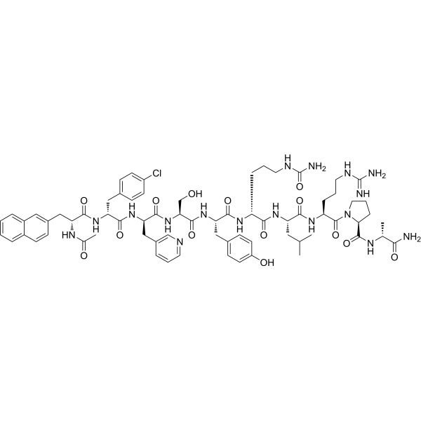
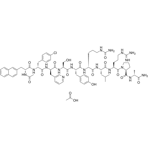
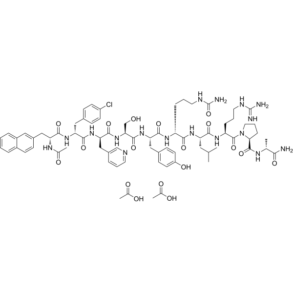
Cetrorelix (SB-75) is a competitive gonadotropin-releasing hormone receptor (GnRHR) antagonist with a Kd of 0.19 nM. Cetrorelix inhibits the secretion of luteinizing hormone (LH) and follicle-stimulating hormone (FSH), thereby suppressing the secretion of downstream sex steroids. Cetrorelix prevents ovulation, induces reversible reproductive shutdown and azoospermia, reduces ovarian fibrosis, improves superovulation outcomes, and inhibits chemotherapy-induced mitochondria-dependent apoptosis. Cetrorelix is applicable for fertility assistance research.
연구목적의 판매만을 진행합니다. 환자를 대상으로 한 판매는 하지 않습니다.
- Purity : 99.82%
- CAS No.: 120287-85-6
- 화학식: C70H92ClN17O14
- 분자량:1431.04
-
보관:
Sealed storage, away from moisture.
Powder -80°C, 2 years , -20°C, 1 year* In solvent : -80°C, 6 months; -20°C, 1 month (sealed storage, away from moisture)

To place orders, for customer services and technical support, please contact: MedChemExpress USA
Tel: 609-228-6898 E-mail: [email protected] [email protected]


-
 Biological Activity
-
Chemical Information
-
용액&용해도
- Protocol
- 순도&문서
- References
-
 Help & FAQs


Publications Citing Use of MedChemExpress (MCE) Cetrorelix
More

Biological Activity
제품 설명
Cellular Effect
|
Cell Line

|
Type | Value | Description | References |
|---|---|---|---|---|
| HEK293 | IC50 |
4.2 nM
Compound: 1 (Cetrorelix)
|
Antagonism of human GnHR receptor, determined in a reporter gene assay in HEK293 cells
Antagonism of human GnHR receptor, determined in a reporter gene assay in HEK293 cells
|
[PMID: 11462984] |
In Vitro
Cetrorelix induces minimal histamine release from rat mast cells, with an ED50 far above pharmacologically relevant plasma concentrations[3].
Cetrorelix does not inhibit EGF-stimulated proliferation of human granulosa cells at pharmacologically relevant concentrations, with inhibitory effects only seen at ~100-fold higher concentrations[3].
MedChemExpress (MCE) has not independently confirmed the accuracy of these methods. They are for reference only.
In Vivo
Cetrorelix (5 μg/day; i.p.; daily; 20 days) alone has no detectable effect on ovarian granulosa cell apoptosis, hormone levels, or mitochondrial function in healthy female Sprague-Dawley rats[1].
Cetrorelix (5 µg/kg; i.p.; once every 3 days or daily; 1 week) significantly increases the number of normal superovulated oocytes in aged female C57BL/6J mice (to 9.8 and 8.7 oocytes, respectively), reduces ovarian fibrosis, and maintains normal oocyte fertilization and fetal development rates[2].
Cetrorelix (~10 μg/kg; s.c.; single dose) immediately suppresses plasma LH by >80% in male castrated rats, with full recovery within 24 hours[3].
Cetrorelix (250-1250 μg/kg; s.c.; single dose) immediately suppresses LH in male castrated cynomolgus monkeys, with suppression lasting for at least 96 hours[3].
MedChemExpress (MCE) has not independently confirmed the accuracy of these methods. They are for reference only.
-
Animal Model:Sprague-Dawley (female, 250 g)[1]
-
Dosage:5 μg/day (pretreatment); 5 μg/day + 20 mg/kg/day (combined with alkylating antineoplastic agent)
-
Administration:i.p.; daily; 10 days (pretreatment); i.p.; daily; 10 days (combined)
-
Result:Reduced granulosa cell apoptotic index from 13.6% to 3.1%.
Restored serum estradiol levels from ~80 pg/mL to ~160 pg/mL.
Preserved normal granulosa cell ultrastructure including intact mitochondria.
Reversed alkylating antineoplastic agent-induced mitochondrial depolarization, resulting in a green fluorescence intensity of 15.36, red fluorescence intensity of 23.16, and a red/green ratio of 1.51.
Inhibited alkylating antineoplastic agent-induced cytochrome c release from mitochondria to cytoplasm.
Suppressed alkylating antineoplastic agent-induced increases in Bax-positive cells from 90% to 25%, caspase-3-positive cells from 85% to 30%, and cytochrome c-positive cells from 80% to 30%.
Restored alkylating antineoplastic agent-reduced Bcl-2-positive cells from 40% to 70%.
-
Animal Model:Sprague-Dawley (female, 250 g)[1]
-
Dosage:5 μg/day
-
Administration:i.p.; daily; 20 days
-
Result:Did not alter granulosa cell apoptotic index (2.8%, similar to control's 2.5%).
Did not change serum estradiol or progesterone levels.
Did not affect granulosa cell ultrastructure, mitochondrial membrane potential, cytochrome c distribution between mitochondria and cytoplasm, or expression levels of Bax, Bcl-2, caspase-3, or cytochrome c.
-
Animal Model:C57BL/6J (female, 52 weeks old, aged)[2]
-
Dosage:5 µg/kg (3 doses over 1 week); 5 µg/kg (7 doses over 1 week)
-
Administration:i.p.; once every 3 days (3 doses over 1 week); daily (7 doses over 1 week)
-
Result:Increased mean total oocyte count to 13.2 (3-dose group) and 11.4 (7-dose group), with normal oocyte counts increased to 9.8 (74.2% normal rate) and 8.7 (76.3% normal rate), respectively, compared to controls (P<0.05).
Achieved fertilization rates of 96.9% (3-dose group) and 88.5% (7-dose group), with birth rates of 58.3% and 51.8%, respectively, comparable to control values.
Reduced ovarian fibrosis, with phalloidin-positive area percentage decreased compared to untreated aged controls (P<0.05).
Showed no significant difference in ovary weight compared to control aged mice.
-
Animal Model:Male castrated rats[3]
-
Dosage:2.5 μg/rat (~10 μg/kg)
-
Administration:s.c.; single dose
-
Result:Suppressed plasma LH concentrations immediately, reaching a nadir at 4 hours with an >80% fall in LH concentrations.
Returned serum LH to normal levels within 24 hours.
-
Animal Model:Male castrated cynomolgus monkeys (*Macaca fascicularis*)[3]
-
Dosage:250 μg/kg; 625 μg/kg; 1250 μg/kg
-
Administration:s.c.; single dose
-
Result:Suppressed LH concentrations immediately across all doses, reaching a nadir at ~12 hours post-injection.
Maintained LH concentrations suppressed for at least 96 hours.
Showed no LH rebound within the 96-hour interval at any dose.
- Fibrosis/Collagen Morphometry
- Reproductive and Developmental Toxicity Study
- Collagen: Sirius Red Staining
- Detection of Apoptosis
- TUNEL staining for apoptotic DNA fragmentation
- Annexin V plus membrane-impermeant dye apoptosis staining
- Research Protocol for Endocrine Diseases
- Connective Tissue: Masson's Trichrome/Collagen Trichrome Staining
- Apoptosis Solutions
Clinical Trial
| NCT Number | Sponsor | Condition | Start Date |
Phase
|
|---|---|---|---|---|
| NCT01329991 | Plexxikon| | 2011-05 | PHASE1 |


Chemical Information
-
CAS No. 120287-85-6
-
Appearance Solid
-
분자량 1431.04
-
화학식 C70H92ClN17O14
-
Color White to off-white
-
Synonyms
SB-75
-
선적
Room temperature in continental US; may vary elsewhere.
-
보관
Sealed storage, away from moisture
Powder -80°C 2 years -20°C 1 year * In solvent : -80°C, 6 months; -20°C, 1 month (sealed storage, away from moisture)


Publications (2)
-
Journal Impact Factor
-
Most Recent
-
Commun Biol
Diurnal testosterone oscillations gate drug delivery via phase separation of ZO-1 at the blood-testis barrier in mice. [Abstract]2026 Jun 1. PMID: 42219451 -
Gen Comp Endocrinol
Neuromodulatory effects of GnRH on the caudal neurosecretory Dahlgren cells in female olive flounder. [Abstract]2021 Jun 1:307:113754. PMID: 33711313


용액&용해도
In Vitro:
DMSO : 25 mg/mL (17.47 mM; Need ultrasonic; Hygroscopic DMSO has a significant impact on the solubility of product, please use newly opened DMSO)
Please refer to the solubility information to select the appropriate solvent. Once prepared, please aliquot and store the solution to prevent product inactivation from repeated freeze-thaw cycles.
Storage method and period of stock solution: -80°C, 6 months; -20°C, 1 month (sealed storage, away from moisture). When stored at -80°C, please use it within 6 months. When stored at -20°C, please use it within 1 month.
Please refer to the solubility information to select the appropriate solvent. Once prepared, please aliquot and store the solution to prevent product inactivation from repeated freeze-thaw cycles.
Storage method and period of stock solution: -80°C, 6 months; -20°C, 1 month (sealed storage, away from moisture). When stored at -80°C, please use it within 6 months. When stored at -20°C, please use it within 1 month.
Concentration (start) × Volume (start) = Concentration (final) × Volume (final)


순도&문서
-
Data Sheet (290 KB)
-
SDS (393 KB)
- English - EN (393 KB)
- Français - FR (393 KB)
- Deutsch - DE (393 KB)
- Norwegian - NO (393 KB)
- Español - ES (393 KB)
- Swedish - SV (393 KB)
- Italian - IT (393 KB)
- Korean - KR (393 KB)
- Portuguese - PT (393 KB)
-
Handling Instructions (2659 KB)
References
[1]. Zhao XJ, et al. GnRH antagonist cetrorelix inhibits mitochondria-dependent apoptosis triggered by chemotherapy in granulosa cells of rats. Gynecologic oncology. 2010 Jul;118(1):69-75. [Content Brief]
[2]. Kanda A, et al. Effect of Cetrorelix administration on ovarian stimulation in aged mice. Experimental animals. 2021 Feb 06;70(1):31-36. [Content Brief]
[3]. Reissmann T, et al. The LHRH antagonist cetrorelix: a review. Human reproduction update. 2000;6(4):322-31. [Content Brief]


Complete Stock Solution Preparation Table
Please refer to the solubility information to select the appropriate solvent. Once prepared, please aliquot and store the solution to prevent product inactivation from repeated freeze-thaw cycles.
Storage method and period of stock solution: -80°C, 6 months; -20°C, 1 month (sealed storage, away from moisture). When stored at -80°C, please use it within 6 months. When stored at -20°C, please use it within 1 month.
| Optional Solvent | Concentration Solvent Mass | 1 mg | 5 mg | 10 mg | 25 mg |
|---|---|---|---|---|---|
| DMSO | 1 mM | 0.6988 mL | 3.4940 mL | 6.9879 mL | 17.4698 mL |
| 5 mM | 0.1398 mL | 0.6988 mL | 1.3976 mL | 3.4940 mL | |
| 10 mM | 0.0699 mL | 0.3494 mL | 0.6988 mL | 1.7470 mL | |
| 15 mM | 0.0466 mL | 0.2329 mL | 0.4659 mL | 1.1647 mL |


Cetrorelix Related Classifications
HY-P0009 Related Classifications
 Powered by Bioz
Keywords