



CID 16020046
Based on 2 publication(s) in Google Scholar
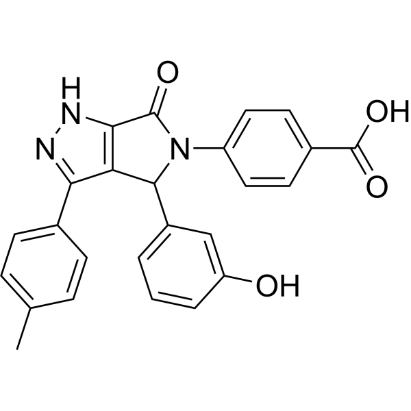
CID 16020046 is a potent and selective GPR55 antagonist and inhibits GPR55 constitutive activity with an IC50 of 0.15 μM. CID 16020046 inhibits GPR55-mediated Ca2+ signaling and GPR55-mediated ERK1/2 phosphorylation. CID 16020046 reduces wound healing in endothelial cells and is involved in the regulation of platelet function.
Nur für Forschungszwecke. Wir verkaufen nicht an Patienten.
- Reinheit : 99.81%
- CAS. Nr.: 834903-43-4
- Formel: C25H19N3O4
- Molecular Weight:425.44
-
Speicherung:Powder -20°C, 3 years , 4°C, 2 years ; In solvent -80°C, 2 years , -20°C, 1 year

To place orders, for customer services and technical support, please contact: MedChemExpress USA
Tel: 609-228-6898 E-mail: [email protected] [email protected]


-
 Biologische Aktivität
-
Chemical Information
-
Lösungsmittel & Löslichkeit
- Protokoll
- Reinheit & Dokumentation
- Verweise
-
 Help & FAQs


-
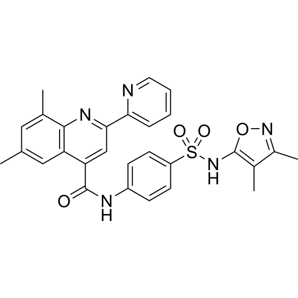
HY-110125
ML-193GPR55 Antagonist -
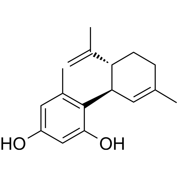
HY-107541
O-1602GPR55 Agonist -
HY-116461
ML-184GPR55 Agonist -
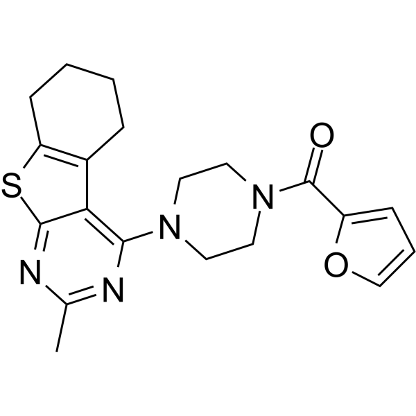
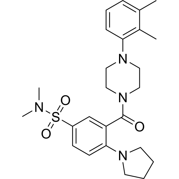
HY-122246
ML192GPR55 Antagonist -

HY-16697R
CID 16020046 (Standard)GPR55 Antagonist -
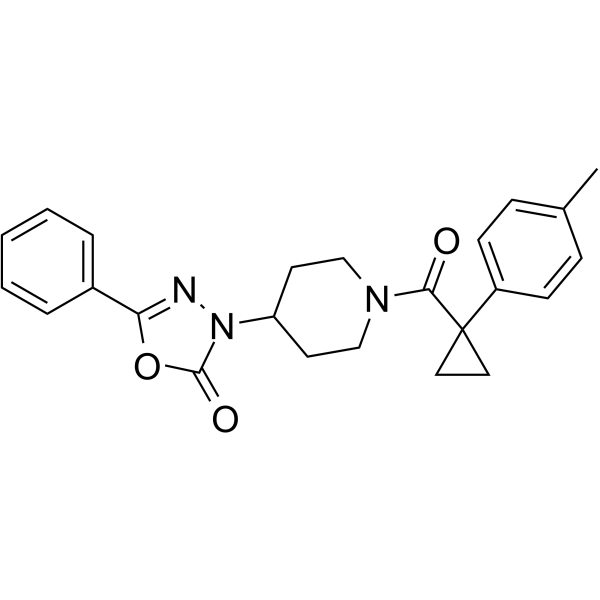
HY-111083
ML191GPR55 Antagonist -
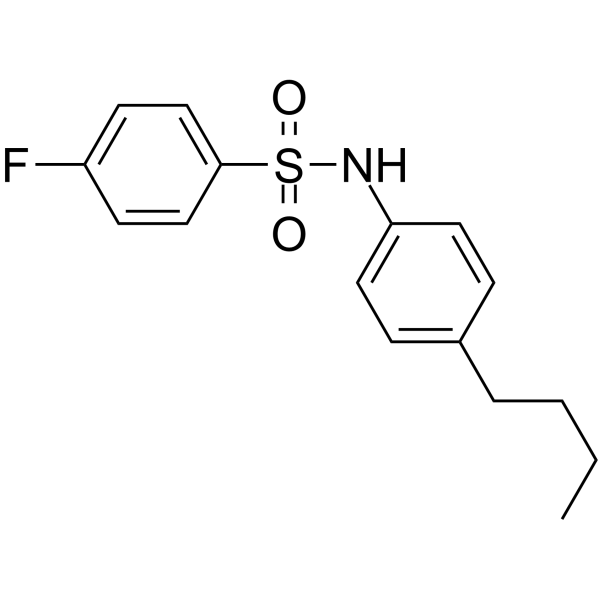
HY-101906
DC260126GPR40 antagonist -
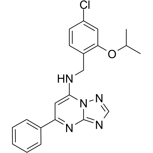
HY-12664
AF64394GPR3 Inverse Agonist -
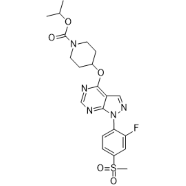
HY-15565
APD668GPR119 Agonist -
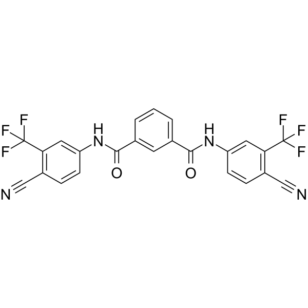
HY-114165
DJ-V-159Androgen Receptor Agonist -

HY-107541R
O-1602 (Standard)GPR55 Agonist -
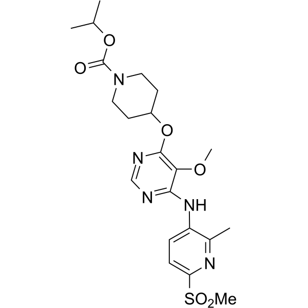
HY-15566
APD597GPR119 Agonist
Publications Citing Use of MedChemExpress (MCE) CID 16020046
More

Biologische Aktivität
Beschreibung
In Vitro
CID 16020046 has weak activities close for inhibition of the acetylcholinesterase (pIC50=4.4), antagonism of the m-opioid receptor (pIC50=4.6), and blockade of KCNH2, the hERG channel (pIC50=4.6) 6 in human embryonic kidney (HEK)-G protein–coupled receptor 55 (GPR55) cells[1].
CID 16020046 (2.5 μM; for ≥25 minutes) significantly inhibits the lysophosphatidylinositol (LPI; 2.5 μM) induced ERK1/2 phosphorylation. CID 16020046 alone fails to induce intracellular Ca2+ release in HEK-GPR55, HEKCB1 cells and shows no ERK1/2 phosphorylation[1].
Pretreatment with CID16020046 (0.01, 0.1, 1, 10 μM) leads to a concentration-dependent decrease in GPR55-mediated NFAT activation, NF-kB activation, and SRE induction in response to 1 μM LPI or GSK319197A in HEKGPR55 and HEK-CB1 cells[1].
CID16020046 (2.5 μM) antagonizes GPR55-mediated activation and nuclear translocation of transcription factors but has no effect on CB1-mediated CREB activation[1].
Pretreatment CID16020046 (1 μM) abolished the LPI-induced stimulation of wound healing in HMVEC-Ls[1].
MedChemExpress (MCE) has not independently confirmed the accuracy of these methods. They are for reference only.
-
Cell Line:HEK-CB1 and HEK-CB2 cells[1]
-
Concentration:2.5 μM
-
Incubation Time:For ≥25 minutes
-
Result:Significantly inhibited the LPI (2.5 μM) induced ERK1/2 phosphorylation.
Treatment alone showed no ERK1/2 phosphorylation and did not alter WIN55,212-2 (2.5 μM) induced ERK1/2 phosphorylation in HEK-CB1 and HEK-CB2 cells.


Chemical Information
-
CAS. Nr. 834903-43-4
-
Appearance Solid
-
Molecular Weight 425.44
-
Formel C25H19N3O4
-
Color White to off-white
-
SMILES
O=C(O)C1=CC=C(N(C2C3=CC=CC(O)=C3)C(C4=C2C(C5=CC=C(C)C=C5)=NN4)=O)C=C1
-
Versand
Room temperature in continental US; may vary elsewhere.
-
Speicherung
Powder -20°C 3 years 4°C 2 years In solvent -80°C 2 years -20°C 1 year


Publications (2)
-
Journal Impact Factor
-
Most Recent
-
Autophagy
Cannabidiol inhibits human glioma by induction of lethal mitophagy through activating TRPV4. [Abstract]2021 Nov;17(11):3592-3606. PMID: 33629929 -
Biochem Pharmacol
The C-terminal region regulates the constitutive signaling activity of GPR119: G protein recruitment. [Abstract]2025 Nov:241:117095. PMID: 40617490


Lösungsmittel & Löslichkeit
In Vitro:
DMSO : 100 mg/mL (235.05 mM; Need ultrasonic; Hygroscopic DMSO has a significant impact on the solubility of product, please use newly opened DMSO)
Please refer to the solubility information to select the appropriate solvent. Once prepared, please aliquot and store the solution to prevent product inactivation from repeated freeze-thaw cycles.
Storage method and period of stock solution: -80°C, 2 years; -20°C, 1 year. When stored at -80°C, please use it within 2 years. When stored at -20°C, please use it within 1 year.
Please refer to the solubility information to select the appropriate solvent. Once prepared, please aliquot and store the solution to prevent product inactivation from repeated freeze-thaw cycles.
Storage method and period of stock solution: -80°C, 2 years; -20°C, 1 year. When stored at -80°C, please use it within 2 years. When stored at -20°C, please use it within 1 year.
Konzentration (Stammlösung) × Volumen (Stammlösung) = Konzentration (Ziellösung) × Volumen (Ziellösung)
In Vivo:
Select the appropriate dissolution method based on your experimental animal and administration route.
- For the following dissolution methods, please ensure to first prepare a clear stock solution using an In Vitro approach and then sequentially add co-solvents:
- To ensure reliable experimental results, the clarified stock solution can be appropriately stored based on storage conditions. As for the working solution for In Vivo experiments, it is recommended to prepare freshly and use it on the same day.
- The percentages shown for the solvents indicate their volumetric ratio in the final prepared solution. If precipitation or phase separation occurs during preparation, heat and/or sonication can be used to aid dissolution.
Add each solvent one by one: 10% DMSO 40% PEG300 5% Tween-80 45% Saline
Solubility: ≥ 2.5 mg/mL (5.88 mM); Clear solution
This protocol yields a clear solution of ≥ 2.5 mg/mL (saturation unknown).
Taking 1 mL working solution as an example, add 100 μL DMSO stock solution (25.0 mg/mL) to 400 μL PEG300, and mix evenly; then add 50 μL Tween-80 and mix evenly; then add 450 μL Saline to adjust the volume to 1 mL.
Preparation of Saline: Dissolve 0.9 g sodium chloride in ddH₂O and dilute to 100 mL to obtain a clear Saline solution.
Add each solvent one by one: 10% DMSO 90% (20% SBE-β-CD in Saline)
Solubility: ≥ 2.5 mg/mL (5.88 mM); Clear solution
This protocol yields a clear solution of ≥ 2.5 mg/mL (saturation unknown).
Taking 1 mL working solution as an example, add 100 μL DMSO stock solution (25.0 mg/mL) to 900 μL 20% SBE-β-CD in Saline, and mix evenly.
Preparation of 20% SBE-β-CD in Saline (4°C, storage for one week): 2 g SBE-β-CD powder is dissolved in 10 mL Saline, completely dissolve until clear.
For the following dissolution methods, please prepare the working solution directly:
It is recommended to prepare fresh solutions and use them promptly within a short period of time.
The percentages shown for the solvents indicate their volumetric ratio in the final prepared solution. If precipitation or phase separation occurs during preparation, heat and/or sonication can be used to aid dissolution.
Add each solvent one by one: 20% PEG300 80% Saline
Solubility: 25 mg/mL (58.76 mM); Suspended solution; Need ultrasonic
In Vivo Dissolution Calculator
Please enter the basic information of animal experiments:
-
-
-
-
Recommended: Prepare an additional quantity of animals to account for potential losses during experiments.
Please enter your animal formula composition:
-
%DMSO +
Recommended: Keep the proportion of DMSO in working solution below 2% if your animal is weak.
-
%+
-
+%Tween-80 + +
-
%Saline +
The co-solvents required include: DMSO, . All of co-solvents are available by MedChemExpress (MCE). , Tween 80. All of co-solvents are available by MedChemExpress (MCE).
Working solution concentration: 0.22 mg/mL
Method for preparing stock solution: mg drug dissolved in μL DMSO. Stock solution concentration: mg/mL.
1. Take μL DMSO stock solution;
2. Add μL .
μL , mix evenly;
3. Then add μL Tween 80, mix evenly;
4. Then add μL
Please ensure that the stock solution in the first step is dissolved to a clear state, and add co-solvents in sequence. You can use ultrasonic heating (ultrasonic cleaner, recommended frequency 20-40 kHz), vortexing, etc. to assist dissolution.


Reinheit & Dokumentation
-
Data Sheet (281 KB)
-
SDS (393 KB)
- English - EN (393 KB)
- Français - FR (393 KB)
- Deutsch - DE (393 KB)
- Norwegian - NO (393 KB)
- Español - ES (393 KB)
- Swedish - SV (393 KB)
- Italian - IT (393 KB)
- Korean - KR (393 KB)
- Portuguese - PT (393 KB)
-
Handling Instructions (2659 KB)
Verweise


Complete Stock Solution Preparation Table
Please refer to the solubility information to select the appropriate solvent. Once prepared, please aliquot and store the solution to prevent product inactivation from repeated freeze-thaw cycles.
Storage method and period of stock solution: -80°C, 2 years; -20°C, 1 year. When stored at -80°C, please use it within 2 years. When stored at -20°C, please use it within 1 year.
| Optional Solvent | Concentration Solvent Mass | 1 mg | 5 mg | 10 mg | 25 mg |
|---|---|---|---|---|---|
| DMSO | 1 mM | 2.3505 mL | 11.7525 mL | 23.5051 mL | 58.7627 mL |
| 5 mM | 0.4701 mL | 2.3505 mL | 4.7010 mL | 11.7525 mL | |
| 10 mM | 0.2351 mL | 1.1753 mL | 2.3505 mL | 5.8763 mL | |
| 15 mM | 0.1567 mL | 0.7835 mL | 1.5670 mL | 3.9175 mL | |
| 20 mM | 0.1175 mL | 0.5876 mL | 1.1753 mL | 2.9381 mL | |
| 25 mM | 0.0940 mL | 0.4701 mL | 0.9402 mL | 2.3505 mL | |
| 30 mM | 0.0784 mL | 0.3918 mL | 0.7835 mL | 1.9588 mL | |
| 40 mM | 0.0588 mL | 0.2938 mL | 0.5876 mL | 1.4691 mL | |
| 50 mM | 0.0470 mL | 0.2351 mL | 0.4701 mL | 1.1753 mL | |
| 60 mM | 0.0392 mL | 0.1959 mL | 0.3918 mL | 0.9794 mL | |
| 80 mM | 0.0294 mL | 0.1469 mL | 0.2938 mL | 0.7345 mL | |
| 100 mM | 0.0235 mL | 0.1175 mL | 0.2351 mL | 0.5876 mL |


CID 16020046 Related Classifications
HY-16697 Related Classifications
 Powered by Bioz