



ω-Hexatoxin-Hv1a
ω-Hexatoxin-Hv1a (ω-ACTX-Hv1; ω-Atracotoxin-HV1) is an orally active insecticidal neurotoxin containing an inhibitor cystine knot motif and a selective calcium channel inhibitor. ω-Hexatoxin-Hv1a blocks L-type voltage-dependent Ca2+ channels and reduces intracellular calcium ion concentration, thereby decreasing apoptosis, necroptosis and oxidative stress, and promoting cell recovery and energy level elevation. ω-Hexatoxin-Hv1a causes larval paralysis and death by impairing neurotransmission in the central nervous system of insects. It shows high injectable toxicity against insects of multiple orders, but exhibits weak oral toxicity. ω-Hexatoxin-Hv1a is widely applicable to studies related to ischemia-reperfusion injury, atopic dermatitis, and ischemic injury of cardiomyocytes and neurons.
Nur für Forschungszwecke. Wir verkaufen nicht an Patienten.
- CAS. Nr.: 193981-10-1
- Formel: C162H247N49O61S6
- Molecular Weight:4049.38
-
Speicherung:
Please store the product under the recommended conditions in the Certificate of Analysis.

To place orders, for customer services and technical support, please contact: MedChemExpress USA
Tel: 609-228-6898 E-mail: [email protected] [email protected]


-
 Biologische Aktivität
-
Chemical Information
- Protokoll
- Reinheit & Dokumentation
- Verweise
-
 Help & FAQs


Alle Calcium Channel Isoform-spezifische Produkte anzeigen
More

Biologische Aktivität
Beschreibung
IC50 & Target
|
L-type calcium channel |
In Vitro
ω-Hexatoxin-Hv1a (10-50 nM; 3 h of reperfusion) inhibits ischemia/reperfusion-induced apoptosis, suppresses ischemia/reperfusion-induced necrosis, alleviates intracellular calcium overload, and promotes the recovery of cell index in CHO-K1 epithelial cells[1].
Recombinant ω-Hexatoxin-Hv1a (100-200 ng) retains immunoreactivity after fusion with GNA, which is confirmed by Western blotting analysis of 200 ng and 100 ng loaded samples of the intact Hv1a/GNA fusion protein[2].
ω-Hexatoxin-Hv1a (10-50 nM; 18 h) blocks AC-1001 H3-induced apoptosis in CHO-K1 cells in a dose-dependent manner in vitro. At 50 nM (18 h incubation), it reduces the apoptosis level to that of the control group, while 10 nM used alone does not alter the baseline apoptosis level[3].
MedChemExpress (MCE) has not independently confirmed the accuracy of these methods. They are for reference only. Further protocols information, click here.
-
Cell Line:CHO-K1 Chinese hamster ovary epithelial cells
-
Concentration:10 nM; 50 nM
-
Incubation Time:3 h (reperfusion)
-
Result:Maintained CHO-K1 cell apoptosis levels at the normal condition level at 50 nM.
Showed no effect on apoptosis at 10 nM.
Maintained necrosis levels at the normal condition level at both 10 nM and 50 nM.
Reduced intracellular calcium ion concentration during reperfusion to levels correlating with reduced apoptosis and necrosis at both 10 nM and 50 nM.
Caused the cell index to return to pre-reperfusion values within 1 h of reperfusion start at both 10 nM and 50 nM, compared to the no-toxin group which returned to normal levels only after 5 h.
-
Cell Line:CHO-K1
-
Concentration:10 nM (alone); 10-50 nM (co-treated with AC-1001 H3 peptide)
-
Incubation Time:18 h (all conditions)
-
Result:Exhibited apoptosis level statistically identical to the control group when used alone at 10 nM for 18 h.
Decreased apoptosis level compared to AC-1001 H3 peptide alone when co-treated at 10 nM for 18 h.
Showed a dose-dependent reduction in AC-1001 H3-induced apoptosis at 10, 30, 50 nM for 18 h, with 50 nM resulting in apoptosis level statistically indistinguishable from the control group.
In Vivo
ω-Hexatoxin-Hv1a (9.6 μg per larva; p.o.; daily; 4 days) alone via daily sucrose droplets has no insecticidal activity against fifth stadium Mamestra brassicae larvae[2].
MedChemExpress (MCE) has not independently confirmed the accuracy of these methods. They are for reference only.
-
Animal Model:Mamestra brassicae (fifth stadium, 40-70 mg)[2]
-
Dosage:46 μg/g insect; 92 μg/g insect; 184 μg/g insect
-
Administration:injection
-
Result:Caused 20% mortality at 72 hours post-injection at 46 μg/g insect.
Caused 80% mortality at 72 hours post-injection at 92 μg/g insect.
Caused 90% mortality at 72 hours post-injection at 184 μg/g insect.
Induced paralysis in larvae injected with doses of 46 μg/g insect and above.
Significantly reduced survival compared to controls (P<0.001).
-
Animal Model:Mamestra brassicae (fifth stadium)[2]
-
Dosage:9.6 μg per larva (daily, total 38.4 μg per larva)
-
Administration:p.o.; daily; 4 days
-
Result:Showed no reduction in larval growth compared to the BSA control group.
Showed no reduction in survival compared to the BSA control group.
Resulted in all larvae surviving to pupation, with no evidence of reduced feeding or paralysis.


Chemical Information
-
CAS. Nr. 193981-10-1
-
Molecular Weight 4049.38
-
Formel C162H247N49O61S6
-
Synonyms
ω-ACTX-Hv1; ω-Atracotoxin-HV1
-
Sequence
Ser-Pro-Thr-Cys-Ile-Pro-Ser-Gly-Gln-Pro-Cys-Pro-Tyr-Asn-Glu-Asn-Cys-Cys-Ser-Gln-Ser-Cys-Thr-Phe-Lys-Glu-Asn-Glu-Asn-Gly-Asn-Thr-Val-Lys-Arg-Cys-Asp (Disulfide bridge:Cys4-Cys18, Cys11-Cys22, Cys17-Cys36)
-
Sequence Shortening
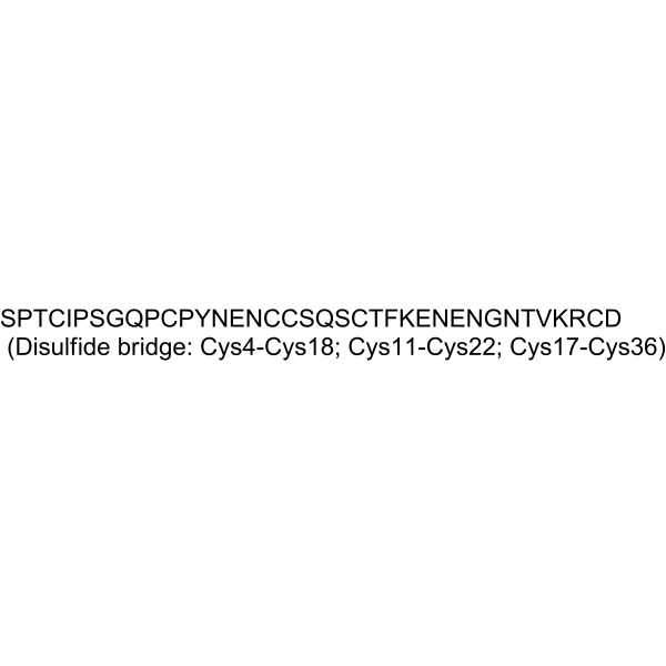
SPTCIPSGQPCPYNENCCSQSCTFKENENGNTVKRCD (Disulfide bridge:Cys4-Cys18, Cys11-Cys22, Cys17-Cys36)
-
Versand
Room temperature in continental US; may vary elsewhere.
-
Speicherung
Please store the product under the recommended conditions in the Certificate of Analysis.

Protokoll
-
Apoptosis
Apoptosis, also called programmed cell death, is generally characterized by distinct morphological characteristics.
-
TUNEL staining for apoptotic DNA fragmentation
TUNEL staining detects DNA strand breaks by using terminal deoxynucleotidyl transferase to add labeled nucleotides to exposed 3′-OH DNA termini, generating either microscopic staining in fixed cells or tissue sections, or fluorescence/cytometric signal in cell suspensions. TUNEL positivity reflects DNA fragmentation but should not be interpreted alone as definitive apoptosis, because TUNEL can also label necrotic, autolytic, mechanically damaged, or DNA-repair-associated DNA breaks.
-
Annexin V plus membrane-impermeant dye apoptosis staining
Annexin V-based apoptosis assays rely on the detection of phosphatidylserine (PS) externalization from the inner leaflet of the plasma membrane to the outer leaflet, an early biochemical hallmark of apoptosis. Fluorescently labeled Annexin V binds PS in a calcium-dependent manner, enabling identification of early apoptotic cells by flow cytometry or fluorescence microscopy. When combined with a membrane-impermeant DNA-binding dye (e. g. , propidium iodide), this approach allows discrimination between viable (Annexin V−/dye−), early apoptotic (Annexin V+/dye−), and late apoptotic or necrotic (Annexin V+/dye+) cell populations by assessing membrane integrity and PS exposure.
-
Cell recovery
Cell recovery is the opposite process of cell cryopreservation, that is, the process of cell recovery and growth, which is to re-culture the cells frozen in liquid nitrogen or -70℃ refrigerator after thawing.
-
ROS/oxidative-stress fluorescent staining
ROS/oxidative-stress fluorescent staining uses cell-permeant fluorogenic probes that become fluorescent after oxidation inside cells or tissues; commonly used examples include DCFH-DA/DCFDA for broad cellular oxidant detection, DHE for superoxide-related signal detection, MitoSOX for mitochondrial superoxide-related signal detection, and CellROX probes for oxidative-stress-associated fluorescence readouts. The assay detects probe oxidation rather than a single ROS species unless the probe and analysis method have been chemically validated for that species. DCFH-DA enters cells, is deacetylated by intracellular esterases to DCFH, and produces fluorescent DCF after oxidation, so the readout is used as an operational measure of total cellular oxidative stress rather than a species-specific ROS measurement. DHE and MitoSOX can report superoxide-related oxidation, but red fluorescence alone can include non-specific ethidium-like oxidation products; HPLC or optimized spectral approaches are
-
TPA/Croton Oil Ear Edema and Dermatitis
The TPA (12-O-tetradecanoylphorbol-13-acetate) and croton oil-induced mouse ear edema model is a well-established acute cutaneous inflammation system used to evaluate topical anti-inflammatory activity by measuring edema formation, neutrophil infiltration, vascular permeability, and cytokine-mediated skin responses in vivo. The inflammatory response is triggered by topical application of phorbol esters (TPA) or croton oil constituents, leading to rapid activation of protein kinase C signaling, leukocyte recruitment, and increased vascular permeability, which can be quantified by ear thickness, weight, dye extravasation, and biochemical markers such as myeloperoxidase (MPO) activity and pro-inflammatory mediators in ear tissue homogenates. This model is widely used for screening anti-inflammatory agents, where reductions in edema and inflammatory biomarkers reflect suppression of acute dermal inflammation and immune cell infiltration. Histological evaluation typically confirms epidermal
-
Calcium Spark Assay
Calcium sparks are localized, transient increases in intracellular calcium concentration ([Ca2+]i) that occur in cardiac myocytes and represent elementary events underlying excitation-contraction coupling. These events are generated by the coordinated opening of clusters of ryanodine receptors (RyRs) on the sarcoplasmic reticulum membrane, leading to a brief release of Ca2+ into the cytosol. The detection and analysis of calcium sparks provide insights into the mechanisms of calcium handling and signaling in cardiac cells. Imaging techniques using fluorescent calcium indicators such as Fluo-3 are employed to visualize these subcellular calcium transients with high spatial and temporal resolution. The protocol is based on established methodologies described in primary literature for both experimental measurement and automated analysis of calcium sparks.
-
Apoptosis Solutions
Apoptosis is a regulated, generally non-lytic cell-death pathway that removes unwanted, damaged, infected, or abnormal cells through coordinated morphological changes, caspase activation, DNA fragmentation, and membrane remodeling. The intrinsic apoptosis pathway is controlled mainly by mitochondrial outer membrane permeabilization, BCL-2 family proteins, cytochrome c release, apoptosome formation, caspase-9 activation, and downstream executioner caspase-3/7 activation. The extrinsic apoptosis pathway is initiated by death receptors such as Fas, TNFR, and TRAIL receptors, which recruit adaptor proteins and activate caspase-8 before engaging executioner caspases or mitochondrial amplification through BID cleavage. Apoptosis is linked to many phenotypes, including cancer cell killing, tissue homeostasis, immune regulation, neurodegeneration, infection response, and treatment-induced cytotoxicity; unresolved questions include how apoptosis interacts with necroptosis, pyroptosis, ferroptos
-
Repeated-Dose Oral Toxicity Study
A repeated-dose oral toxicity study evaluates systemic toxic effects after daily oral exposure to a test substance for a defined period, commonly 28 days, 6 weeks, 90 days, or 13 weeks in rodent studies. The readout is generated by integrating mortality, clinical signs, body-weight change, food and water intake, functional or behavioral observations, hematology, serum biochemistry, urinalysis, organ weights, necropsy, and histopathology to identify dose-related adverse effects, target organs, and the no-observed-adverse-effect level (NOAEL).
-
Ca2+ Staining Technique
Ca2+ staining is an experimental technique that utilizes specific fluorescent probes (such as Fluo-4 AM, Fura-2, etc.) to qualitatively or quantitatively detect dynamic changes in intracellular Ca2+ concentrations; this is achieved by monitoring the changes in fluorescent signals generated when these probes bind to free intracellular calcium ions. The underlying principle relies primarily on the presence of chelating groups within the probe's molecular structure that possess high affinity for calcium ions.
-
Standard Mammalian Cell Line Thawing and Recovery
Standard mammalian cell line thawing and recovery restores cryopreserved cells to active culture by rapidly warming frozen cells, diluting or removing cryoprotectant, and assessing whether cells regain viability, attachment, proliferation, and expected function. Cryopreservation commonly uses permeating cryoprotectants such as DMSO to reduce freeze-thaw injury, but DMSO exposure can also produce dose- and time-dependent cytotoxicity, so post-thaw recovery should minimize unnecessary exposure while preserving cell recovery.


Reinheit & Dokumentation
Verweise
[1]. Iurova EV, et al. Effect of the Peptide Calcium Channel Blocker ω-hexatoxin-Hv1a on Cell Death during Ischemia/Reperfusion in vitro. Sovrem Tekhnologii Med. 2023;15(1):21-27. [Content Brief]
[2]. Fitches EC, et al. Fusion to snowdrop lectin magnifies the oral activity of insecticidal ω-Hexatoxin-Hv1a peptide by enabling its delivery to the central nervous system. PLoS One. 2012;7(6):e39389. [Content Brief]
[3]. Iurova E, et al. Arthropod toxins inhibiting Ca2+ and Na+ channels prevent AC-1001 H3 peptide-induced apoptosis. J Pept Sci. 2021;27(1):e3288. [Content Brief]

Calculators
Konzentration (Stammlösung) × Volumen (Stammlösung) = Konzentration (Ziellösung) × Volumen (Ziellösung)


ω-Hexatoxin-Hv1a Related Classifications
HY-P5142 Related Classifications
 Powered by Bioz
Keywords
- ω-Hexatoxin-Hv1a
- 193981-10-1
- ω-ACTX-Hv1
- ω-Atracotoxin-HV1
- Insecticide
- Calcium Channel
- Apoptosis
- Necroptosis
- voltage-gated calcium channels
- mammalian voltage-gated calcium channels
- insect voltage-gated calcium channels
- CHO-K1 cells
- atopic dermatitis
- insect larvae
- epithelial cells
- central nervous system ganglionic neural transmission
- ischemic and reperfusion injury
- inhibitor cystine knot motif
- Inhibitor
- inhibitor
- inhibit