



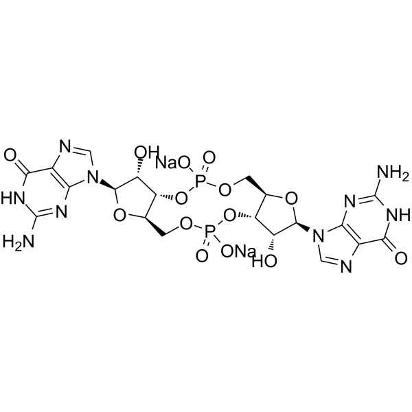
Cyclic-di-GMP disodium
Based on 9 publication(s) in Google Scholar
Cyclic-di-GMP disodium is a STING agonist and a bacterial second messenger that coordinates different aspects of bacterial growth and behavior, including motility, virulence, biofilm formation, and cell cycle progression. Cyclic-di-GMP disodium has anti-cancer cell proliferation activity and also induces elevated CD4 receptor expression and cell cycle arrest. Cyclic-di-GMP disodium can be used in cancer research.
For research use only. We do not sell to patients.
- Purity : 98.78%
- CAS No.: 2222132-40-1
- Formula: C20H22N10Na2O14P2
- Molecular Weight:734.37
-
Storage:
-20°C, sealed storage, away from moisture and light, under nitrogen
* In solvent : -80°C, 6 months; -20°C, 1 month (sealed storage, away from moisture and light, under nitrogen)

To place orders, for customer services and technical support, please contact: MedChemExpress USA
Tel: 609-228-6898 E-mail: [email protected] [email protected]


-
 Biological Activity
-
Chemical Information
-
Solvent & Solubility
- Protocols
- Purity & Documentation
- References
-
 Help & FAQs


-
HY-12326A
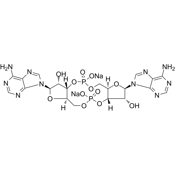
c-di-AMP disodiumSTING Agonist -
HY-12512
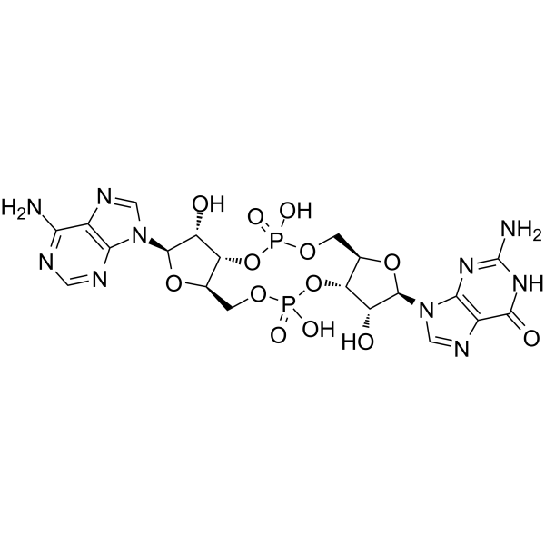
cGAMPSTING Activator -
HY-110385
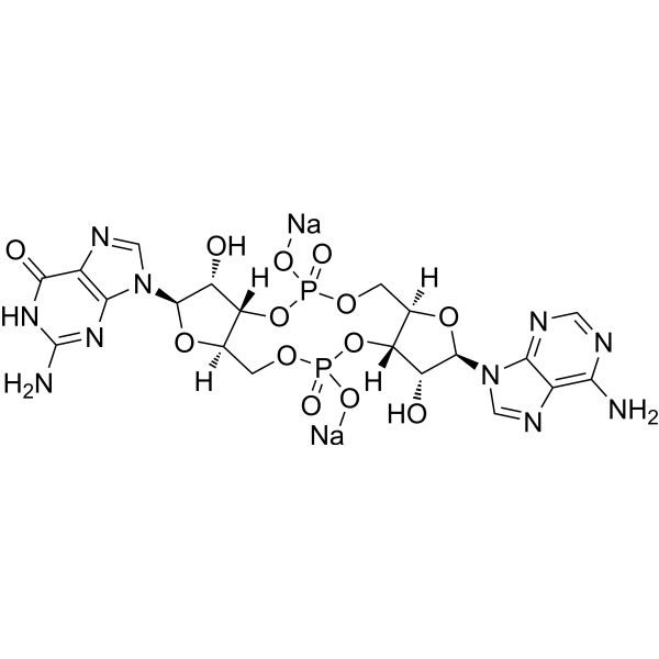
cGAMP disodiumSTING Activator -
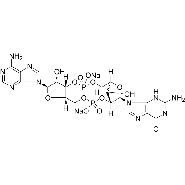
HY-100564A
2',3'-cGAMP sodiumSTING Agonist -
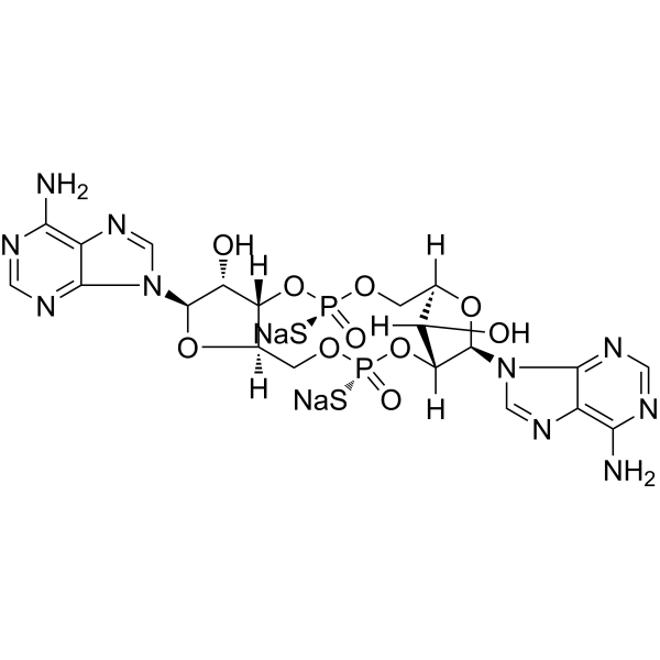
HY-12885A
2’3’-c-di-AM(PS)2 (Rp,Rp) disodium saltSTING Agonist -

HY-18739
Phorbol 12-myristate 13-acetatePKC Activator -
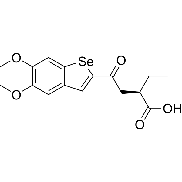
HY-151264
BSP16STING Agonist -
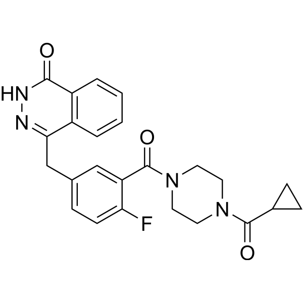
HY-10162
OlaparibPARP Inhibitor -

HY-Y1891
Tween 80 -
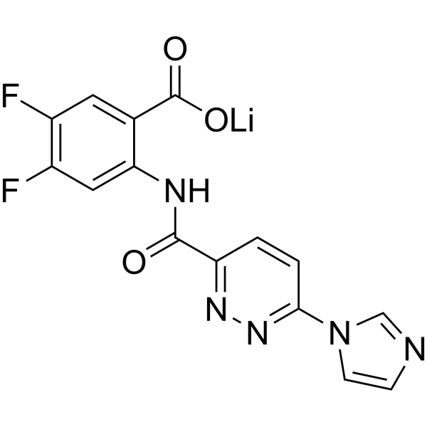
HY-131454
SR-717STING Agonist -
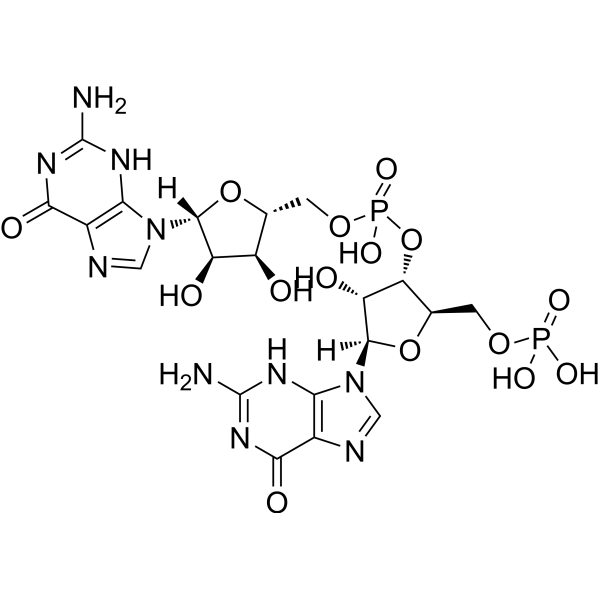
HY-137662
5'-Phosphoguanylyl-(3',5')-guanosinec-di-GMP Degradate -
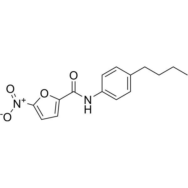
HY-138682
STING-IN-2STING Inhibitor
Publications Citing Use of MedChemExpress (MCE) Cyclic-di-GMP disodium
More- Cell. 2026 Mar 19;189(6):1748-1767.e26. [Abstract]
- Gut Microbes. 2022 Jan-Dec;14(1):2119055. [Abstract]
- Nucleic Acids Res. 2025 Oct 28;53(20):gkaf1069. [Abstract]
- NPJ Biofilms Microbiomes. 2024 Jun 20;10(1):51. [Abstract]
- J Transl Med. 2025 Mar 4;23(1):271. [Abstract]
- Int J Biol Macromol. 2024 Sep 14;280(Pt 1):135660. [Abstract]
- Clin Transl Med. 2024 Aug;14(8):e1744. [Abstract]
- Mbio. 2021 Oct 26;12(5):e0119221. [Abstract]
- J Biol Chem. 2025 Dec 6;302(1):111021. [Abstract]
 Customer Validation & Images

-
WB
-
Histological Imaging/Staining
-
In Vivo Efficacy Study
-
Bio/Physico-chemical Assay
-
Gel Electrophoresis
All Endogenous Metabolite Isoforms
More

Biological Activity
Description
IC50 & Target
In Vitro
Cyclic-di-GMP disodium (0.5-50 μM; 5 days) inhibits proliferation of human colon cancer cells[1].
Cyclic-di-GMP disodium (0.5-50 μM; 5 days) specifically elevates CD4 expression in Jurkat cells[2].
Cyclic-di-GMP disodium (0.5-50 μM; 5 days) induces cell cycle arrest at the S-phase in Jurkat cells[2].
MedChemExpress (MCE) has not independently confirmed the accuracy of these methods. They are for reference only. Further protocols information, click here.
-
Cell Line:H508 cells
-
Concentration:0.5-50 µM
-
Incubation Time:5 days
-
Result:Reduced basal H508 cell proliferation by approx 15%, even inhibited acetylcholine- and EGF-induced cell proliferation.
-
Cell Line:Jurkat cells
-
Concentration:50 µM
-
Incubation Time:24 h
-
Result:Specifically induced of CD4 (no effect on the expression of CD8), with a 6.3-fold upregulation over control and in a dose-dependent manner.
-
Cell Line:Jurkat cells
-
Concentration:50 µM
-
Incubation Time:24 h
-
Result:Increased the percentage of cells in S-phase by 79%, with almost complete disappearance of G2/M-phase cells which decreased by 93%.
In Vivo
MedChemExpress (MCE) has not independently confirmed the accuracy of these methods. They are for reference only.
-
Animal Model:C57BL/6 (B6) mice (8- to 10-week-old)[3].
-
Dosage:100 µg/per
-
Administration:Intravenous injection; two sequential vaccinations 9 days apart; combine with TriVax.
-
Result:Significantly higher numbers of antigen-specific CD8 T cells when combined with TriVax. (TriVax consisted of a mixture of 120 μg Pam-hgp100, 100 µg hgp100 or 100 µg Ova, 50 or 25 μg anti-CD40 antibody, and 25 μg Poly-IC).
Enhanced the anti-tumor activity of TriVax.


Chemical Information
-
CAS No. 2222132-40-1
-
Appearance Solid
-
Molecular Weight 734.37
-
Formula C20H22N10Na2O14P2
-
Color Off-white to light yellow
-
SMILES
O[C@@H]([C@H](N1C=NC2=C1N=C(N)NC2=O)O3)[C@H](OP(OC[C@@H]4[C@H]([C@H]([C@H](N5C=NC6=C5N=C(N)NC6=O)O4)O)O7)(O[Na])=O)[C@H]3COP7(O[Na])=O
-
Synonyms
c-di-GMP disodium; cyclic diguanylate disodium; 5GP-5GP disodium
-
Structure Classification
-
Initial Source
-
Shipping
Room temperature in continental US; may vary elsewhere.
-
Storage
-20°C, sealed storage, away from moisture and light, under nitrogen
* In solvent : -80°C, 6 months; -20°C, 1 month (sealed storage, away from moisture and light, under nitrogen)


Publications (9)
-
Journal Impact Factor
-
Most Recent
-
Cell
2026 Mar 19;189(6):1748-1767.e26. PMID: 41643675 -
Gut Microbes
Gut microbiota modulate radiotherapy-associated antitumor immune responses against hepatocellular carcinoma Via STING signaling. [Abstract]2022 Jan-Dec;14(1):2119055. PMID: 36093568 -
Nucleic Acids Res
Cyclic-di-GMP interferes with DNA-MucR-DNA bridging to derepress genes targeted by the xenogeneic silencer MucR. [Abstract]2025 Oct 28;53(20):gkaf1069. PMID: 41148150 -
NPJ Biofilms Microbiomes
2024 Jun 20;10(1):51. PMID: 38902226 -
J Transl Med
Photothermal therapy combined with a STING agonist induces pyroptosis, and gasdermin D could be a new biomarker for guiding the treatment of pancreatic cancer. [Abstract]2025 Mar 4;23(1):271. PMID: 40038726
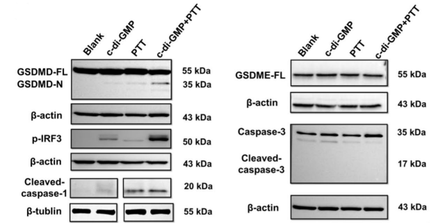
Cyclic-di-GMP disodium purchased from MedChemExpress. Usage Cited in: J Transl Med. 2025 Mar 4;23(1):271. [Abstract]
Cyclic-di-GMP disodium (c-di-GMP; 200 μg/mL; 50 μL; intratumoural injection; day 0 and day 2) combined with photothermal therapy (PTT) increased the protein expression of GSDMD-N, cleaved caspase-3, and phosphorylated-IRF3 (p-IRF3) in C57BL/6 N mice (Pan02-luc cells).
Cyclic-di-GMP disodium purchased from MedChemExpress. Usage Cited in: J Transl Med. 2025 Mar 4;23(1):271. [Abstract]
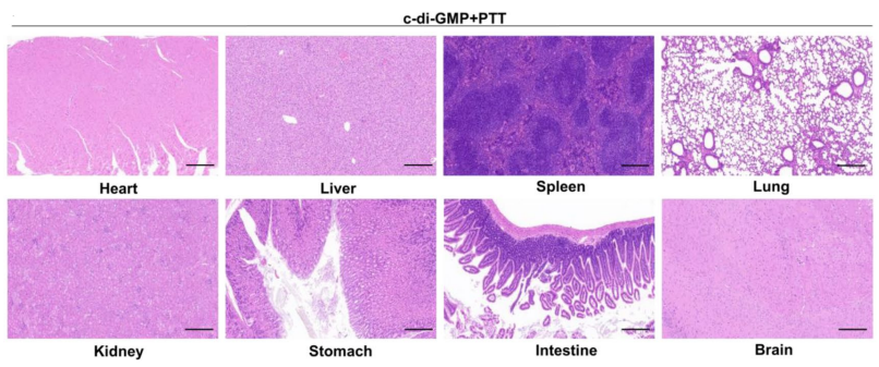
Cyclic-di-GMP disodium (c-di-GMP; 200 μg/mL; 50 μL; intratumoural injection; day 0 and day 2) combined with photothermal therapy (PTT) indicated the good biocompatibility in C57BL/6 N mice (Pan02-luc cells) by H&E staining of the major organs.
Cyclic-di-GMP disodium purchased from MedChemExpress. Usage Cited in: J Transl Med. 2025 Mar 4;23(1):271. [Abstract]
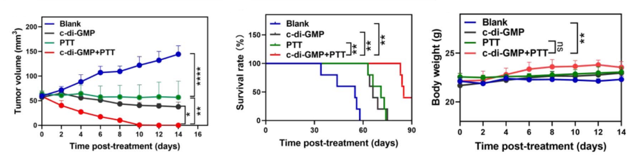
Cyclic-di-GMP disodium (c-di-GMP; 200 μg/mL; 50 μL; intratumoural injection; day 0 and day 2) showed antitumour effect in C57BL/6 N mice (Pan02-luc cells).
-
Int J Biol Macromol
C-di-GMP@ZIF-8 nanocomposite injectable hydrogel based on modified chitosan and hyaluronic acid for infected wound healing by activating STING signaling. [Abstract]2024 Sep 14;280(Pt 1):135660. PMID: 39284469 -
Clin Transl Med
2024 Aug;14(8):e1744. PMID: 39166890 -
Mbio
Global Transcriptional Repression of Diguanylate Cyclases by MucR1 Is Essential for Sinorhizobium-Soybean Symbiosis. [Abstract]2021 Oct 26;12(5):e0119221. PMID: 34700374 -
J Biol Chem
Dynamic control of bacterial antiphage defense through the CdnG-Cap5 cyclic oligonucleotide-based antiphage pathway in Vibrio cholerae. [Abstract]2025 Dec 6;302(1):111021. PMID: 41360261
Cyclic-di-GMP disodium purchased from MedChemExpress. Usage Cited in: J Biol Chem. 2025 Dec 6;302(1):111021. [Abstract]
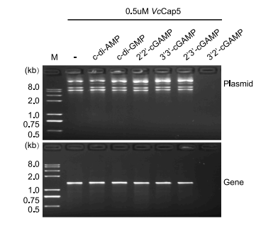
agarose gel electrophoresis analysis of DNA degradation assays. Nuclease activity assays were performed by incubating 0.5 μM VcCap5 with 20 ng/μl DNA in the presence of 0.5 μM of various cyclic dinucleotides (Cyclic-di-GMP disodium; c-di-GMP).
Cyclic-di-GMP disodium purchased from MedChemExpress. Usage Cited in: J Biol Chem. 2025 Dec 6;302(1):111021. [Abstract]
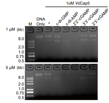
second messenger-dependent activation of VcCap5 DNA cleavage. Nuclease activity assays were performed by incubating 1 µM VcCap5 with 10 ng/µl DNA in the presence of 1 µM or 5 µM of various cyclic dinucleotides (Cyclic-di-GMP disodium; c-di-GMP).
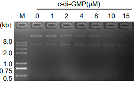
Cyclic-di-GMP disodium purchased from MedChemExpress. Usage Cited in: J Biol Chem. 2025 Dec 6;302(1):111021. [Abstract]
dose-response analysis of VcCap5 activity with increasing concentrations of cyclic dinucleotides. The nuclease assay mixture contained 1 µM VcCap5, 10 ng/µl DNA, and cyclic dinucleotide (Cyclic-di-GMP disodium; c-di-GMP) over a concentration range of 0.1 to 15 µM.


Solvent & Solubility
In Vitro:
H2O : 160 mg/mL (217.87 mM; Need ultrasonic)
Please refer to the solubility information to select the appropriate solvent. Once prepared, please aliquot and store the solution to prevent product inactivation from repeated freeze-thaw cycles.
Storage method and period of stock solution: -80°C, 6 months; -20°C, 1 month (sealed storage, away from moisture and light, under nitrogen). When stored at -80°C, please use it within 6 months. When stored at -20°C, please use it within 1 month.
* Note: If you choose water as the stock solution, please dilute it to the working solution, then filter and sterilize it with a 0.22 μm filter before use.
Please refer to the solubility information to select the appropriate solvent. Once prepared, please aliquot and store the solution to prevent product inactivation from repeated freeze-thaw cycles.
Storage method and period of stock solution: -80°C, 6 months; -20°C, 1 month (sealed storage, away from moisture and light, under nitrogen). When stored at -80°C, please use it within 6 months. When stored at -20°C, please use it within 1 month.
* Note: If you choose water as the stock solution, please dilute it to the working solution, then filter and sterilize it with a 0.22 μm filter before use.
Concentration (start) × Volume (start) = Concentration (final) × Volume (final)
In Vivo:
For the following dissolution methods, please prepare the working solution directly:
It is recommended to prepare fresh solutions and use them promptly within a short period of time.
The percentages shown for the solvents indicate their volumetric ratio in the final prepared solution. If precipitation or phase separation occurs during preparation, heat and/or sonication can be used to aid dissolution.
Add each solvent one by one: PBS
Solubility: 50 mg/mL (68.09 mM); Clear solution; Need ultrasonic

Protocols
-
RNA extraction experimental
By lysing cells, releasing RNA, and removing impurities such as proteins and DNA, high-purity RNA products are finally obtained. The commonly used traditional method is the guanidine isothiocyanate/phenol/chloroform method (Trizol), which is suitable for a variety of animal materials including animal tissues, microorganisms, cultured cells, etc., and most plant materials.
-
BrdU Incorporation Assay
Bromodeoxyuridine (BrdU) incorporation assay is based on the principle that BrdU, a thymidine analog, is incorporated into newly synthesized DNA during the S phase of the cell cycle, thereby serving as a marker of DNA replication and cellular proliferation. Incorporated BrdU can be detected using anti-BrdU antibodies following DNA denaturation, enabling visualization or quantification of proliferating cells through immunochemical detection methods such as immunofluorescence or immunohistochemistry.
-
Somatic Cell Culture
A method of simulating the in vivo environment in vitro to maintain the cell growth, differentation and main functions.
-
CFSE Dye Dilution Proliferation Assay
The CFSE (carboxyfluorescein diacetate succinimidyl ester) dye dilution proliferation assay is based on the covalent labeling of intracellular proteins by a cell-permeant fluorescent dye that becomes fluorescent upon intracellular ester cleavage and then is stably retained within cells. As labeled cells divide, the dye is partitioned equally between daughter cells, resulting in a stepwise halving of fluorescence intensity that can be quantified by flow cytometry to determine the number of cell divisions undergone by each cell population. This fluorescence dilution approach enables quantitative tracking of lymphocyte proliferation at the single-cell level over multiple rounds of division. CFSE-based proliferation analysis has been widely applied to measure antigen-driven lymphocyte expansion in vitro, where discrete fluorescence peaks correspond to successive cell divisions and allow reconstruction of proliferative history within heterogeneous populations.
-
Dye-dilution cell tracking and proliferation staining
Dye-dilution cell tracking assays quantify cell proliferation by covalently labeling intracellular proteins with a stable fluorescent dye that is equally partitioned between daughter cells during mitosis, resulting in stepwise halving of fluorescence intensity with each cell division as measured by flow cytometry histograms. Carboxyfluorescein diacetate succinimidyl ester (CFSE) is a prototypical dye that diffuses into cells, is enzymatically converted into a fluorescent compound, and then covalently binds intracellular amine groups, producing long-lived fluorescence suitable for tracking multiple rounds of division in vitro and in vivo. Successive generations of dividing cells form discrete peaks of decreasing fluorescence intensity, enabling estimation of proliferation history, precursor frequency, and division index within heterogeneous populations. Alternative dyes such as CellTrace Violet (CTV) and far-red membrane dyes (e. g. , PKH26) follow the same dilution principle but differ
-
Protocol for Cell Cycle
Cell-cycle analysis by flow cytometry measures DNA content in single cells to estimate the fraction of cells in G0/G1, S, and G2/M phases. Propidium iodide intercalates into DNA, and after RNA removal with RNase, fluorescence intensity reflects cellular DNA content: 2N cells are assigned to G0/G1, cells between 2N and 4N to S phase, and 4N cells to G2/M. DNA-content analysis alone cannot reliably separate G0 from G1 or G2 from M. Ki-67 can distinguish quiescent G0 cells from cycling cells, EdU or BrdU incorporation marks active DNA synthesis in S phase, and phospho-histone H3 staining identifies mitotic cells within the 4N population.
-
CCK-8/WST-8 Cell Proliferation Assay
The CCK-8/WST-8 assay is based on the reduction of the water-soluble tetrazolium salt WST-8 to a water-soluble formazan product by cellular dehydrogenases in metabolically active cells, where the generated formazan amount is proportional to the number of living cells and is quantified by measuring absorbance in the visible range, providing a colorimetric readout for cell viability and proliferation assessment. This class of tetrazolium-based assays improves upon earlier MTT-based systems by producing a water-soluble formazan, eliminating the need for organic solubilization steps and enabling direct spectrophotometric measurement in culture medium.
-
Cell Counting-Based Growth Curve Assay
Cell counting-based growth curve assays quantify cell proliferation by directly measuring changes in viable cell number over time using manual or automated counting methods such as hemocytometer-based counting or instrument-assisted cell enumeration, enabling construction of growth curves that reflect population expansion dynamics in response to culture conditions. A widely used approach is trypan blue exclusion with hemocytometer counting, where membrane-compromised (non-viable) cells take up the dye, allowing discrimination between viable and non-viable cells while simultaneously enabling total cell number quantification. Repeated sampling across time points allows estimation of proliferation rate, growth phases, and comparative growth kinetics between experimental conditions.
-
Colony Formation (Clonogenic) Assay
The clonogenic (colony formation) assay measures the ability of a single cell to retain reproductive viability and form a macroscopic colony, typically defined as a cluster derived from one progenitor cell after a defined growth period. This assay is widely used to evaluate cell survival after exposure to ionizing radiation or cytotoxic treatments and is considered a standard method in radiation biology for generating dose-response relationships of reproductive cell death. Colony formation reflects long-term proliferative capacity rather than short-term metabolic activity, and survival is quantified by comparing treated versus untreated conditions based on colony number and derived survival fractions.
-
Cell Viability Determination by MTT Colorimetric Assay
The following protocol uses the MTT colorimetric assay as a classic literature-established method for assessing cell viability/metabolic activity in cultured mammalian cells. MTT[3-(4,5-dimethylthiazol-2-yl)-2,5-diphenyltetrazolium bromide] is reduced by metabolically active cells to a colored formazan product; the amount of formazan is quantified spectrophotometrically and provides an indirect measure of metabolically active viable cells. Importantly, MTT reduction reflects cellular oxidoreductase/metabolic activity rather than an absolute direct count of living cells, so changes in cellular metabolism can alter the signal independently of cell number.
-
EdU Incorporation Assay (Click Chemistry-Based DNA Synthesis Measurement)
The EdU incorporation assay measures DNA synthesis by adding the thymidine analog 5-ethynyl-2′-deoxyuridine to cells or tissues, where it is incorporated into newly synthesized DNA during S phase. Incorporated EdU is detected by copper-catalyzed azide-alkyne cycloaddition, in which a fluorescent azide covalently reacts with the ethynyl group on EdU, allowing S-phase cells to be detected by fluorescence microscopy, flow cytometry, or high-content imaging. EdU detection does not require DNA denaturation or anti-BrdU antibody access, which preserves sample structure and improves compatibility with immunostaining and multiparameter cytometry compared with BrdU-based detection. EdU can be cytotoxic in a cell-type- and exposure-dependent manner, so pulse duration, concentration, and continuous-labeling designs should be validated for each cell type.
-
Ki-67 Immunostaining Proliferation Assay
Ki-67 immunostaining measures the growth fraction of a cell population by detecting Ki-67, a nuclear antigen present in proliferating cells and absent in quiescent G0 cells. The readout is the percentage of Ki-67-positive nuclei among total counted cells, commonly called the Ki-67 labeling index or proliferation index.
-
Flow cytometric DNA-content cell-cycle staining
Flow cytometric DNA-content cell-cycle staining measures the fluorescence intensity of DNA-bound fluorochromes in single cells or nuclei to estimate DNA content distributions, allowing assignment of populations to G0/G1, S, and G2/M phases by DNA histogram deconvolution. Propidium iodide (PI) intercalates into DNA, and PI fluorescence is proportional to cellular DNA content when staining is performed under conditions that make DNA accessible and minimize non-DNA signal. Cells with G2/M DNA content are expected to show approximately twice the fluorescence intensity of G0/G1 cells, while S-phase cells occupy intermediate fluorescence values. PI-based DNA-content analysis can also detect cells with fractional DNA content, often reported as sub-G1, when DNA fragmentation and extraction during staining reduce retained DNA signal in apoptotic cells. DAPI is an alternative DNA fluorochrome for univariate DNA-content analysis, while bivariate approaches combining DNA content with proliferation
-
PCNA Immunodetection Proliferation Assay
PCNA immunodetection measures proliferative activity by detecting proliferating cell nuclear antigen, a nuclear protein associated with DNA polymerase δ function and DNA replication. The assay readout is the proportion of PCNA-positive nuclei among total counted cells, but PCNA labeling is not identical to BrdU labeling because PCNA can mark late G1/early S-associated replication competence and may persist beyond active DNA synthesis depending on fixation and extraction conditions.
-
Protocol for Cell Counting and Cell Density Analysis
Cell counting and cell-density analysis estimate the number of cells in a known volume or field area. Manual hemocytometer counting uses a chamber of defined geometry to convert counted cells into cells/mL, while automated counters and image-analysis workflows detect cell objects from optical, brightfield, fluorescence, impedance, or digital-image features. Trypan blue viability counting is based on dye exclusion: viable cells with intact membranes exclude dye, while non-viable cells with compromised membranes stain blue. The readout is total cell density, viable-cell density, dead-cell density, and percent viability. Cell density can also be estimated from microscopy images by counting objects per image area, from flow cytometry using calibrated volume or reference particles, or from in situ microscopy in bioreactors after calibration against reference methods such as hemocytometer or flow cytometry.
-
Aerobic Bacterial Batch Culture on Broth/Agar
Aerobic bacterial batch culture grows a closed inoculated population in broth or on agar without continuous medium replacement; growth readouts include turbidity/OD for total suspended biomass and colony-forming units for viable cells able to form colonies on agar. OD-based growth curves reflect light scattering by cells, but OD is instrument-, pathlength-, species-, cell-size-, and density-dependent, so OD should be calibrated or interpreted alongside viable counts when quantitative cell density is required.
-
MTT Cell Proliferation Assay
The MTT assay is a colorimetric endpoint assay for estimating viable cell number, cell growth, cytotoxicity, or cell activation in cultured mammalian cells. Living cells reduce the yellow tetrazolium salt MTT into purple/blue formazan, while dead cells do not generate the same signal; the resulting color can be quantified with a multiwell spectrophotometer. MTT reduction is commonly interpreted as a readout of metabolic activity that often correlates with viable cell number, but it should not be treated as a direct cell-counting method unless the assay is optimized for the cell type and experimental condition. Studies show that MTT reduction can involve mitochondrial and non-mitochondrial reducing systems, and formazan may accumulate in intracellular lipid droplets rather than simply marking mitochondria.
-
Fungal Biofilm Culture
Fungal biofilm culture is an in vitro method for growing surface-attached fungal communities, most commonly Candida albicans, on abiotic substrates such as polystyrene wells, silicone elastomer, or polymethylmethacrylate; the assay models adhesion, proliferation, filamentation, extracellular-matrix-associated maturation, and dispersion. Biofilm output can be read by optical density at 600 nm for adherent biomass, XTT reduction for metabolic activity, CFU recovery for viable attached or dispersed cells, and microscopy for architecture.


Purity & Documentation
-
Data Sheet (284 KB)
-
SDS (254 KB)
- English - EN (254 KB)
- Français - FR (254 KB)
- Deutsch - DE (254 KB)
- Norwegian - NO (254 KB)
- Español - ES (254 KB)
- Swedish - SV (254 KB)
- Italian - IT (254 KB)
- Korean - KR (254 KB)
- Portuguese - PT (254 KB)
-
Handling Instructions (2659 KB)
References
[1]. Karaolis DK, et al. 3',5'-Cyclic diguanylic acid (c-di-GMP) inhibits basal and growth factor-stimulated human colon cancer cell proliferation. Biochem Biophys Res Commun. 2005 Apr 1;329(1):40-5. [Content Brief]
[2]. Steinberger O, et al. Elevated expression of the CD4 receptor and cell cycle arrest are induced in Jurkat cells by treatment with the novel cyclic dinucleotide 3',5'-cyclic diguanylic acid. FEBS Lett. 1999 Feb 5;444(1):125-9. [Content Brief]
[3]. Wang Z, et al. STING activator c-di-GMP enhances the anti-tumor effects of peptide vaccines in melanoma-bearing mice. Cancer Immunol Immunother. 2015 Aug;64(8):1057-66. [Content Brief]
[4]. Jenal U, et al. Cyclic di-GMP: second messenger extraordinaire. Nat Rev Microbiol. 2017 May;15(5):271-284. [Content Brief]


Complete Stock Solution Preparation Table
Please refer to the solubility information to select the appropriate solvent. Once prepared, please aliquot and store the solution to prevent product inactivation from repeated freeze-thaw cycles.
Storage method and period of stock solution: -80°C, 6 months; -20°C, 1 month (sealed storage, away from moisture and light, under nitrogen). When stored at -80°C, please use it within 6 months. When stored at -20°C, please use it within 1 month.
| Optional Solvent | Concentration Solvent Mass | 1 mg | 5 mg | 10 mg | 25 mg |
|---|---|---|---|---|---|
| H2O | 1 mM | 1.3617 mL | 6.8086 mL | 13.6171 mL | 34.0428 mL |
| 5 mM | 0.2723 mL | 1.3617 mL | 2.7234 mL | 6.8086 mL | |
| 10 mM | 0.1362 mL | 0.6809 mL | 1.3617 mL | 3.4043 mL | |
| 15 mM | 0.0908 mL | 0.4539 mL | 0.9078 mL | 2.2695 mL | |
| 20 mM | 0.0681 mL | 0.3404 mL | 0.6809 mL | 1.7021 mL | |
| 25 mM | 0.0545 mL | 0.2723 mL | 0.5447 mL | 1.3617 mL | |
| 30 mM | 0.0454 mL | 0.2270 mL | 0.4539 mL | 1.1348 mL | |
| 40 mM | 0.0340 mL | 0.1702 mL | 0.3404 mL | 0.8511 mL | |
| 50 mM | 0.0272 mL | 0.1362 mL | 0.2723 mL | 0.6809 mL | |
| 60 mM | 0.0227 mL | 0.1135 mL | 0.2270 mL | 0.5674 mL | |
| 80 mM | 0.0170 mL | 0.0851 mL | 0.1702 mL | 0.4255 mL | |
| 100 mM | 0.0136 mL | 0.0681 mL | 0.1362 mL | 0.3404 mL |
* Note: If you choose water as the stock solution, please dilute it to the working solution, then filter and sterilize it with a 0.22 μm filter before use.


Cyclic-di-GMP disodium Related Classifications
HY-110382 Related Classifications
 Powered by Bioz