



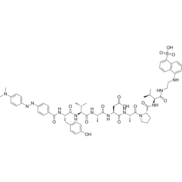
Dabcyl-YVADAPV-EDANS
Dabcyl-YVADAPV-EDANS is a fluorogenic interleukin-1β converting enzyme (ICE) substrate for measuring ICE-like protease activity. Dabcyl-YVADAPV-EDANS detects fluorescence at 360 nm excitation and 480 nm emission wavelengths. ICE-like protease is a critical mediator of K+ deprivation-induced apoptosis of cerebellar granule neurons.
For research use only. We do not sell to patients.
- CAS No.: 161877-70-9
- Formula: C61H76N12O14S
- Molecular Weight:1233.39
-
Storage:
Please store the product under the recommended conditions in the Certificate of Analysis.

To place orders, for customer services and technical support, please contact: MedChemExpress USA
Tel: 609-228-6898 E-mail: [email protected] [email protected]


-
 Biological Activity
-
Chemical Information
- Protocols
- Purity & Documentation
- References
-
 Help & FAQs


-
HY-W011063
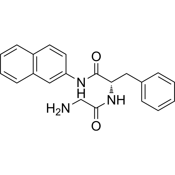
Gly-Phe-β-naphthylamideCathepsin C Substrate -
HY-16658B
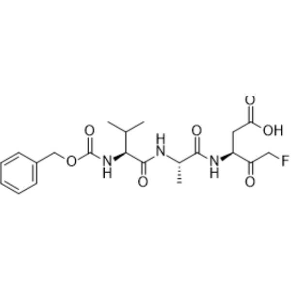
Z-VAD-FMKPan-Caspase Inhibitor -
HY-12305
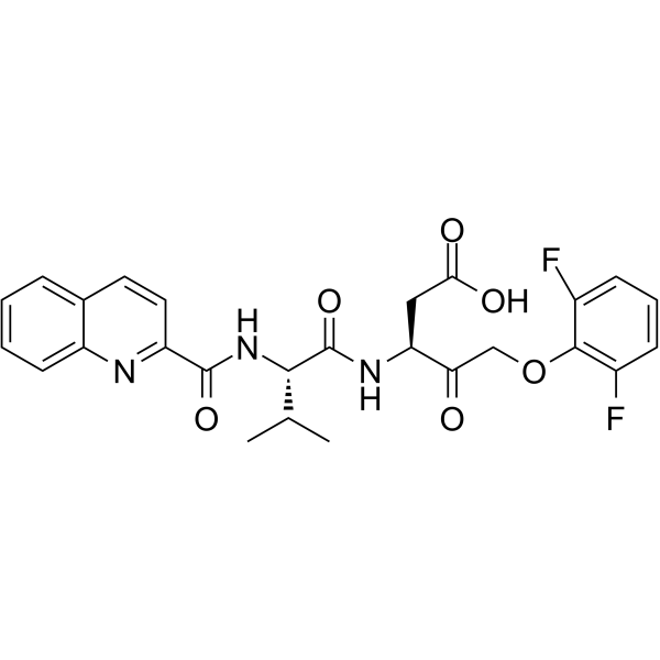
Q-VD-OPhPan-caspase Inhibitor -
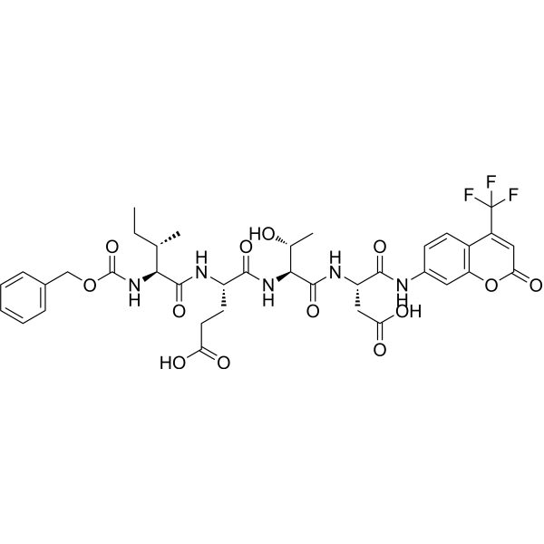
HY-P2208
Z-IETD-AFCCaspase-8 Fluorescence Substrate -
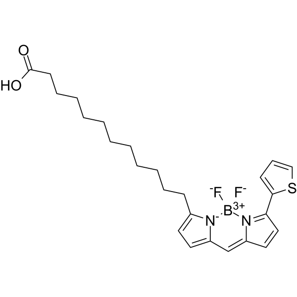
HY-138226
BODIPY 558/568 C12Lipid Droplets Probe -
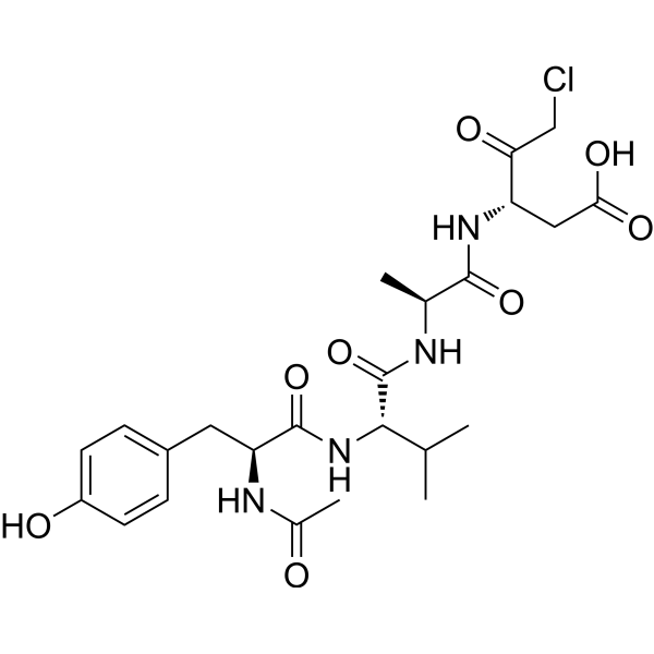
HY-16990
Ac-YVAD-cmkCaspase-1 Inhibitor -

HY-DY1061
BODIPY 558/568 C12 (solution)Lipid Droplets Probe -
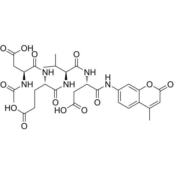
HY-P1003
Ac-DEVD-AMCCaspase-3/7 Fluorescent Substrate -
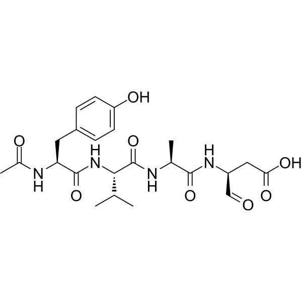
HY-120019
Ac-YVAD-CHOICE Inhibitor -
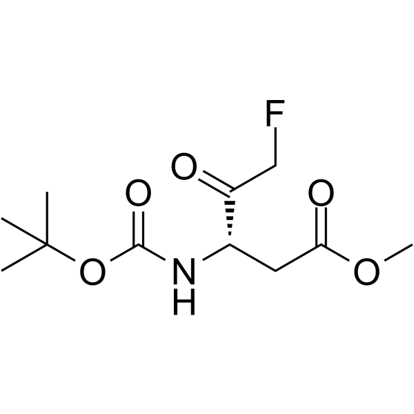
HY-103348
Boc-Asp(OMe)-fluoromethyl ketoneCaspase Inhibitor -
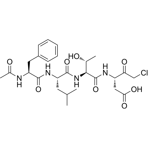
HY-111675
Ac-FLTD-CMKCaspase Inhibitor -
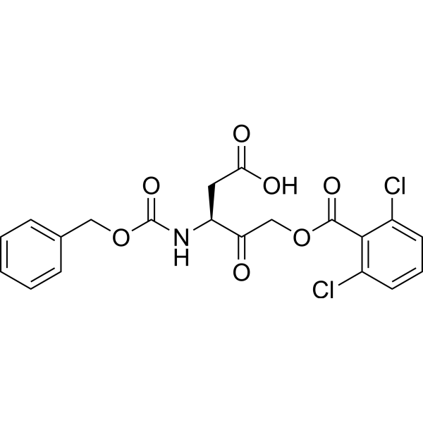
HY-113953
Z-Asp-CH2-DCBCaspase Inhibitor


Biological Activity
Description
In Vitro
Guide (The following is our recommended protocol. This protocol is only a guide and should be modified according to your specific needs).
ICE activity assay methods[1]:
(1) Cerebellar granule neurons were seeded in 24-well plates at a density of 2.1*105 cells/cm2.
(2) After the switch to medium containing low (5 mM) or high (25 mM) concentrations of KCl at DIV 8, cerebellar granule neurons were washed with Locke’s buffer and made permeable by 0.03% Digitonin (HY-N4000).
(3) After 10 min, DABCYL-YVADAPV-EDANS (20 μM) was added.
(4) Fluorescence was determined in 10 min intervals for 1 h using 360 nm excitation and 480 nm emission wavelengths in the 24-well plates.
(5) The fluorometric intensity peaked at 20 min. This time point was used for statistical analysis.
MedChemExpress (MCE) has not independently confirmed the accuracy of these methods. They are for reference only. Further protocols information, click here.


Chemical Information
-
CAS No. 161877-70-9
-
Molecular Weight 1233.39
-
Formula C61H76N12O14S
-
Shipping
Room temperature in continental US; may vary elsewhere.
-
Storage
Please store the product under the recommended conditions in the Certificate of Analysis.

Protocols
-
Apoptosis
Apoptosis, also called programmed cell death, is generally characterized by distinct morphological characteristics.
-
TUNEL staining for apoptotic DNA fragmentation
TUNEL staining detects DNA strand breaks by using terminal deoxynucleotidyl transferase to add labeled nucleotides to exposed 3′-OH DNA termini, generating either microscopic staining in fixed cells or tissue sections, or fluorescence/cytometric signal in cell suspensions. TUNEL positivity reflects DNA fragmentation but should not be interpreted alone as definitive apoptosis, because TUNEL can also label necrotic, autolytic, mechanically damaged, or DNA-repair-associated DNA breaks.
-
Annexin V plus membrane-impermeant dye apoptosis staining
Annexin V-based apoptosis assays rely on the detection of phosphatidylserine (PS) externalization from the inner leaflet of the plasma membrane to the outer leaflet, an early biochemical hallmark of apoptosis. Fluorescently labeled Annexin V binds PS in a calcium-dependent manner, enabling identification of early apoptotic cells by flow cytometry or fluorescence microscopy. When combined with a membrane-impermeant DNA-binding dye (e. g. , propidium iodide), this approach allows discrimination between viable (Annexin V−/dye−), early apoptotic (Annexin V+/dye−), and late apoptotic or necrotic (Annexin V+/dye+) cell populations by assessing membrane integrity and PS exposure.
-
Apoptosis Solutions
Apoptosis is a regulated, generally non-lytic cell-death pathway that removes unwanted, damaged, infected, or abnormal cells through coordinated morphological changes, caspase activation, DNA fragmentation, and membrane remodeling. The intrinsic apoptosis pathway is controlled mainly by mitochondrial outer membrane permeabilization, BCL-2 family proteins, cytochrome c release, apoptosome formation, caspase-9 activation, and downstream executioner caspase-3/7 activation. The extrinsic apoptosis pathway is initiated by death receptors such as Fas, TNFR, and TRAIL receptors, which recruit adaptor proteins and activate caspase-8 before engaging executioner caspases or mitochondrial amplification through BID cleavage. Apoptosis is linked to many phenotypes, including cancer cell killing, tissue homeostasis, immune regulation, neurodegeneration, infection response, and treatment-induced cytotoxicity; unresolved questions include how apoptosis interacts with necroptosis, pyroptosis, ferroptos
-
Cerebellar Granule Neuron Culture
Cerebellar granule neuron culture is a primary neuronal culture method in which postnatal rodent cerebella are dissected, meninges are removed, tissue is enzymatically and mechanically dissociated, and neurons are plated on poly-cation-coated surfaces in medium that supports neuronal attachment, maturation, neurite-network formation, and biochemical or imaging analysis. Cultured CGNs are used to study neuronal development, survival, apoptosis, differentiation, neurotransmitter release, and toxin-sensitive synaptic proteins; mature cultures develop dense neuritic networks, neuronal activity, glutamate release, and neuronal marker expression. A common survival paradigm uses depolarizing extracellular potassium: CGNs maintained in high potassium medium survive and differentiate, whereas switching mature cultures from 25 mM KCl to 5 mM KCl induces apoptotic death that can be used as a readout of activity-dependent neuronal survival.


Purity & Documentation
References

Calculators
Concentration (start) × Volume (start) = Concentration (final) × Volume (final)


Dabcyl-YVADAPV-EDANS Related Classifications
HY-P2628 Related Classifications
 Powered by Bioz