



Datopotamab deruxtecan (solution)
Based on 3 publication(s) in Google Scholar
Datopotamab deruxtecan (DS-1062) solution is a TROP2-targeted antibody-drug conjugate (ADC) with human TROP2 Kd of 0.74 nmol/L. Datopotamab deruxtecan solution consists of the antibody Datopotamab (HY-P99843) and the toxic molecule-linker conjugate Deruxtecan (HY-13631E). Datopotamab deruxtecan solution binds TROP2, triggers internalization and lysosomal trafficking and releases DXd topoisomerase I inhibitor payload. Datopotamab deruxtecan solution disrupts DNA function, induces DNA damage, apoptosis, and bystander killing of tumor microenvironment cells. Datopotamab deruxtecan solution can be used in research related to triple-negative breast cancer, gastric cancer, and non-small cell lung cancer.
For research use only. We do not sell to patients.
- Purity : 99.98%
- CAS No.: 2238831-60-0
- Molecular Weight:149000 (average)
-
Storage:
-80°C, protect from light


To place orders, for customer services and technical support, please contact: MedChemExpress USA
Tel: 609-228-6898 E-mail: [email protected] [email protected]


-
 Biological Activity
-
Chemical Information
- Protocols
- Purity & Documentation
- References
-
 Help & FAQs


-

HY-132254
Sacituzumab govitecanTrop-2 ADC -

HY-132254A
Sacituzumab govitecan (solution)Trop-2 ADC -

HY-138298A
Trastuzumab deruxtecanHER2 ADC -

HY-P99843
DatopotamabAnti-TROP2 Antibody -

HY-P99045
SacituzumabAnti-TROP-2 Antibody -

HY-138298
Trastuzumab deruxtecan (solution)HER2 ADC -

HY-164789
Sacituzumab tirumotecanTROP2 ADC -
HY-13704
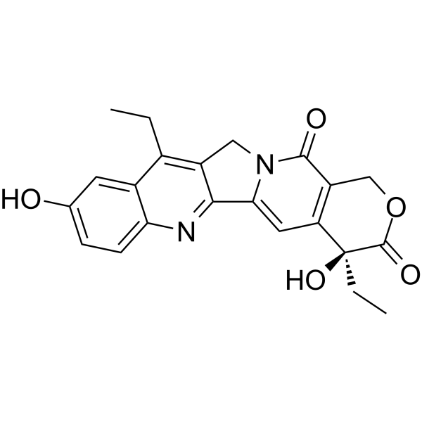
SN-38Topoisomerase I Inhibitor -

HY-141601
Telisotuzumab vedotinAnti-c-MET ADC -

HY-P99016A
Enfortumab vedotin-ejfvNectin-4 ADC -

HY-P99813
Patritumab deruxtecanHER3-Directed ADC -

HY-P9921
Trastuzumab emtansineAntibody-drug Conjugate
Publications Citing Use of MedChemExpress (MCE) Datopotamab deruxtecan (solution)
More Customer Validation & Images

-
WB
-
IF
-
In Vivo Efficacy Study
-
In Vivo Imaging
-
In Vivo Efficacy Study
All Topoisomerase Isoforms
More

Biological Activity
Description
IC50 & Target
[1]|
Topoisomerase I |
In Vitro
Datopotamab deruxtecan solution binds specifically to human TROP2[2].
Datopotamab deruxtecan solution binds with similar affinity to human and cynomolgus monkey TROP2, and does not bind to rat or mouse TROP2[2].
Datopotamab deruxtecan (100 μg/mL; 21 days) solution is stable in human, cynomolgus monkey, rat, and mouse plasma, with minimal DXd release (1.4%-5.5%) after 21 days of incubation at 37°C[2].
Datopotamab deruxtecan (0.32-1000 nmol/L; 6 days) solution potently inhibits the growth of TROP2-high human cancer cell lines (FaDu, BxPC-3, Caov-3, NCI-N87, CFPAC-1, HCC1806, COR-L23) with GI50 values of 0.48-7.8 nM, but does not inhibit TROP2-low cell lines (LK-2, Calu-6)[2].
Datopotamab deruxtecan (10 μg/mL) solution efficiently internalizes into TROP2-expressing human cancer cell lines (BxPC-3, Caov-3, CFPAC-1, COR-L23, FaDu, HCC1806, NCI-N87) with an average internalization rate of 68.5% after 3 hours of incubation at 37°C[2].
Datopotamab deruxtecan (100 nM; 24 h) solution releases DXd in amounts that correlate positively with cell-surface TROP2 expression levels after 24 hours of incubation with TROP2-expressing human cancer cell lines[2].
Datopotamab deruxtecan (10 μg/mL; 15 min-3 h) solution internalizes time-dependently into BxPC-3 human pancreatic cancer cells, traffics to perinuclear regions, and colocalizes with the lysosomal marker LAMP-2[2].
Datopotamab deruxtecan (6-72 h) solution time-dependently induces DNA damage (via phosphorylation of H2AX, KAP1, Chk1) and apoptosis (via cleavage of PARP and caspase-3) in NCI-N87 human gastric cancer cells[2].
MedChemExpress (MCE) has not independently confirmed the accuracy of these methods. They are for reference only. Further protocols information, click here.
-
Cell Line:BxPC-3 human pancreatic cancer cells
-
Concentration:10 μg/mL
-
Incubation Time:15 min; 30 min; 1 h; 3 h
-
Result:Internalized time-dependently.
Aggregated in perinuclear regions.
Partly colocalized with the lysosomal marker LAMP-2.
In Vivo
Datopotamab deruxtecan (10 mg/kg; i.v.; single dose) solution induces potent tumor growth inhibition (TGI = 85-100%) in TROP2-high NSCLC cell line-derived xenografts, with no activity in TROP2-low models[2].
Datopotamab deruxtecan (10 mg/kg; i.v.; single dose) solution induces potent tumor growth inhibition (TGI = 77-98%) in TROP2-high NSCLC patient-derived xenografts[2].
Datopotamab deruxtecan (20-200 mg/kg; i.v.; once every 3 weeks for 3 months) solution is well tolerated in rats at doses up to 200 mg/kg with no life-threatening toxicity, and the STD10 is greater than 200 mg/kg[2].
Datopotamab deruxtecan (10-80 mg/kg; i.v.; once every 3 weeks for 3 months) solution has an HNSTD of 10 mg/kg in cynomolgus monkeys, with severe pulmonary toxicity observed at doses ≥30 mg/kg[2].
MedChemExpress (MCE) has not independently confirmed the accuracy of these methods. They are for reference only.
-
Animal Model:CAnN.Cg-Foxn1nu/CrlCrlj (female, athymic nude, subcutaneous inoculation of 1×10⁷ NCI-N87 human gastric cancer cells to establish a gastric cancer xenograft model)[2]
-
Dosage:10 mg/kg
-
Administration:i.v.; single dose
-
Result:Induced significant tumor growth inhibition with a TGI of 96% (P < 0.001).
Accumulated DXd in tumor tissue, with measurable concentrations detected up to day 21 post-administration.
Observed time-dependent phosphorylation of the DNA damage marker γH2AX in tumors through day 7 post-administration.
Caused no obvious body weight loss or unforeseen events.
-
Animal Model:CAnN.Cg-Foxn1nu/CrlCrlj (female, athymic nude, subcutaneous inoculation of human NSCLC cell lines: 6×10⁶ Calu-3 cells, 5×10⁶ NCI-H2170 cells, 5×10⁶ HCC827 cells, 1×10⁶ LK-2 cells, 6×10⁶ Calu-6 cells, 5×10⁶ EBC-1 cells, to establish a lung cancer xenograft model)[2]
-
Dosage:10 mg/kg
-
Administration:i.v.; single dose
-
Result:Induced significant tumor growth inhibition in TROP2-high NSCLC models: Calu-3 (TROP2 H-score 145) with TGI of 85% (P < 0.001), NCI-H2170 (TROP2 H-score 115) with TGI of 95% (P < 0.001), HCC827 (TROP2 H-score 220) with TGI of 90% (P < 0.001), and EBC-1 (TROP2 H-score 133) with TGI of 100% (P < 0.001).
Caused no tumor growth inhibition in TROP2-low models (LK-2, TROP2 H-score 20; Calu-6, TROP2 H-score 0).
Caused no obvious body weight loss or unforeseen events.
-
Animal Model:Hsd:Athymic Nude-Foxn1nu (female, athymic nude, subcutaneous inoculation of patient-derived tumor fragments from NSCLC patients to establish a lung cancer xenograft model)[2]
-
Dosage:10 mg/kg
-
Administration:i.v.; single dose
-
Result:Induced significant tumor growth inhibition in all TROP2-high NSCLC patient-derived xenograft models: CTG-0163 (TROP2 H-score 262) with TGI of 77% (P < 0.001), CTG-0838 (TROP2 H-score 163) with TGI of 98% (P < 0.001), and CTG-1014 (TROP2 H-score 252) with TGI of 95% (P < 0.001).
Caused no obvious body weight loss or unforeseen events.
-
Animal Model:Crl:CD(SD) (male and female, healthy)[2]
-
Dosage:20 mg/kg; 60 mg/kg; 200 mg/kg
-
Administration:i.v.; once every 3 weeks; 3 months
-
Result:Caused no deaths or life-threatening toxicities at any dose tested.
Induced body weight decrease at ≥60 mg/kg.
Caused decreased white blood cells, reticulocytes, neutrophils, lymphocytes, albumin, and increased urea nitrogen at 200 mg/kg.
Induced target organ toxicities: thymus changes at ≥20 mg/kg; kidney, intestine, and incisor changes at ≥60 mg/kg; and lung, skin, bone marrow, spleen, reproductive organ changes at 200 mg/kg.
Determined the severely toxic dose in 10% of animals (STD10) was >200 mg/kg.
-
Animal Model:(male and female, healthy)[2]
-
Dosage:10 mg/kg; 30 mg/kg; 80 mg/kg
-
Administration:i.v.; once every 3 weeks; 3 months
-
Result:Caused no deaths at any dose tested.
Induced severe pulmonary toxicity (cell infiltration, edema, fibrosis) at ≥30 mg/kg (1 animal at 30 mg/kg, 2 animals at 80 mg/kg).
Induced body weight decrease at ≥30 mg/kg.
Induced target organ toxicities: intestinal changes at ≥10 mg/kg; lung, cornea, skin, thymus, liver changes at ≥30 mg/kg; and kidney, joint cartilage changes at 80 mg/kg.
Showed reversibility of skin and corneal lesions (except pigmentation) after treatment withdrawal.
Determined the highest non-severely toxic dose (HNSTD) to be 10 mg/kg based on pulmonary findings.
Clinical Trial
| NCT Number | Sponsor | Condition | Start Date |
Phase

|
|---|---|---|---|---|
| NCT01329991 | Plexxikon| | 2011-05 | PHASE1 |


Chemical Information
-
CAS No. 2238831-60-0
-
Appearance Liquid
-
Molecular Weight 149000 (average)
-
Color Colorless to light yellow
-
SMILES
[Datopotamab deruxtecan (solution)]
-
Synonyms
DS-1062 (solution); Dato-DXd (solution)
-
Shipping
Shipping with dry ice.
-
Storage
-80°C, protect from light


Publications (3)
-
Journal Impact Factor
-
Most Recent
-
-
Anal Chem
Preclinical Evaluation of a Trop2-Targeted Peptide Probe for PET Imaging of Triple-Negative Breast Cancer. [Abstract]2025 Oct 28;97(42):23598-23608. PMID: 41105928
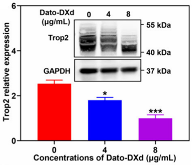
Datopotamab deruxtecan (solution) purchased from MedChemExpress. Usage Cited in: Anal Chem. 2025 Oct 28;97(42):23598-23608. [Abstract]
Western blotting analysis of the expression level of Trop2 in MDA-MB-468 cells treated with different concentrations of Dato-DXd (0-8 μg/mL; 48 h).
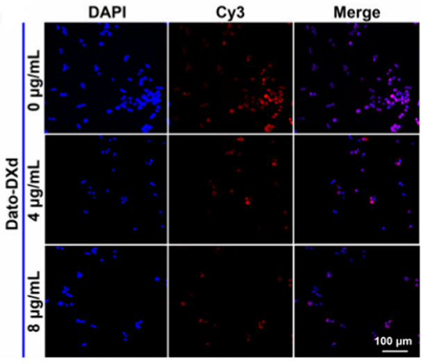
Datopotamab deruxtecan (solution) purchased from MedChemExpress. Usage Cited in: Anal Chem. 2025 Oct 28;97(42):23598-23608. [Abstract]
Immunofluorescence analysis of Trop2 expression in MDA-MB-468 cells treated with different concentrations of Dato-DXd (0-8 μg/mL; 48 h).
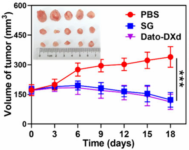
Datopotamab deruxtecan (solution) purchased from MedChemExpress. Usage Cited in: Anal Chem. 2025 Oct 28;97(42):23598-23608. [Abstract]
Time course of tumor volume of MDA-MB-468 tumor-bearing mice treated with PBS, SG (10 mg/kg, tail vein injection), and Dato-DXd (10 mg/kg, tail vein injection).
Datopotamab deruxtecan (solution) purchased from MedChemExpress. Usage Cited in: Anal Chem. 2025 Oct 28;97(42):23598-23608. [Abstract]
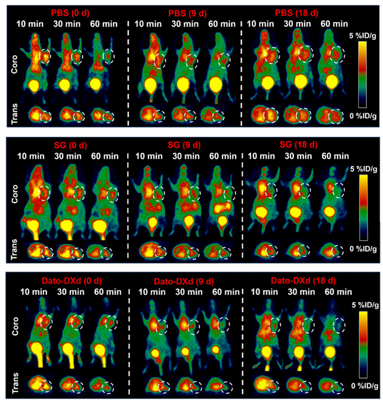
MicroPET imaging of MDA-MB-468 tumor-bearing mice at different time postinjection of [68Ga]Ga-NOTA-PEG2-WP8 during PBS, SG (10 mg/kg, tail vein injection) and Dato-DXd (10 mg/kg, tail vein injection) treatment for 0, 9, and 18 days, respectively. White dotted line circles indicate the tumor locations.
Datopotamab deruxtecan (solution) purchased from MedChemExpress. Usage Cited in: Anal Chem. 2025 Oct 28;97(42):23598-23608. [Abstract]
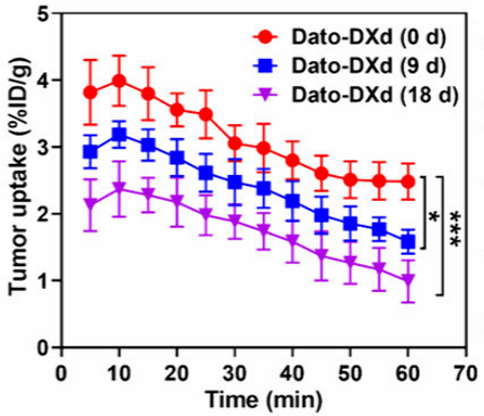
Time-activity curve of tumor uptake of [68Ga]Ga-NOTA-PEG2-WP8 in MDA-MB-468 xenografts during Dato-DXd (10 mg/kg, tail vein injection) treatment.
-

Protocols
-
Multiplex immunofluorescence IHC
Multiplex immunofluorescence IHC detects multiple protein biomarkers in one tissue section by sequential antibody staining, HRP-mediated tyramide fluorophore deposition, heat-mediated antibody stripping, nuclear counterstaining, multispectral imaging, spectral unmixing, and digital cell phenotyping; TSA deposits fluorophore near the antigen so the fluorescence signal remains after primary and secondary antibodies are removed, enabling repeated staining cycles, including with antibodies from the same host species. Classic FFPE tumor immune-profiling applications use panels such as CD3, CD8, CD68/CD163, FOXP3, PD-1, PD-L1, pancytokeratin, Ki67, and DAPI to identify tumor cells, immune-cell subsets, checkpoint-marker expression, co-expression phenotypes, cell density, and spatial relationships in the tumor microenvironment.
-
Apoptosis
Apoptosis, also called programmed cell death, is generally characterized by distinct morphological characteristics.
-
TUNEL staining for apoptotic DNA fragmentation
TUNEL staining detects DNA strand breaks by using terminal deoxynucleotidyl transferase to add labeled nucleotides to exposed 3′-OH DNA termini, generating either microscopic staining in fixed cells or tissue sections, or fluorescence/cytometric signal in cell suspensions. TUNEL positivity reflects DNA fragmentation but should not be interpreted alone as definitive apoptosis, because TUNEL can also label necrotic, autolytic, mechanically damaged, or DNA-repair-associated DNA breaks.
-
Annexin V plus membrane-impermeant dye apoptosis staining
Annexin V-based apoptosis assays rely on the detection of phosphatidylserine (PS) externalization from the inner leaflet of the plasma membrane to the outer leaflet, an early biochemical hallmark of apoptosis. Fluorescently labeled Annexin V binds PS in a calcium-dependent manner, enabling identification of early apoptotic cells by flow cytometry or fluorescence microscopy. When combined with a membrane-impermeant DNA-binding dye (e. g. , propidium iodide), this approach allows discrimination between viable (Annexin V−/dye−), early apoptotic (Annexin V+/dye−), and late apoptotic or necrotic (Annexin V+/dye+) cell populations by assessing membrane integrity and PS exposure.
-
Multiplex immunohistochemistry
Multiplex immunohistochemistry (mIHC), also known as tyramide dignal amplification (TSA), is an enzymatic detection method that uses horseradish peroxidase (HRP) to perform high-density in-situ labeling of target proteins or nucleic acids.
-
Apoptosis Solutions
Apoptosis is a regulated, generally non-lytic cell-death pathway that removes unwanted, damaged, infected, or abnormal cells through coordinated morphological changes, caspase activation, DNA fragmentation, and membrane remodeling. The intrinsic apoptosis pathway is controlled mainly by mitochondrial outer membrane permeabilization, BCL-2 family proteins, cytochrome c release, apoptosome formation, caspase-9 activation, and downstream executioner caspase-3/7 activation. The extrinsic apoptosis pathway is initiated by death receptors such as Fas, TNFR, and TRAIL receptors, which recruit adaptor proteins and activate caspase-8 before engaging executioner caspases or mitochondrial amplification through BID cleavage. Apoptosis is linked to many phenotypes, including cancer cell killing, tissue homeostasis, immune regulation, neurodegeneration, infection response, and treatment-induced cytotoxicity; unresolved questions include how apoptosis interacts with necroptosis, pyroptosis, ferroptos
-
Lysosome and acidic-vesicle live-cell staining
Lysosome and acidic-vesicle live-cell staining detects acidic intracellular compartments by using membrane-permeant acidotropic probes that accumulate in low-pH vesicles, including lysosomes, late endosomes, autolysosomes, and acidic phagosomes. LysoTracker staining is commonly used as an intensity-based readout of acidic lysosomal compartment abundance or enlargement, while acridine orange produces green fluorescence in less concentrated compartments and red fluorescence after concentration-dependent accumulation in acidic vesicular organelles. Loss or reduction of acridine-orange red signal can be used as a readout of lysosomal membrane permeabilization or reduced acidic-vesicle integrity. This protocol is designed for live cultured cells and can be adapted for fluorescence microscopy, high-content imaging, plate-reader readout, or flow cytometry when the selected literature supports the readout. Because these dyes report acidotropic accumulation rather than lysosome identity alone,
-
Breast Cancer Modeling
Breast cancer is a heterogeneous cancer, and it has been distinguished into four subtypes: luminal A, luminal B, HER2-positive and basal-like. Molecular mutations, epigenetic alterations, hormone exposure and immune microenvironment are related to the progression of breast cancer.
-
Genotoxicity/Mutagenicity Study
The bacterial reverse mutation assay detects point mutations that restore amino-acid prototrophy in auxotrophic Salmonella typhimurium or Escherichia coli tester strains; after exposure to a test article, mutagenic activity is read out as an increased number of revertant colonies on minimal agar compared with the vehicle control. The assay uses tester strains with different mutation targets so that base-substitution and frameshift mutagens can be detected, and testing is performed with and without exogenous mammalian metabolic activation because some chemicals require biotransformation to become mutagenic.
-
Research Protocol for Cancer Immunology
Cancer immunology studies how the immune system recognizes, suppresses, edits, or fails to eliminate malignant cells through tumor antigen release, antigen presentation, T-cell priming, immune trafficking, tumor-cell killing, and feedback inhibition in the tumor microenvironment. The cancer-immunity cycle links tumor antigenicity, dendritic-cell priming, CD8+ T-cell infiltration, cytotoxic function, and immune-checkpoint regulation to tumor rejection or immune escape. Immune-checkpoint pathways such as PD-1/PD-L1 and CTLA-4 suppress antitumor T-cell activity and can be therapeutically blocked, but many tumors remain resistant because of poor antigen presentation, weak T-cell infiltration, suppressive myeloid cells, regulatory T cells, and tumor-intrinsic immune-exclusion programs. Unresolved questions include which immune-cell states predict response, how tumor-intrinsic pathways exclude immune cells, how myeloid suppression limits checkpoint blockade, and which combination strategies


Purity & Documentation
-
Data Sheet (284 KB)
-
SDS (252 KB)
- English - EN (252 KB)
- Français - FR (252 KB)
- Deutsch - DE (252 KB)
- Norwegian - NO (252 KB)
- Español - ES (252 KB)
- Swedish - SV (252 KB)
- Italian - IT (252 KB)
- Korean - KR (252 KB)
- Portuguese - PT (252 KB)
-
Handling Instructions (2659 KB)
References
[1]. Bardia A, et al. Datopotamab Deruxtecan in Advanced or Metastatic HR+/HER2- and Triple-Negative Breast Cancer: Results From the Phase I TROPION-PanTumor01 Study. J Clin Oncol. 2024;42(19):2281-2294. [Content Brief]
[2]. Okajima D, et al. Datopotamab Deruxtecan, a Novel TROP2-directed Antibody-drug Conjugate, Demonstrates Potent Antitumor Activity by Efficient Drug Delivery to Tumor Cells. Mol Cancer Ther. 2021;20(12):2329-2340. [Content Brief]

Calculators
Concentration (start) × Volume (start) = Concentration (final) × Volume (final)


Datopotamab deruxtecan (solution) Related Classifications
HY-141598 Related Classifications
 Powered by Bioz