



DB2313 tetrahydrochloride
Based on 7 publication(s) in Google Scholar
DB2313 tetrahydrochloride is a potent transcription factor PU.1 inhibitor with an apoptosis of 14 nM. DB2313 tetrahydrochloride disrupts the interaction of PU.1 with target gene promoters. DB2313 tetrahydrochloride induces apoptosis of acute myeloid leukemia (AML) cells, and has anticancer effects.
For research use only. We do not sell to patients.
- CAS No.: 2170606-75-2
- Formula: C42H45Cl4FN8O2
- Molecular Weight:854.67
-
Storage:
Please store the product under the recommended conditions in the Certificate of Analysis.


To place orders, for customer services and technical support, please contact: MedChemExpress USA
Tel: 609-228-6898 E-mail: [email protected] [email protected]


-
 Biological Activity
-
Chemical Information
- Protocols
- Purity & Documentation
- References
-
 Help & FAQs


Publications Citing Use of MedChemExpress (MCE) DB2313 tetrahydrochloride
More- Ann Rheum Dis. 2024 Aug 20:ard-2024-226067. [Abstract]
- Nat Commun. 2026 Feb 24;17(1):3123. [Abstract]
- Nat Commun. 2025 Aug 4;16(1):7134. [Abstract]
- Adv Sci (Weinh). 2025 Jun;12(21):e2415205. [Abstract]
- Chem Biol Interact. 2024 Dec 1:404:111262. [Abstract]
- J Craniofac Surg. 2026 Mar 2. [Abstract]
- McGill University. 2026.
 Customer Validation & Images

-
RT-PCR
-
ELISA
-
Flow Cytometry
-
Flow Cytometry
-
Histological Imaging/Staining


Biological Activity
Description


Chemical Information
-
CAS No. 2170606-75-2
-
Molecular Weight 854.67
-
Formula C42H45Cl4FN8O2
-
SMILES
FC1=C(COC2=CC=C(C3=NC4=CC=C(C(NC(C)C)=N)C=C4N3)C=C2)C=CC=C1COC5=CC=C(C6=NC7=CC=C(C(NC(C)C)=N)C=C7N6)C=C5.Cl.Cl.Cl.Cl
-
Shipping
Room temperature in continental US; may vary elsewhere.
-
Storage
Please store the product under the recommended conditions in the Certificate of Analysis.


Publications (7)
-
Journal Impact Factor
-
Most Recent
-
Ann Rheum Dis
2024 Aug 20:ard-2024-226067. PMID: 39164066
DB2313 tetrahydrochloride purchased from MedChemExpress. Usage Cited in: Ann Rheum Dis. 2024 Aug 20:ard-2024-226067. [Abstract]
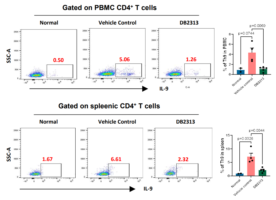
DB2313 (0.3 mg; i.p.; once every 3 d) downregulated IL-9 expression and the proportions of Th9 cells in the spleen and PBMCs of collagen-induced arthritis (CIA) mice.
DB2313 tetrahydrochloride purchased from MedChemExpress. Usage Cited in: Ann Rheum Dis. 2024 Aug 20:ard-2024-226067. [Abstract]
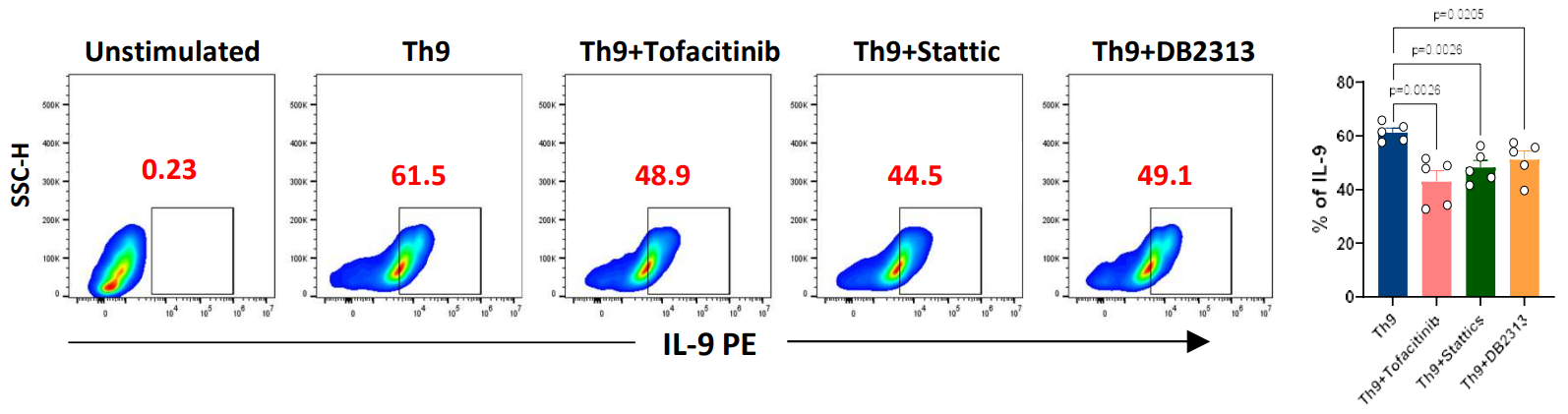
PU.1 inhibitor DB2313 (0.3 mg; i.p.; once every 3 d) suppressed IL-9 release from Th9 cells of collagen-induced arthritis (CIA) mice.
DB2313 tetrahydrochloride purchased from MedChemExpress. Usage Cited in: Ann Rheum Dis. 2024 Aug 20:ard-2024-226067. [Abstract]
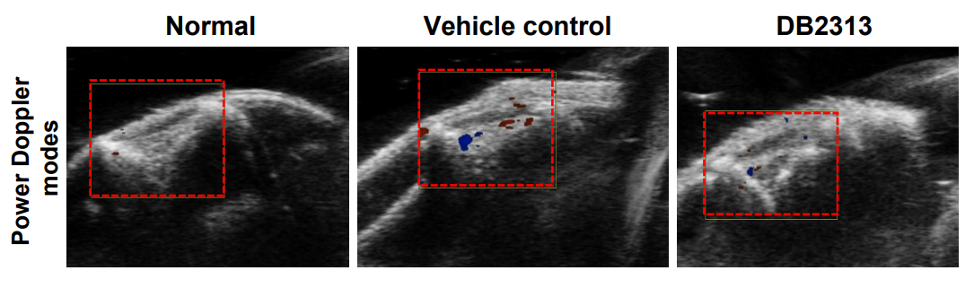
Effect of PU.1 inhibitor DB2313 (0.3 mg; i.p.; once every 3 d) on joint neovascularization in collagen-induced arthritis (CIA) mice.
-
Nat Commun
DNA-PK-mediated phosphorylation of STAT6 establishes a non-canonical type 2 immunity axis to prevent macrophage senescence. [Abstract]2026 Feb 24;17(1):3123. PMID: 41735318 -
Nat Commun
Unveiling genetic signatures of immune response in immune-related diseases through single-cell eQTL analysis across diverse conditions. [Abstract]2025 Aug 4;16(1):7134. PMID: 40759647
DB2313 tetrahydrochloride purchased from MedChemExpress. Usage Cited in: Nat Commun. 2025 Aug 4;16(1):7134. [Abstract]
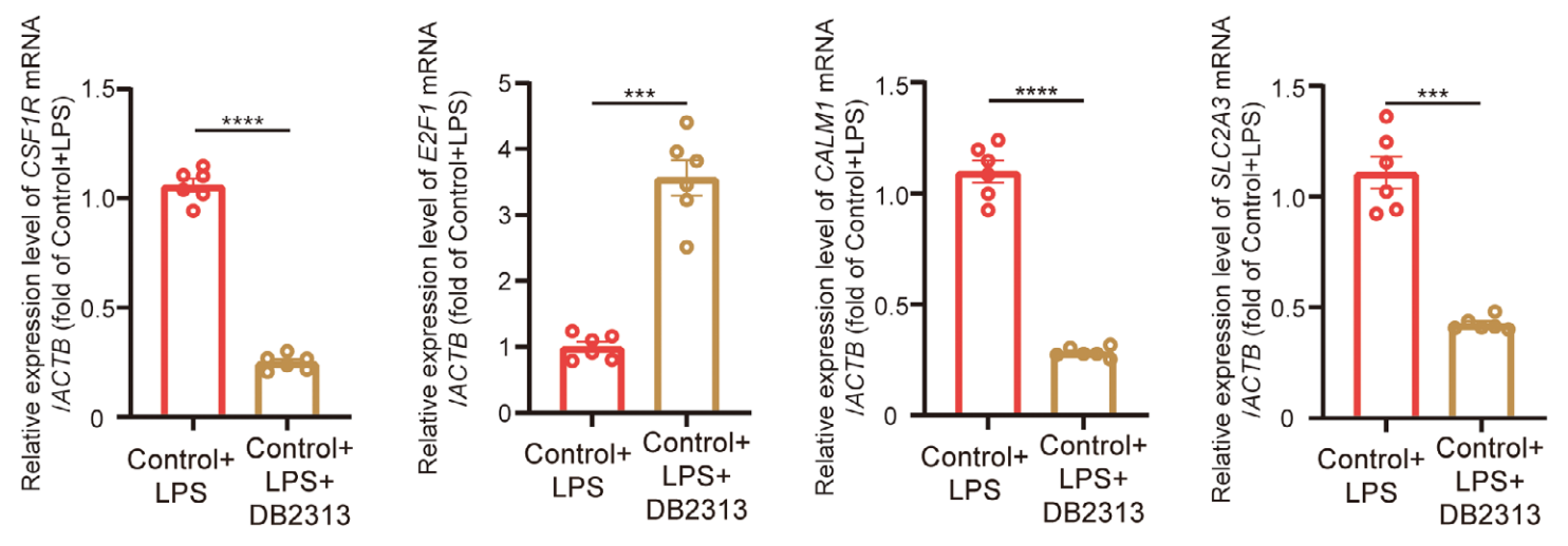
DB2313 (660 nM) inhibited the transcription of CSF1R, CALM1, and SLC2A3 while promoting E2F in neutrophils.
DB2313 tetrahydrochloride purchased from MedChemExpress. Usage Cited in: Nat Commun. 2025 Aug 4;16(1):7134. [Abstract]
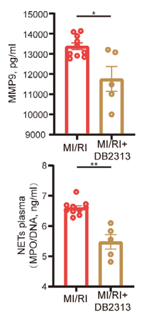
DB2313 (17 mg/kg; i.p.) decreased serum levels of MMP9 and MPO/DNA‐NETs in the murine MI/RI model.
-
Adv Sci (Weinh)
MMP9High Neutrophils are Critical Mediators of Neutrophil Extracellular Traps Formation and Myocardial Ischemia/Reperfusion Injury. [Abstract]2025 Jun;12(21):e2415205. PMID: 40151877 -
Chem Biol Interact
4-Hydroxybenzoic acid restrains Nlrp3 inflammasome priming and activation via disrupting PU.1 DNA binding activity and direct antioxidation. [Abstract]2024 Dec 1:404:111262. PMID: 39389441 -
J Craniofac Surg
PU.1 Inhibition Attenuates Neuroinflammation and Pyroptosis After Traumatic Brain Injury by Modulating Microglial Polarization. [Abstract]2026 Mar 2. PMID: 41769894 -

Protocols
-
Nuclear Protein Extraction (High-Salt/Hypotonic Fractionation)
The high-salt/hypotonic fractionation method for nuclear protein extraction is based on the differential solubility of cellular components. Cytoplasmic proteins are extracted first using a hypotonic buffer that causes cell swelling and membrane rupture, followed by centrifugation to separate the cytoplasmic supernatant from the nuclear pellet. The nuclear pellet is then subjected to high-salt extraction (e. g. , 0. 4 M (NH4)2SO4 or 1 M NaCl) to solubilize tightly bound nuclear matrix proteins, including transcription factors, histones, and structural proteins associated with chromatin and the nuclear scaffold. This approach allows for the isolation of both soluble cytoplasmic proteins and salt-resistant nuclear proteins while minimizing cross-contamination.
-
Apoptosis
Apoptosis, also called programmed cell death, is generally characterized by distinct morphological characteristics.
-
TUNEL staining for apoptotic DNA fragmentation
TUNEL staining detects DNA strand breaks by using terminal deoxynucleotidyl transferase to add labeled nucleotides to exposed 3′-OH DNA termini, generating either microscopic staining in fixed cells or tissue sections, or fluorescence/cytometric signal in cell suspensions. TUNEL positivity reflects DNA fragmentation but should not be interpreted alone as definitive apoptosis, because TUNEL can also label necrotic, autolytic, mechanically damaged, or DNA-repair-associated DNA breaks.
-
Annexin V plus membrane-impermeant dye apoptosis staining
Annexin V-based apoptosis assays rely on the detection of phosphatidylserine (PS) externalization from the inner leaflet of the plasma membrane to the outer leaflet, an early biochemical hallmark of apoptosis. Fluorescently labeled Annexin V binds PS in a calcium-dependent manner, enabling identification of early apoptotic cells by flow cytometry or fluorescence microscopy. When combined with a membrane-impermeant DNA-binding dye (e. g. , propidium iodide), this approach allows discrimination between viable (Annexin V−/dye−), early apoptotic (Annexin V+/dye−), and late apoptotic or necrotic (Annexin V+/dye+) cell populations by assessing membrane integrity and PS exposure.
-
Apoptosis Solutions
Apoptosis is a regulated, generally non-lytic cell-death pathway that removes unwanted, damaged, infected, or abnormal cells through coordinated morphological changes, caspase activation, DNA fragmentation, and membrane remodeling. The intrinsic apoptosis pathway is controlled mainly by mitochondrial outer membrane permeabilization, BCL-2 family proteins, cytochrome c release, apoptosome formation, caspase-9 activation, and downstream executioner caspase-3/7 activation. The extrinsic apoptosis pathway is initiated by death receptors such as Fas, TNFR, and TRAIL receptors, which recruit adaptor proteins and activate caspase-8 before engaging executioner caspases or mitochondrial amplification through BID cleavage. Apoptosis is linked to many phenotypes, including cancer cell killing, tissue homeostasis, immune regulation, neurodegeneration, infection response, and treatment-induced cytotoxicity; unresolved questions include how apoptosis interacts with necroptosis, pyroptosis, ferroptos


Purity & Documentation
References
[1]. Iléana Antony-Debré, et al. Pharmacological inhibition of the transcription factor PU.1 in leukemia. J Clin Invest. 2017 Dec 1;127(12):4297-4313. [Content Brief]
[2]. Zhang S, Zhao S, Qi Y, et al. SPI1-induced downregulation of FTO promotes GBM progression by regulating pri-miR-10a processing in an m6A-dependent manner. Mol Ther Nucleic Acids. 2022;27:699-717. [Content Brief]

Calculators
Concentration (start) × Volume (start) = Concentration (final) × Volume (final)


DB2313 tetrahydrochloride Related Classifications
HY-124629A Related Classifications
 Powered by Bioz