



DBCO-PEG4-MMAF

Based on 1 Customer Validation
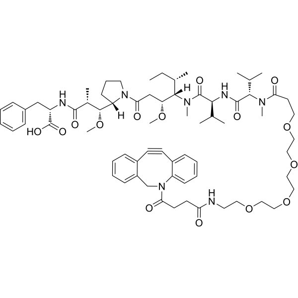
DBCO-PEG4-MMAF is a agent-linker conjugate for ADC with potent antitumor activity by using the tubulin polymerization inhibitor, MMAF, linked via the linker DBCO-PEG4. DBCO-PEG4-MMAF is a click chemistry reagent, it contains a DBCO group that can undergo strain-promoted alkyne-azide cycloaddition (SPAAC) with molecules containing Azide groups.
For research use only. We do not sell to patients.
- Purity : 99.54%
- CAS No.: 2360411-65-8
- Formula: C69H99N7O15
- Molecular Weight:1266.56
-
Storage:Powder -20°C, 3 years
* The compound is unstable in solutions, freshly prepared is recommended.

To place orders, for customer services and technical support, please contact: MedChemExpress USA
Tel: 609-228-6898 E-mail: [email protected] [email protected]


-
 Biological Activity
-
Chemical Information
-
Solvent & Solubility
- Protocols
- Purity & Documentation
-
 Help & FAQs


-
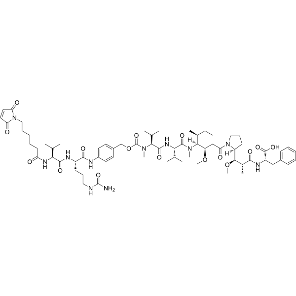
HY-112786
MC-Val-Cit-PAB-MMAFDrug-Linker Conjugate for ADC -
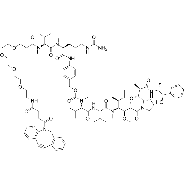
HY-136314
DBCO-PEG4-VC-PAB-MMAEDrug-Linker Conjugate for ADC -
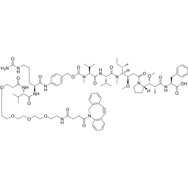
HY-130990
DBCO-PEG4-Val-Cit-PAB-MMAFDrug-Linker Conjugates for ADC -
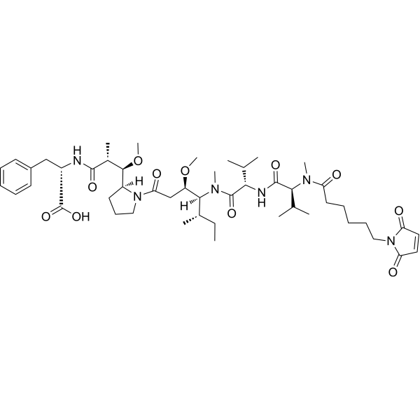
HY-15578
McMMAFADC Drug-Linker -
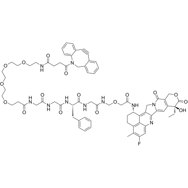
HY-134723
DBCO-PEG4-GGFG-DxdDrug-Linker Conjugates for ADC -
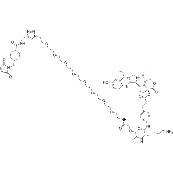
HY-128946
CL2A-SN-38DNA Topoisomerase I Inhibitor -
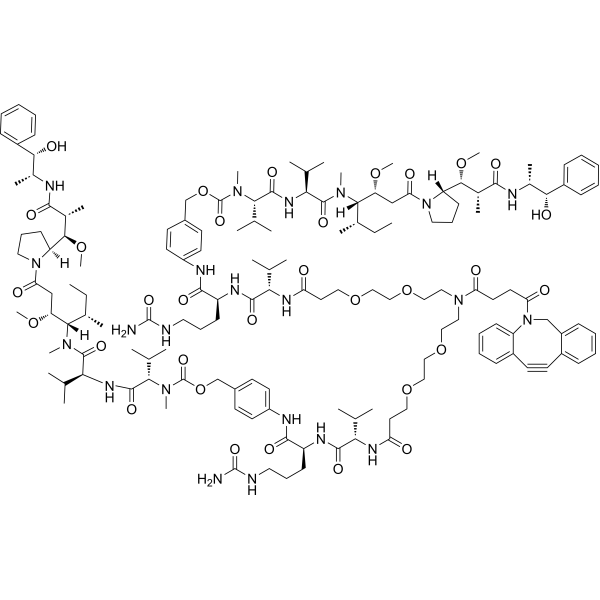
HY-126690
DBCO-(PEG2-VC-PAB-MMAE)2Drug-Linker Conjugates for ADC -
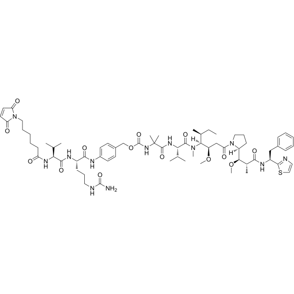
HY-128955
MC-VC-PABC-Aur0101Drug-Linker Conjugates for ADC -
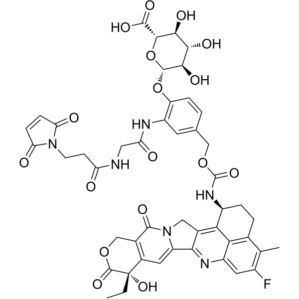
HY-153179
Mal-Gly-PAB-Exatecan-D-glucuronic acidADC Intermediate Compound -
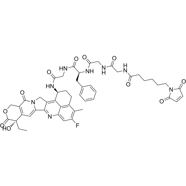
HY-114233
MC-GGFG-ExatecanDrug-Linker Conjugate for ADC -
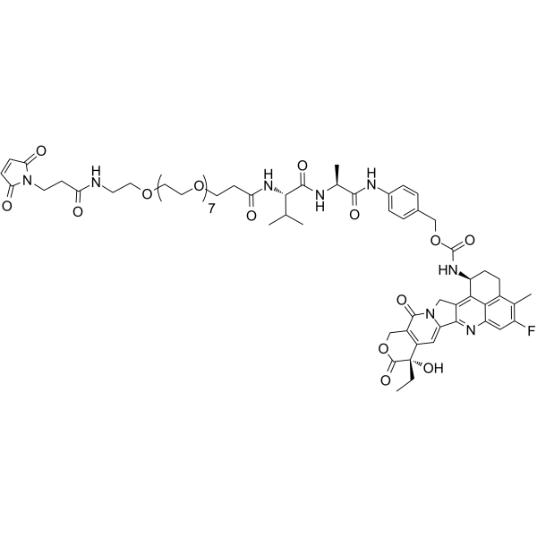
HY-147271
Mal-PEG8-Val-Ala-PAB-ExatecanAnti-Nectin-4 ADC Linker -
HY-130989
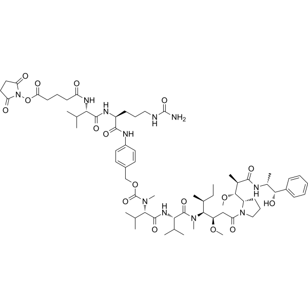
SuO-Glu-Val-Cit-PAB-MMAEADC Linker
All Drug-Linker Conjugates for ADC Isoforms
More

Biological Activity
Description
IC50 & Target
|
Auristatin |
In Vitro
ADCs are comprised of an antibody to which is attached an ADC cytotoxin through an ADC linker.
MedChemExpress (MCE) has not independently confirmed the accuracy of these methods. They are for reference only. Further protocols information, click here.


Chemical Information
-
CAS No. 2360411-65-8
-
Appearance Solid
-
Molecular Weight 1266.56
-
Formula C69H99N7O15
-
Color White to off-white
-
SMILES
O=C(N1C2=CC=CC=C2C#CC3=CC=CC=C3C1)CCC(NCCOCCOCCOCCOCCC(N(C)[C@@H](C(C)C)C(N[C@@H](C(C)C)C(N(C)[C@]([C@@H](C)CC)([H])[C@H](OC)CC(N(CCC4)[C@]4([H])[C@H](OC)[C@@H](C)C(N[C@H](C(O)=O)CC5=CC=CC=C5)=O)=O)=O)=O)=O)=O
-
Shipping
Room temperature in continental US; may vary elsewhere.
-
Storage
Powder -20°C 3 years * The compound is unstable in solutions, freshly prepared is recommended.


Solvent & Solubility
In Vitro:
DMSO : ≥ 100 mg/mL (78.95 mM; Hygroscopic DMSO has a significant impact on the solubility of product, please use newly opened DMSO)
* "≥" means soluble, but saturation unknown.
Please refer to the solubility information to select the appropriate solvent. The compound is unstable in solutions, freshly prepared is recommended.
Please refer to the solubility information to select the appropriate solvent. The compound is unstable in solutions, freshly prepared is recommended.
Concentration (start) × Volume (start) = Concentration (final) × Volume (final)
In Vivo:
Select the appropriate dissolution method based on your experimental animal and administration route.
- For the following dissolution methods, please ensure to first prepare a clear stock solution using an In Vitro approach and then sequentially add co-solvents:
- To ensure reliable experimental results, the clarified stock solution can be appropriately stored based on storage conditions. As for the working solution for In Vivo experiments, it is recommended to prepare freshly and use it on the same day.
- The percentages shown for the solvents indicate their volumetric ratio in the final prepared solution. If precipitation or phase separation occurs during preparation, heat and/or sonication can be used to aid dissolution.
Add each solvent one by one: 10% DMSO 40% PEG300 5% Tween-80 45% Saline
Solubility: 5 mg/mL (3.95 mM); Suspended solution; Need ultrasonic
This protocol yields a suspended solution of 5 mg/mL. Suspended solution can be used for oral and intraperitoneal injection.
Taking 1 mL working solution as an example, add 100 μL DMSO stock solution (50.0 mg/mL) to 400 μL PEG300, and mix evenly; then add 50 μL Tween-80 and mix evenly; then add 450 μL Saline to adjust the volume to 1 mL.
Preparation of Saline: Dissolve 0.9 g sodium chloride in ddH₂O and dilute to 100 mL to obtain a clear Saline solution.
Add each solvent one by one: 10% DMSO 90% (20% SBE-β-CD in Saline)
Solubility: 5 mg/mL (3.95 mM); Suspended solution; Need ultrasonic
This protocol yields a suspended solution of 5 mg/mL. Suspended solution can be used for oral and intraperitoneal injection.
Taking 1 mL working solution as an example, add 100 μL DMSO stock solution (50.0 mg/mL) to 900 μL 20% SBE-β-CD in Saline, and mix evenly.
Preparation of 20% SBE-β-CD in Saline (4°C, storage for one week): 2 g SBE-β-CD powder is dissolved in 10 mL Saline, completely dissolve until clear.
In Vivo Dissolution Calculator
Please enter the basic information of animal experiments:
-
-
-
-
Recommended: Prepare an additional quantity of animals to account for potential losses during experiments.
Please enter your animal formula composition:
-
%DMSO +
Recommended: Keep the proportion of DMSO in working solution below 2% if your animal is weak.
-
%+
-
+%Tween-80 + +
-
%Saline +
The co-solvents required include: DMSO, . All of co-solvents are available by MedChemExpress (MCE). , Tween 80. All of co-solvents are available by MedChemExpress (MCE).
Working solution concentration: 0.22 mg/mL
Method for preparing stock solution: mg drug dissolved in μL DMSO. Stock solution concentration: mg/mL. * The compound is unstable in solutions, freshly prepared is recommended.
1. Take μL DMSO stock solution;
2. Add μL .
μL , mix evenly;
3. Then add μL Tween 80, mix evenly;
4. Then add μL
Please ensure that the stock solution in the first step is dissolved to a clear state, and add co-solvents in sequence. You can use ultrasonic heating (ultrasonic cleaner, recommended frequency 20-40 kHz), vortexing, etc. to assist dissolution.

Protocols
-
EdU Incorporation Assay (Click Chemistry-Based DNA Synthesis Measurement)
The EdU incorporation assay measures DNA synthesis by adding the thymidine analog 5-ethynyl-2′-deoxyuridine to cells or tissues, where it is incorporated into newly synthesized DNA during S phase. Incorporated EdU is detected by copper-catalyzed azide-alkyne cycloaddition, in which a fluorescent azide covalently reacts with the ethynyl group on EdU, allowing S-phase cells to be detected by fluorescence microscopy, flow cytometry, or high-content imaging. EdU detection does not require DNA denaturation or anti-BrdU antibody access, which preserves sample structure and improves compatibility with immunostaining and multiparameter cytometry compared with BrdU-based detection. EdU can be cytotoxic in a cell-type- and exposure-dependent manner, so pulse duration, concentration, and continuous-labeling designs should be validated for each cell type.
-
Subcutaneous Cell-Line-Derived Xenograft
Subcutaneous cell-line-derived xenograft (CDX) models are established by implanting cultured human cancer cell lines into immunodeficient mice, where the injected cells form localized tumors that can be monitored in vivo as a measure of tumorigenic potential, growth kinetics, and treatment response. These models are widely used in oncology research because they allow reproducible tumor formation and enable comparative assessment of tumor growth between different cell lines or genetic manipulations in a controlled in vivo microenvironment. Subcutaneous implantation of cancer cells in immunodeficient mice is a standard approach for evaluating tumor growth behavior and therapeutic response across multiple cancer types, including prostate, esophageal, pancreatic, and colon cancer models.


Purity & Documentation
-
Data Sheet (251 KB)
-
SDS (252 KB)
- English - EN (252 KB)
- Français - FR (252 KB)
- Deutsch - DE (252 KB)
- Norwegian - NO (252 KB)
- Español - ES (252 KB)
- Swedish - SV (252 KB)
- Italian - IT (252 KB)
- Korean - KR (252 KB)
- Portuguese - PT (252 KB)
-
Handling Instructions (2659 KB)


Complete Stock Solution Preparation Table
Please refer to the solubility information to select the appropriate solvent. The compound is unstable in solutions, freshly prepared is recommended.
| Optional Solvent | Concentration Solvent Mass | 1 mg | 5 mg | 10 mg | 25 mg |
|---|---|---|---|---|---|
| DMSO | 1 mM | 0.7895 mL | 3.9477 mL | 7.8954 mL | 19.7385 mL |
| 5 mM | 0.1579 mL | 0.7895 mL | 1.5791 mL | 3.9477 mL | |
| 10 mM | 0.0790 mL | 0.3948 mL | 0.7895 mL | 1.9739 mL | |
| 15 mM | 0.0526 mL | 0.2632 mL | 0.5264 mL | 1.3159 mL | |
| 20 mM | 0.0395 mL | 0.1974 mL | 0.3948 mL | 0.9869 mL | |
| 25 mM | 0.0316 mL | 0.1579 mL | 0.3158 mL | 0.7895 mL | |
| 30 mM | 0.0263 mL | 0.1316 mL | 0.2632 mL | 0.6580 mL | |
| 40 mM | 0.0197 mL | 0.0987 mL | 0.1974 mL | 0.4935 mL | |
| 50 mM | 0.0158 mL | 0.0790 mL | 0.1579 mL | 0.3948 mL | |
| 60 mM | 0.0132 mL | 0.0658 mL | 0.1316 mL | 0.3290 mL |


DBCO-PEG4-MMAF Related Classifications
HY-133492 Related Classifications
 Powered by Bioz