



5(6)-FITC
Based on 2 publication(s) in Google Scholar
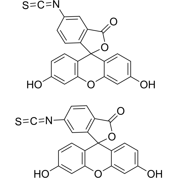
5(6)-FITC (Fluorescein 5(6)-isothiocyanate) is an amine-reactive derivative of a fluorescent dye, characterized by high absorbance and excellent fluorescence quantum yield. The isothiocyanate group of FITC can react with various functional groups on proteins, including amines, thiols, imidazoles, tyrosines and carbonyls, enabling the labeling of proteins such as antibodies and lectins. 5(6)-FITC has a wide range of applications, including flow cytometry, immunofluorescence, protease assays and conjugation. The maximum excitation/emission wavelengths are 492/518 nm.
For research use only. We do not sell to patients.
- Purity : 97.59%
- CAS No.: 27072-45-3
- Formula: C21H11NO5S
- Molecular Weight:389.38
-
Storage:
-20°C, protect from light
* The compound is unstable in solutions, freshly prepared is recommended.

To place orders, for customer services and technical support, please contact: MedChemExpress USA
Tel: 609-228-6898 E-mail: [email protected] [email protected]


-
 Biological Activity
-
Chemical Information
-
Solvent & Solubility
- Protocols
- Purity & Documentation
- References
-
 Help & FAQs


-
HY-66019
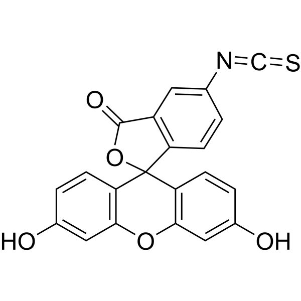
FITCFluorochrome -

HY-15763
ErastinFerroptosis Inducer -
HY-D1048
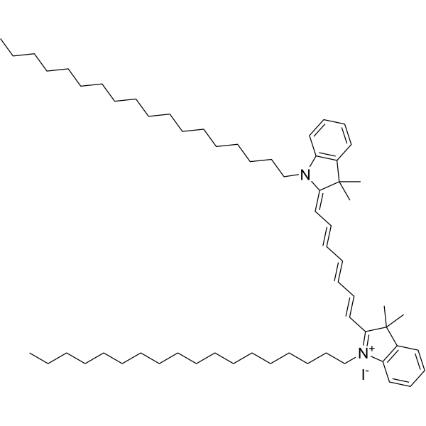
DiRMembrane Dye -
HY-D0940
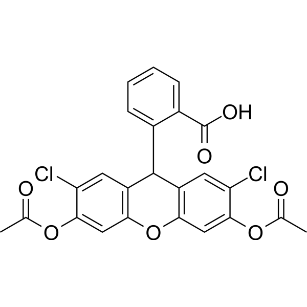
H2DCFDAROS-Sensitive Probe -
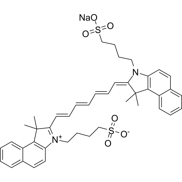
HY-D0711
Indocyanine greenFluorescent Agent -

HY-DY1007
FITC (solution)Fluorochrome -
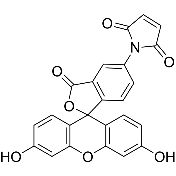
HY-D0098
Fluorescein-5-maleimideFluorescent Dye -
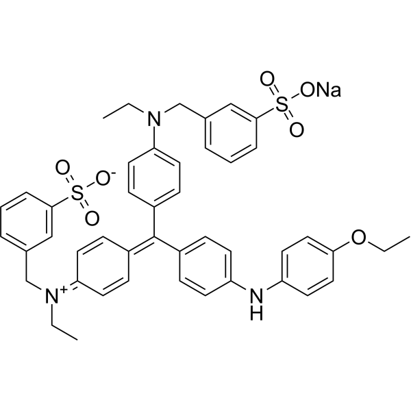
HY-D0232
Brilliant Blue R250Anionic Dye -
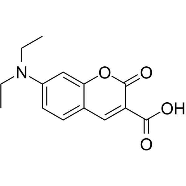
HY-D0067
7ACC1Monocarboxylate Transporter Inhibitor -
HY-D0027
7-Amino-4-methylcoumarinFluorescent Probe -
HY-139109
IR-783Anticancer Dye -
HY-D0025
7-Amino-4-methylcoumarin-3-acetic acidFluorescent Protein Labeling Agent
Publications Citing Use of MedChemExpress (MCE) 5(6)-FITC
More

Biological Activity
Description
Cellular Effect
|
Cell Line

|
Type | Value | Description | References |
|---|---|---|---|---|
| L1210 | IC50 |
700 μM
Compound: FITC
|
Cytotoxicity against mouse L1210 cells after 24 hrs by WST8 assay
Cytotoxicity against mouse L1210 cells after 24 hrs by WST8 assay
|
[PMID: 18951028] |
In Vitro
Guidelines (The following is our recommended protocol. This protocol only provides guidance and should be modified according to your specific needs).
Preparation of stock solution[1]
Prepare a 1 mg/mL 5(6)-FITC stock solution in acetone or DMSO and store at 0°C.
Preparation of working solution: Prepare a 2.5 pg/mL solution in phosphate-buffered saline (PBS) and adjust the pH to 7.8.
5(6)-FITC for flow cytometry detection[1]
(1) Cell preparation: Wash cells from solid nutrient agar plates with 10 mL PBS, centrifuge at 2000 g for 30 min, then resuspend in 10 mL 70% ethanol and incubate at 4°C.
(2) Staining: Collect 3×10⁵ cells by centrifugation, resuspend in 2.5 pg/mL 5(6)-FITC, and stain at 4°C for 45 min.
(3) Detection: Analyze the proportion of green fluorescence using a flow cytometer.
5(6)-FITC for protease assay[2]
(1) Dissolve 1 g casein in 100 mL solution containing 50 mM calcium carbonate and 150 mM sodium chloride at pH 9.5. The solution may first be heated to boiling and then cooled to room temperature.
(2) Add 40 mg 5(6)-FITC and stir gently at room temperature for 1 h.
(3) Remove free 5(6)-FITC by dialysis, first against 2 L water containing activated charcoal at 4°C, then against 50 mM Tris buffer at pH 8.5, and finally against 50 mM Tris buffer at pH 7.2.
(4) Adjust the protein concentration to 0.5% using 50 mM Tris buffer at pH 7.2. Aliquot the substrate into 5 mL portions and store frozen at -20°C, stable for up to 1 year.
(5) Determine the number of FITC residues per casein molecule (0.65 FITC residues per molecule) by measuring substrate absorbance at 490 nm and protein content using the Lowry method.
(6) Perform reactions using FITC-casein. The reaction mixture contains 10 μL enzyme, 20 μL assay buffer, and 20 μL 0.5% FITC-casein.
(7) Chymotrypsin and trypsin: assay buffer is 100 mM Tris buffer at pH 7.8 containing 10 mM CaCl2; or 100 mM phosphate buffer at pH 7.8 containing 150 mM NaCl.
(8) Subtilisin: assay buffer is 20 mM phosphate buffer at pH 7.6 containing 150 mM NaCl.
(9) Thermolysin: assay buffer is 100 mM Tris buffer at pH 7.8 containing 10 mM CaCl2.
(10) Elastase: assay buffer is 20 mM borate buffer at pH 8.8 containing 150 mM NaCl.
(11) After thorough mixing, incubate the solution in a shaking water bath at 37°C for 5 min to 24 h. For incubations longer than 3 h, add 0.2% sodium azide to prevent bacterial growth.
(12) Terminate the reaction by adding 120 μL 5% trichloroacetic acid (TCA) and mixing thoroughly. Leave the tubes at room temperature for at least 1 h, then store overnight at 4°C. Precipitate TCA-insoluble proteins by centrifugation for 5 min. Remove 60 μL supernatant and dilute to 400 μL or 3 mL with 500 mM Tris buffer at pH 8.5, mixing thoroughly to ensure the entire sample reaches the appropriate pH.
(13) Fluorescence measurements are performed with excitation wavelengths of 365 nm or 490 nm and an emission wavelength of 525 nm. Use the average of replicate measurements. Calculate the correlation coefficient r and the standard error S of the regression line using single values.
(14) Cleavage of FITC-casein by trypsin, subtilisin, chymotrypsin, elastase, and thermolysin is linearly related to enzyme concentration.
Immunofluorescence detection of FITC-labeled antibodies on platelets[3]
(1) Preparation of platelet-rich plasma (PRP): Centrifuge blood at 180 × g for 8 min at 22°C to isolate platelet-rich plasma.
(2) Platelet collection: Centrifuge PRP at 1800 × g for 10 min at 22°C to collect platelets. Contamination by red and white blood cells is less than 0.5%.
(3) Platelet washing: Wash platelets twice with PBS by centrifugation at 1800 × g for 10 min at 22°C, then resuspend in PBS.
(4) Purification of human IgG: Human IgG is purified from normal human serum by DEAE-cellulose chromatography.
(5) Use FITC-labeled goat anti-human IgG antibody: total protein concentration 6.2 mg/mL, fluorescein/protein molar ratio 2.6, and specific antibody concentration 1.6 mg/mL.
(6) Add FITC-labeled goat anti-human IgG antibody to 0.5 mL platelet suspension containing 8.0 × 10⁸ cells/mL to a final concentration of 1.0 × 10⁻⁷ M. After incubation at 22°C for 20 min, wash platelets twice with PBS and resuspend in PBS. Then add SDS (1.5 mg/mL), and determine bound anti-human IgG antibody by fluorescence measurement with excitation at 493 nm and emission at 516 nm.
5(6)-FITC conjugation with PAMAM G4[4]
(1) Dissolve 30 μL (3 mg) PAMAM-G4 dendrimer in 10 wt% methanol solution and add to 250 μL 0.1 M phosphate buffer (pH 8.5).
(2) Incubate the mixture with 1 mg 5(6)-FITC dissolved in 60 μL dry DMSO at room temperature for 1 h.
(3) Purify FITC-conjugated PAMAM-G4 dendrimers by gel filtration using a PD-10 desalting column containing Sephadex G25. Use 0.1 M phosphate buffer (pH 8.5) as the mobile phase and collect 0.5 mL fractions.
Notes
1. 5(6)-FITC is sensitive to light and moisture. Prepare 5(6)-FITC solutions immediately before use and discard any unused portion.
2. Low concentrations of sodium azide (≤3 mM or 0.02%) or thimerosal (≤0.02 mM or 0.01%) do not significantly interfere with protein labeling; however, 20-50% glycerol reduces labeling efficiency.
3. Avoid using buffers containing primary amines (e.g., Tris, glycine) or ammonium ions, as they compete with proteins for labeling reactions.
4. This product is intended for scientific research use by professionals only and is not intended for clinical diagnosis or treatment, food, or pharmaceutical use.
5. For your safety and health, please wear a lab coat and disposable gloves during operation.
MedChemExpress (MCE) has not independently confirmed the accuracy of these methods. They are for reference only. Further protocols information, click here.
Emission (Em)
525
Excitation (Ex)
488


Chemical Information
-
CAS No. 27072-45-3
-
Appearance Solid
-
Molecular Weight 389.38
-
Formula C21H11NO5S
-
Color Yellow to orange
-
SMILES
OC1=CC=C(C2(O3)C(C=C(N=C=S)C=C4)=C4C3=O)C(OC5=C2C=CC(O)=C5)=C1.OC6=CC=C(C7(O8)C(C=CC(N=C=S)=C9)=C9C8=O)C(OC%10=C7C=CC(O)=C%10)=C6
-
Synonyms
Fluorescein 5(6)-isothiocyanate; Fluorescein isothiocyanate 5- and 6- isomers
-
Shipping
Room temperature in continental US; may vary elsewhere.
-
Storage
-20°C, protect from light
* The compound is unstable in solutions, freshly prepared is recommended.


Publications (2)
-
Journal Impact Factor
-
Most Recent
-
-
Cell Prolif
2020 Jan;53(1):e12721. PMID: 31782850


Solvent & Solubility
In Vitro:
DMSO : ≥ 156 mg/mL (400.64 mM; Hygroscopic DMSO has a significant impact on the solubility of product, please use newly opened DMSO)
* "≥" means soluble, but saturation unknown.
Please refer to the solubility information to select the appropriate solvent. The compound is unstable in solutions, freshly prepared is recommended.
Please refer to the solubility information to select the appropriate solvent. The compound is unstable in solutions, freshly prepared is recommended.
Concentration (start) × Volume (start) = Concentration (final) × Volume (final)

Protocols
-
Flow Cytometry
Flow cytometry (FC) is a technique for high-speed, step-by-step quantitative analysis and sorting of single cells or other biological particles in a suspension by detecting labeled fluorescent signals.
-
Flow cytometric DNA-content cell-cycle staining
Flow cytometric DNA-content cell-cycle staining measures the fluorescence intensity of DNA-bound fluorochromes in single cells or nuclei to estimate DNA content distributions, allowing assignment of populations to G0/G1, S, and G2/M phases by DNA histogram deconvolution. Propidium iodide (PI) intercalates into DNA, and PI fluorescence is proportional to cellular DNA content when staining is performed under conditions that make DNA accessible and minimize non-DNA signal. Cells with G2/M DNA content are expected to show approximately twice the fluorescence intensity of G0/G1 cells, while S-phase cells occupy intermediate fluorescence values. PI-based DNA-content analysis can also detect cells with fractional DNA content, often reported as sub-G1, when DNA fragmentation and extraction during staining reduce retained DNA signal in apoptotic cells. DAPI is an alternative DNA fluorochrome for univariate DNA-content analysis, while bivariate approaches combining DNA content with proliferation
-
Protocol for Phospho-flow cytometry
Phospho-flow cytometry detects intracellular phosphorylated signaling proteins in single cells using phospho-specific antibodies after rapid fixation and permeabilization; the fluorescence intensity reflects phosphorylation state and therefore kinase-pathway activation, inhibition, or drug response in defined cell subsets. Unlike Western blot, phospho-flow preserves single-cell resolution and can measure signaling heterogeneity in cancer cells, primary immune cells, dissociated mouse tumors, macrophages, organoid-derived cells, and drug-screening samples when validated antibodies and fixation/permeabilization conditions are used.
-
Antibody-based immunofluorescence/immunocytochemistry staining
Antibody-based immunofluorescence/immunocytochemistry detects the cellular or subcellular localization of a target antigen by binding a primary antibody to the target and detecting that antibody directly with a fluorophore-conjugated primary antibody or indirectly with a fluorophore-conjugated secondary antibody. Indirect immunofluorescence can amplify signal because multiple secondary antibodies can bind one primary antibody. The assay readout is fluorescence intensity and localization measured by fluorescence or confocal microscopy, and the result reflects antigen distribution only when the antibody has been validated for the target, sample type, fixation condition, and imaging workflow. Antibody specificity must not be assumed from catalog information alone, and appropriate validation or control experiments are required for serious interpretation.
- Immunocytochemistry/Immunofluorescence


Purity & Documentation
-
Data Sheet (292 KB)
-
SDS (645 KB)
- English - EN (645 KB)
- Français - FR (645 KB)
- Deutsch - DE (645 KB)
- Norwegian - NO (645 KB)
- Español - ES (645 KB)
- Swedish - SV (645 KB)
- Italian - IT (645 KB)
- Korean - KR (645 KB)
- Portuguese - PT (645 KB)
-
Handling Instructions (2659 KB)
References
[1]. J S Miller , ET AL. Flow cytometric identification of microorganisms by dual staining with FITC and PI. Cytometry. 1990;11(6):667-75. [Content Brief]
[2]. S S Twining, et al. Fluorescein isothiocyanate-labeled casein assay for proteolytic enzymes.Anal Biochem. 1984 Nov 15;143(1):30-4. [Content Brief]
[3]. K Sugiura, et al. A simple quantitative immunofluorescence assay for FITC-labeled antibody on platelets. J Immunol Methods. 1981;40(2):165-70. [Content Brief]


Complete Stock Solution Preparation Table
Please refer to the solubility information to select the appropriate solvent. The compound is unstable in solutions, freshly prepared is recommended.
| Optional Solvent | Concentration Solvent Mass | 1 mg | 5 mg | 10 mg | 25 mg |
|---|---|---|---|---|---|
| DMSO | 1 mM | 2.5682 mL | 12.8409 mL | 25.6819 mL | 64.2046 mL |
| 5 mM | 0.5136 mL | 2.5682 mL | 5.1364 mL | 12.8409 mL | |
| 10 mM | 0.2568 mL | 1.2841 mL | 2.5682 mL | 6.4205 mL | |
| 15 mM | 0.1712 mL | 0.8561 mL | 1.7121 mL | 4.2803 mL | |
| 20 mM | 0.1284 mL | 0.6420 mL | 1.2841 mL | 3.2102 mL | |
| 25 mM | 0.1027 mL | 0.5136 mL | 1.0273 mL | 2.5682 mL | |
| 30 mM | 0.0856 mL | 0.4280 mL | 0.8561 mL | 2.1402 mL | |
| 40 mM | 0.0642 mL | 0.3210 mL | 0.6420 mL | 1.6051 mL | |
| 50 mM | 0.0514 mL | 0.2568 mL | 0.5136 mL | 1.2841 mL | |
| 60 mM | 0.0428 mL | 0.2140 mL | 0.4280 mL | 1.0701 mL | |
| 80 mM | 0.0321 mL | 0.1605 mL | 0.3210 mL | 0.8026 mL | |
| 100 mM | 0.0257 mL | 0.1284 mL | 0.2568 mL | 0.6420 mL |


5(6)-FITC Related Classifications
HY-15941 Related Classifications
 Powered by Bioz