



Dimethoate
Based on 6 publication(s) in Google Scholar
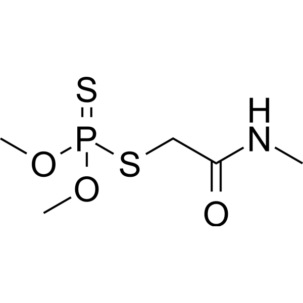
Dimethoate is an organophosphate insecticide and acaricide. Dimethoate is an orally active acetylcholinesterase inhibitor. Dimethoate induces reactive oxygen species (ROS). Dimethoate induces DNA damage and cell apoptosis in vivo. Dimethoate affect immune system in mice.
For research use only. We do not sell to patients.
- Purity : 99.91%
- CAS No.: 60-51-5
- Formula: C5H12NO3PS2
- Molecular Weight:229.26
-
Storage:
4°C, stored under nitrogen
* In solvent : -80°C, 6 months; -20°C, 1 month (stored under nitrogen)

To place orders, for customer services and technical support, please contact: MedChemExpress USA
Tel: 609-228-6898 E-mail: [email protected] [email protected]


-
 Biological Activity
-
Chemical Information
-
Solvent & Solubility
- Protocols
- Purity & Documentation
- References
-
 Help & FAQs



Publications Citing Use of MedChemExpress (MCE) Dimethoate
More

Biological Activity
Description
In Vitro
Dimethoate (0.1-10 μM, 24 h) inhibits cholinesterase (ChE) enzymatic activity in a concentration-dependent manner in pancreatic stellate cells (PSCs)[1].
Dimethoate (10-6-104 μM, 24 h) inhibits carboxylesterase (CbE) in a concentration-dependent manner (IC50 = 0.97 μM) in the PSCs extracts[1].
Dimethoate (0.1-10 μM, 24-96 h) decreases PSCs cell viability[1].
Dimethoate (0.1-10 μM, 1 h) induces reactive oxygen species (ROS) in the mitochondria of PSCs cells[1].
MedChemExpress (MCE) has not independently confirmed the accuracy of these methods. They are for reference only.
-
Cell Line:pancreatic stellate cells (PSCs)
-
Concentration:0.1, 1, 10 μM
-
Incubation Time:24, 48, 72, 96 h
-
Result:Decreased cell viability.
In Vivo
Dimethoate (16 mg/kg; single p.o.) has effects on the immune system of female swiss albino mice[3].
MedChemExpress (MCE) has not independently confirmed the accuracy of these methods. They are for reference only.
-
Animal Model:Female swiss albino mice each weighing 25-30 g[3]
-
Dosage:16 mg/kg
-
Administration:Single oral gavage (p.o.)
-
Result:Showed no significant loss of body weight.
Observed a time depended reduction in spleen weigh.
Suppressed the lymphocyte proliferation responses to T-cell mitogen (Phytohemagglutinin, PHA) (HY-N7038) and B-cell mitogen (Lipopolysaccharides, LPS) (HY-D1056).
The percentage of total and active T cells were significantly decreased.
The total antibody and IgM responses to SRBC's were significantly decreased.
Caused a significant decrease in the number of plaque forming cell (PFC / 106 splenocytes) in a time dependent manner.
-
Animal Model:Rattus norvegicus (Wistar strain; 8-10 weeks old; 120 g; males only)[2]
-
Dosage:20, 40, 60 mg/kg
-
Administration:Intraperitoneal injection (i.p.); 24, 48, 72 h
-
Result:Significant micronuclei induction and metaphase chromosome abnormalities in bone marrow cells indicated positive dose response relationship, confirming its genotoxic and cytotoxic potential.
Showed significant mitotic index decrease.
Decreased mitochondrial membrane potential (MMP), affected the cell cycle, and inflicted DNA damage, which led to cellular apoptosis of leukocytes culminating into immunotoxic effects.


Chemical Information
-
CAS No. 60-51-5
-
Appearance Solid
-
Molecular Weight 229.26
-
Formula C5H12NO3PS2
-
Color White to off-white
-
SMILES
S=P(OC)(SCC(NC)=O)OC
-
Shipping
Room temperature in continental US; may vary elsewhere.
-
Storage
4°C, stored under nitrogen
* In solvent : -80°C, 6 months; -20°C, 1 month (stored under nitrogen)


Publications (6)
-
Journal Impact Factor
-
Most Recent
-
ACS Environ Au
Machine Learning-Assisted Recognition of Environmental Sulfur-Containing Chemicals in Nontargeted Mass Spectrometry Analysis of Inadequate Mass Resolution. [Abstract]2025 Aug 5;5(6):573-582. PMID: 41277996 -
Anal Chem
Exposome-Scale Investigation of Cl-/Br-Containing Chemicals Using High-Resolution Mass Spectrometry, Multistage Machine Learning, and Cloud Computing. [Abstract]2025 Jun 3;97(21):11099-11109. PMID: 40401576 -
Pest Manag Sci
Repurposing isoxazoline insecticides to control Varroa destructor populations in honey bee colonies. [Abstract]2025 Oct 2. PMID: 41039832 -
Insects
Evaluating the Efficacy of Active Ingredients Used in Roach Baits against Small Hive Beetle (Aethina tumida) and Their Safety to Honey Bees (Apis mellifera). [Abstract]2024 Jun 25;15(7):472. PMID: 39057205 -
J Econ Entomol
Seasonal honey bee (Apis mellifera) physiology differentially impacts toxicity to pesticides used to control honey bee pests. [Abstract]2026 Jun 6:toag160. PMID: 42250246 -


Solvent & Solubility
In Vitro:
H2O : 10 mg/mL (43.62 mM; Need ultrasonic)
Please refer to the solubility information to select the appropriate solvent. Once prepared, please aliquot and store the solution to prevent product inactivation from repeated freeze-thaw cycles.
Storage method and period of stock solution: -80°C, 6 months; -20°C, 1 month (stored under nitrogen). When stored at -80°C, please use it within 6 months. When stored at -20°C, please use it within 1 month.
* Note: If you choose water as the stock solution, please dilute it to the working solution, then filter and sterilize it with a 0.22 μm filter before use.
Please refer to the solubility information to select the appropriate solvent. Once prepared, please aliquot and store the solution to prevent product inactivation from repeated freeze-thaw cycles.
Storage method and period of stock solution: -80°C, 6 months; -20°C, 1 month (stored under nitrogen). When stored at -80°C, please use it within 6 months. When stored at -20°C, please use it within 1 month.
* Note: If you choose water as the stock solution, please dilute it to the working solution, then filter and sterilize it with a 0.22 μm filter before use.
Concentration (start) × Volume (start) = Concentration (final) × Volume (final)
In Vivo:
For the following dissolution methods, please prepare the working solution directly:
It is recommended to prepare fresh solutions and use them promptly within a short period of time.
The percentages shown for the solvents indicate their volumetric ratio in the final prepared solution. If precipitation or phase separation occurs during preparation, heat and/or sonication can be used to aid dissolution.
Add each solvent one by one: PBS
Solubility: 10 mg/mL (43.62 mM); Clear solution; Need ultrasonic


Purity & Documentation
-
Data Sheet (278 KB)
-
SDS (480 KB)
- English - EN (480 KB)
- Français - FR (480 KB)
- Deutsch - DE (480 KB)
- Norwegian - NO (480 KB)
- Español - ES (480 KB)
- Swedish - SV (480 KB)
- Italian - IT (480 KB)
- Korean - KR (480 KB)
- Portuguese - PT (480 KB)
-
Handling Instructions (2659 KB)
References
[1]. Martínez-Morcillo S, et al. The organophosphorus pesticide dimethoate decreases cell viability and induces changes in different biochemical parameters of rat pancreatic stellate cells. Toxicol In Vitro. 2019 Feb;54:89-97. [Content Brief]
[2]. Nazam N, et al. Dimethoate Induces DNA Damage and Mitochondrial Dysfunction Triggering Apoptosis in Rat Bone-Marrow and Peripheral Blood Cells. Toxics. 2020 Oct 1;8(4):80. [Content Brief]
[3]. Aly NM, et al. Effect of dimethoate on the immune system of female mice. J Environ Sci Health B. 2000 Jan;35(1):77-86. [Content Brief]


Complete Stock Solution Preparation Table
Please refer to the solubility information to select the appropriate solvent. Once prepared, please aliquot and store the solution to prevent product inactivation from repeated freeze-thaw cycles.
Storage method and period of stock solution: -80°C, 6 months; -20°C, 1 month (stored under nitrogen). When stored at -80°C, please use it within 6 months. When stored at -20°C, please use it within 1 month.
| Optional Solvent | Concentration Solvent Mass | 1 mg | 5 mg | 10 mg | 25 mg |
|---|---|---|---|---|---|
| H2O | 1 mM | 4.3619 mL | 21.8093 mL | 43.6186 mL | 109.0465 mL |
| 5 mM | 0.8724 mL | 4.3619 mL | 8.7237 mL | 21.8093 mL | |
| 10 mM | 0.4362 mL | 2.1809 mL | 4.3619 mL | 10.9047 mL | |
| 15 mM | 0.2908 mL | 1.4540 mL | 2.9079 mL | 7.2698 mL | |
| 20 mM | 0.2181 mL | 1.0905 mL | 2.1809 mL | 5.4523 mL | |
| 25 mM | 0.1745 mL | 0.8724 mL | 1.7447 mL | 4.3619 mL | |
| 30 mM | 0.1454 mL | 0.7270 mL | 1.4540 mL | 3.6349 mL | |
| 40 mM | 0.1090 mL | 0.5452 mL | 1.0905 mL | 2.7262 mL |
* Note: If you choose water as the stock solution, please dilute it to the working solution, then filter and sterilize it with a 0.22 μm filter before use.


Dimethoate Related Classifications
HY-B1946 Related Classifications
 Powered by Bioz