



Mitochondria Dyes

- Formula: C42H43I2N3
- Molecular Weight: 843.62
Mito‑VR2 is a mitochondria-targeted molecular rotor-type viscosity fluorescent probe with high responsiveness to microenvironmental viscosity, which is suitable for synchronous imaging of mitochondrial viscosity during autophagy in living cells. Mito-VR2 achieves specific targeting of mitochondria through its hydrophobic cationic structure, and its fluorescence intensity responds only to microenvironmental viscosity. Mito-VR2 emits almost no fluorescence in low-viscosity media, but its fluorescence emission is significantly enhanced in high-viscosity environments.

- Formula: C62H73N7O5
- Molecular Weight: 996.29
Mito-RhFe is a Fluorescent probe for mitochondrial labile Fe³⁺ monitoring via imaging and flow cytometry. This probe is a rhodamine-based construct with a spirolactam fluorescence signaling group and an N2-hydroxyethyldiethylenetriamine chelator; its delocalized positive charge enables mitochondria-targeting ability in live cells, and it exhibits fine cell membrane permeability. In its native state, it exists in the non-fluorescent spirolactam form, but upon binding to Fe³⁺, it undergoes a ring-opening conversion to the fluorescent rhodamine form, triggering a turn-on fluorescent response; this process is reversible, as the addition of the metal chelator TPEN removes Fe³⁺ and converts the probe back to its non-fluorescent spirolactam form, and re-addition of Fe³⁺ restores fluorescence. The probe shows high selectivity for Fe³⁺ over most other metal cations present in living systems, with a ~90-fold fluorescence enhancement upon binding to 20 equiv of Fe³⁺. Mito-RhFe has excitation/emission wavelengths of Ex/Em = 540/578 nm, with an ~8 nm bathochromic shift in emission upon Fe³⁺ binding, and it can also be excited at 543 nm for confocal imaging with emission detected at 570-620 nm[1].

- Formula: C46H40F6NP
- Molecular Weight: 751.78
TPE-Ph-In is a mitochondria-targeted voltage-sensitive probe that can be used for mitochondrial membrane potential detection, mitochondrial imaging of animal and plant cells and tissues, mouse sperm activity indication, and detection of mitochondrial membrane potential changes associated with early apoptosis. TPE-Ph-In is non-fluorescent in solution, but emits strong fluorescence when aggregated via restriction of intramolecular rotation/movement, and its fluorescence intensity is directly correlated with local probe concentration or mitochondrial membrane potential level. TPE-Ph-In can penetrate cell membranes, target mitochondria through charge-dependent accumulation, has low cytotoxicity and high photostability, and enables super-resolution imaging of plant mitochondrial fission, fusion and cristae structures. The excitation wavelengths of TPE-Ph-In include 450 nm, 488 nm, and 489 nm, while its emission wavelengths include 551-661 nm, 660 nm, and 694 nm. TPE-Ph-In can be used for studies on mitochondrial dysfunction associated with early apoptosis.

- Formula: C31H35ClN2O
- Molecular Weight: 487.08
Spring Red is a fluorescent probe for mitochondrial pH detection (Ex/Em = 530/680 nm) that can be taken up by live cells and mitochondria. The detection mechanism of Spring Red depends on pH changes and mitochondrial membrane potential. Fluorescence intensity increases with elevated pH, and a significant pH-dependent fluorescent response is observed in the pH range of 5.0‑8.0. Spring Red is suitable for long-term live-cell imaging and in vivo imaging, and can be used to monitor dynamic mitochondrial pH changes.

- Formula: C42H36I2N2O2
- Molecular Weight: 854.56
MitoAIE1 is a Fluorescent probe for mitochondrial viscosity detection. MitoAIE1 contains a pyridinium structural unit, which endows it with mitochondria-targeting specificity. The detection mechanism of this probe is based on aggregation-induced emission (AIE): in low-viscosity media such as PBS buffer, intramolecular rotation leads to non-radiative energy dissipation, resulting in only weak fluorescence; however, in high-viscosity environments such as the mitochondrial matrix or the vicinity of the inner mitochondrial membrane, such intramolecular motion is restricted, thereby significantly enhancing its fluorescent signal; in addition, this probe is not interfered by changes in microenvironment polarity and pH. Its emission wavelength is 625 nm, with absorption peaks at 325 nm and 450 nm; when transferred from low-viscosity PBS to high-viscosity 99% glycerol, its fluorescence intensity at 625 nm can be increased by 38-fold. It can be used to monitor changes in mitochondrial viscosity during processes such as Stauroporine (HY-15141)-induced apoptosis and starvation-induced mitophagy in live cells, and has good biocompatibility.

- Formula: C48H42F6N2O7P2
- Molecular Weight: 934.79
Ac-MtFluNox is a mitochondria-targeted Fluorescent probe that can be used for the selective detection of labile Fe (II). Ac-MtFluNox can enter living cells, where intracellular esterases cleave its acetyl protecting group to generate activated MtFluNox. Ac-MtFluNox functions through an Fe (II)-mediated deoxygenation mechanism of its amine N-oxide group, which eliminates the fluorescence quenching effect and oxidizes Fe (II) to Fe (III) to deplete the catalytically active Fe (II) pool. A FITC filter set (excitation 465-500 nm, emission 516-556 nm) is used for live-cell imaging. Ac-MtFluNox can be applied to studies related to myocardial ferroptosis.

- Formula: C31H36N2OSi
- Molecular Weight: 480.72
DMA-SiRd is a fluorescent probe that can be used for crosstalk-free dual-channel imaging of lipid droplets and mitochondria, mapping of lipid microenvironment heterogeneity, and imaging of lipid droplet-mitochondria interactions. DMA-SiRd has two imaging channels: Ex/Em = 633/650-750 nm and Ex/Em = 546/570-620 nm, with two-photon excitation wavelengths of 1040 nm and 1200 nm for brain imaging. DMA-SiRd can be applied to the research of atherosclerosis.

- Formula: C43H44BClF4N2
- Molecular Weight: 711.08
MitoLumina 590 is a mitochondria-targeting fluorescent probe used to label mitochondria in live cells. MitoLumina 590 contains a chloromethyl group that allows for covalent binding to mitochondrial thiol-containing proteins. MitoLumina 590s primarily used for mitochondrial imaging, live-cell mitochondrial tracking, and multicolor fluorescence microscopy (Ex/Em = 589/606 nm).

- Formula: C36H38BClF4N2
- Molecular Weight: 620.96
MitoLumina 750 is a mitochondria-targeting fluorescent probe used to label mitochondria in live cells. MitoLumina 750 contains a chloromethyl group that allows for covalent binding to mitochondrial thiol-containing proteins. MitoLumina 750 is primarily used for mitochondrial imaging, live-cell mitochondrial tracking, and multicolor fluorescence microscopy (Ex/Em = 745/775 nm).

- Formula: C45H46BClF4N2
- Molecular Weight: 737.12
MitoLumina 680 is a mitochondria-targeting fluorescent probe used to label mitochondria in live cells. MitoLumina 680 contains a chloromethyl group that allows for covalent binding to mitochondrial thiol-containing proteins. MitoLumina 680 is primarily used for mitochondrial imaging, live-cell mitochondrial tracking, and multicolor fluorescence microscopy (Ex/Em = 678/700 nm).

- Formula: C32H34BClF4N2
- Molecular Weight: 568.88
MitoLumina 545 is a mitochondria-targeting fluorescent probe used to label mitochondria in live cells. MitoLumina 545 contains a chloromethyl group that allows for covalent binding to mitochondrial thiol-containing proteins. MitoLumina 545 is primarily used for mitochondrial imaging, live-cell mitochondrial tracking, and multicolor fluorescence microscopy (Ex/Em = 540/565 nm).

- Formula: C34H36BClF4N2
- Molecular Weight: 594.92
MitoLumina 640 is a mitochondria-targeting fluorescent probe used to label mitochondria in live cells. MitoLumina 640 contains a chloromethyl group that allows for covalent binding to mitochondrial thiol-containing proteins. MitoLumina 640 is primarily used for mitochondrial imaging, live-cell mitochondrial tracking, and multicolor fluorescence microscopy (Ex/Em = 640/665 nm).

- Formula: C47H48BClF4N2
- Molecular Weight: 763.16
MitoLumina 780 is a mitochondria-targeting fluorescent probe used to label mitochondria in live cells. MitoLumina 780 contains a chloromethyl group that allows for covalent binding to mitochondrial thiol-containing proteins. MitoLumina 780 is primarily used for mitochondrial imaging, live-cell mitochondrial tracking, and multicolor fluorescence microscopy (Ex/Em = 778/805 nm).

- Formula: C26H28F6N3OP
- Molecular Weight: 543.48

- Formula: C32H27N3O
- Molecular Weight: 469.58
Mito-Cu (II) is a mitochondria-targeted fluorescent probe (Ex/Em = 370/450 nM). Mito-Cu (II) specifically accumulates in mitochondria of living cells and enables real-time detection of exogenous Cu2+ within mitochondria of living cells. Mito-Cu (II) achieves "on-off-on" fluorescence switching through sequential exposure to Cu2+ and ethylenediaminetetraacetic acid (EDTA) (HY-Y0682). Its fluorescence is quenched after forming a 1:1 complex with Cu2+, and the fluorescence recovers when Cu2+ is chelated by EDTA.

- Formula: C26H37NP4S5
- Molecular Weight: 647.80
Crisp-17 is a monovalent copper ion probe. Crisp-17 can be used to characterize cellular phenotypes associated with copper metabolism defects. Crisp-17 is applicable to research related to Menkes disease.

- Formula: C26H21Cl2NO4
- Molecular Weight: 482.36
CEMT is a carboxylesterase (CES) substrate and a ratiometric two-photon fluorescent reporter probe. CEMT can be hydrolyzed by CES to generate HMT, which is used for mitochondrial pH sensing. After activation by CES, CEMT exhibits ratiometric fluorescence changes in response to pH variations. CEMT targets and covalently binds to mitochondria, and can avoid leakage during acidification, thus enabling in situ imaging (Ex/Em = 410 nm/550 nm).

- Formula: C31H34Cl2N2O7
- Molecular Weight: 617.52
MI-BP-CC is a mitochondrial-targeting near-infrared fluorescent probe. MI-BP-CC can specifically localize in the mitochondria of living cells. MI-BP-CC visualizes viscosity with an emission wavelength of 722 nm for detecting mitochondrial viscosity.
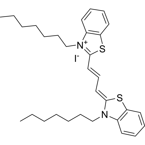
- Formula: C31H41IN2S2
- Molecular Weight: 632.71
3,3′-Diheptylthiacarbocyanine iodide is a cyanine dye for measuring membrane potential. (λex=562 nm,λem=575 nm).
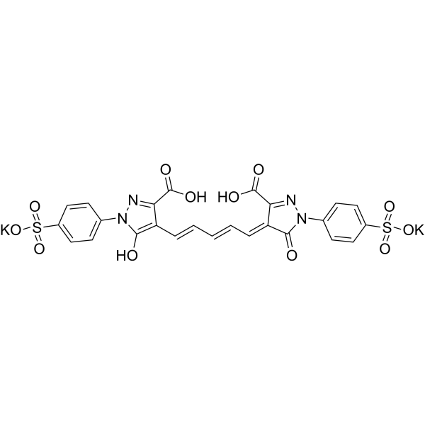
- Formula: C25H16K2N4O12S2
- Molecular Weight: 706.74
Oxonol Blue is a staining dye. Oxonol Blue can be used as a monitor of membrane potential.



