



EGFR-IN-55
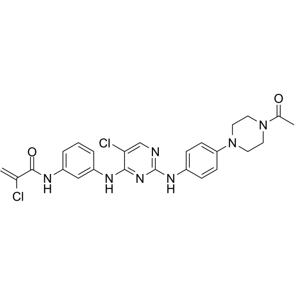
EGFR-IN-55 (Compound 8a) is a potent EGFR inhibitor with IC50 values of 70 nM and 3.9 nM against EGFRWT and EGFRL858R/T790M, respectively. EGFR-IN-55 arrests NCI-H1975 cells in G0/G1 phase and shows anticancer activity.
Para uso exclusivo en investigación. No vendemos a pacientes.
- No. CAS: 2057423-46-6
- Fòrmula: C25H25Cl2N7O2
- Peso molecular:526.42
-
Almacenamiento:
Please store the product under the recommended conditions in the Certificate of Analysis.

To place orders, for customer services and technical support, please contact: MedChemExpress USA
Tel: 609-228-6898 E-mail: [email protected] [email protected]


-
 Actividad biológica
-
Chemical Information
- Protocolo
- Pureza y Documentación
- Referencias
-
 Help & FAQs


Ver todos los productos específicos de isoformas EGFR
More

Actividad biológica
Descripciòn
IC50 & Target
|
EGFRL858R/T790M 3.9 nM (IC50) |
EGFRWT 70 nM (IC50) |
Cellular Effect
|
Cell Line

|
Type | Value | Description | References |
|---|---|---|---|---|
| A-431 | IC50 |
3.14 μM
Compound: 8a
|
Antiproliferation activity against human A431 cells assessed as reduction in cell viability incubated incubated for 48 hrs by MTT assay
Antiproliferation activity against human A431 cells assessed as reduction in cell viability incubated incubated for 48 hrs by MTT assay
|
[PMID: 32912431] |
| COS-7 | IC50 |
10.44 μM
Compound: 8a
|
Cytotoxicity against African green monkey COS7 cells assessed as decrease in cell viability incubated incubated for 48 hrs by MTT assay
Cytotoxicity against African green monkey COS7 cells assessed as decrease in cell viability incubated incubated for 48 hrs by MTT assay
|
[PMID: 32912431] |
| NCI-H1975 | IC50 |
0.75 μM
Compound: 8a
|
Antiproliferation activity against human NCI-H1975 cells expressing EGFR L858R/T790M mutant assessed as reduction in cell viability incubated incubated for 48 hrs by MTT assay
Antiproliferation activity against human NCI-H1975 cells expressing EGFR L858R/T790M mutant assessed as reduction in cell viability incubated incubated for 48 hrs by MTT assay
|
[PMID: 32912431] |
| SW-620 | IC50 |
29.15 μM
Compound: 8a
|
Antiproliferation activity against EGFR-negative human SW620 cells assessed as reduction in cell viability incubated incubated for 48 hrs by MTT assay
Antiproliferation activity against EGFR-negative human SW620 cells assessed as reduction in cell viability incubated incubated for 48 hrs by MTT assay
|
[PMID: 32912431] |


Chemical Information
-
No. CAS 2057423-46-6
-
Peso molecular 526.42
-
Fòrmula C25H25Cl2N7O2
-
SMILES
C=C(Cl)C(NC1=CC=CC(NC2=NC(NC3=CC=C(N4CCN(C(C)=O)CC4)C=C3)=NC=C2Cl)=C1)=O
-
Envío
Room temperature in continental US; may vary elsewhere.
-
Almacenamiento
Please store the product under the recommended conditions in the Certificate of Analysis.


Pureza y Documentación
Referencias

Calculators
Concentration (start) × Volume (start) = Concentration (final) × Volume (final)


EGFR-IN-55 Related Classifications
HY-146132 Related Classifications
 Powered by Bioz