



Epertinib hydrochloride (Standard)
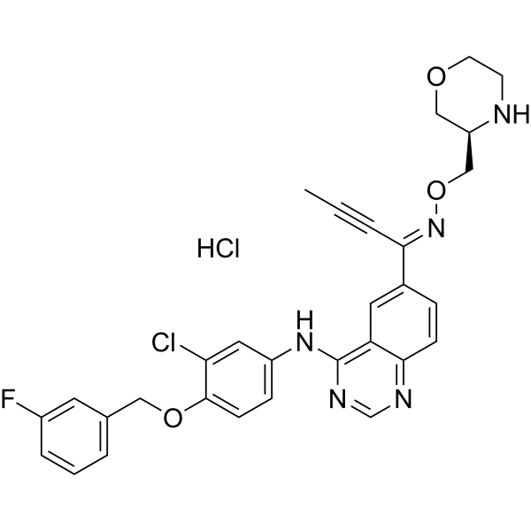
Epertinib hydrochloride (Standard) (S-22611 hydrochloride (Standard)) is the analytical standard of Epertinib (hydrochloride) (HY-107367A). This product is intended for research and analytical applications. Epertinib (S-22611) hydrochloride is a potent, orally active, reversible, and selective tyrosine Kinase inhibitor of EGFR, HER4 and HER2, with IC50s of 1.48 nM, 2.49 nM and 7.15 nM, respectively. Epertinib hydrochloride shows potent antitumor activity. Epertinib (hydrochloride) is a click chemistry reagent, it contains an Alkyne group and can undergo copper-catalyzed azide-alkyne cycloaddition (CuAAc) with molecules containing Azide groups.
For research use only. We do not sell to patients.
- CAS No.: 2071195-74-7
- Formula: C30H28Cl2FN5O3
- Molecular Weight:596.48
-
Storage:
Please store the product under the recommended conditions in the Certificate of Analysis.


To place orders, for customer services and technical support, please contact: MedChemExpress USA
Tel: 609-228-6898 E-mail: [email protected] [email protected]


-
 Biological Activity
-
Chemical Information
- Protocols
- Purity & Documentation
- References
-
 Help & FAQs



All EGFR Isoforms
More

Biological Activity
Description
IC50 & Target
IC50: 1.48 nM (EGFR), 7.15 nM (HER2), 2.49 nM (HER4)[1]


Chemical Information
-
CAS No. 2071195-74-7
-
Molecular Weight 596.48
-
Formula C30H28Cl2FN5O3
-
SMILES
CC#C/C(C1=CC2=C(NC3=CC=C(OCC4=CC=CC(F)=C4)C(Cl)=C3)N=CN=C2C=C1)=N\OC[C@@H]5NCCOC5.Cl
-
Synonyms
S-22611 hydrochloride (Standard)
-
Shipping
Room temperature in continental US; may vary elsewhere.
-
Storage
Please store the product under the recommended conditions in the Certificate of Analysis.

Protocols
-
EdU Incorporation Assay (Click Chemistry-Based DNA Synthesis Measurement)
The EdU incorporation assay measures DNA synthesis by adding the thymidine analog 5-ethynyl-2′-deoxyuridine to cells or tissues, where it is incorporated into newly synthesized DNA during S phase. Incorporated EdU is detected by copper-catalyzed azide-alkyne cycloaddition, in which a fluorescent azide covalently reacts with the ethynyl group on EdU, allowing S-phase cells to be detected by fluorescence microscopy, flow cytometry, or high-content imaging. EdU detection does not require DNA denaturation or anti-BrdU antibody access, which preserves sample structure and improves compatibility with immunostaining and multiparameter cytometry compared with BrdU-based detection. EdU can be cytotoxic in a cell-type- and exposure-dependent manner, so pulse duration, concentration, and continuous-labeling designs should be validated for each cell type.
-
Subcutaneous Cell-Line-Derived Xenograft
Subcutaneous cell-line-derived xenograft (CDX) models are established by implanting cultured human cancer cell lines into immunodeficient mice, where the injected cells form localized tumors that can be monitored in vivo as a measure of tumorigenic potential, growth kinetics, and treatment response. These models are widely used in oncology research because they allow reproducible tumor formation and enable comparative assessment of tumor growth between different cell lines or genetic manipulations in a controlled in vivo microenvironment. Subcutaneous implantation of cancer cells in immunodeficient mice is a standard approach for evaluating tumor growth behavior and therapeutic response across multiple cancer types, including prostate, esophageal, pancreatic, and colon cancer models.


Purity & Documentation
References
[1]. Tanaka Y, et al. Distribution analysis of epertinib in brain metastasis of HER2-positive breast cancer by imaging mass spectrometry and prospect for antitumor activity. Sci Rep. 2018 Jan 10;8(1):343. [Content Brief]
[2]. Tanaka H, et al. Preclinical antitumor activity of S-222611, an oral reversible tyrosine kinase inhibitor of epidermal growth factor receptor and human epidermal growth factor receptor 2. Cancer Sci. 2014 Aug;105(8):1040-8. [Content Brief]

Calculators
Concentration (start) × Volume (start) = Concentration (final) × Volume (final)


Epertinib hydrochloride (Standard) Related Classifications
HY-107367AR Related Classifications
 Powered by Bioz