



Fluorescein-5-maleimide (solution)

Based on 1 Customer Validation
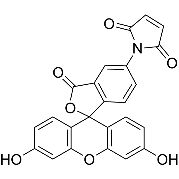
Fluorescein-5-maleimide (solution) (N-(5-Fluoresceinyl)maleimide (solution)) is a fluorescent dye. Fluorescein-5-maleimide can be used to detect the redox state of thiols in eukaryotic cells. Fluorescein-5-maleimide can label peptides and is used to detect negatively charged nanoparticles. Fluorescein-5-maleimide can also label actin to explore its interaction with cardiac myosin-binding protein C (cMyBP-C), which helps in developing small molecule modulators for heart failure. Fluorescein-5-maleimide can screen mutant proteins that contain cysteine residues. The excitation wavelength of Fluorescein-5-maleimide is 494 nm, and the emission wavelength is 519 nm.
Solvent and concentration: DMSO: 10 mM
For research use only. We do not sell to patients.
- CAS No.: 75350-46-8
- Formula: C24H13NO7
- Molecular Weight:427.36
-
Storage:
Please store the product under the recommended conditions in the Certificate of Analysis.


To place orders, for customer services and technical support, please contact: MedChemExpress USA
Tel: 609-228-6898 E-mail: [email protected] [email protected]


-
 Biological Activity
-
Chemical Information
- Protocols
- Purity & Documentation
- References
-
 Help & FAQs


-
HY-D0098
Fluorescein-5-maleimideFluorescent Dye -
HY-126793
2′,7′-Dichlorofluorescein diacetateFluorescent Probe -
HY-D0069
CPMFluorescent Dye -
HY-139109
IR-783Anticancer Dye -
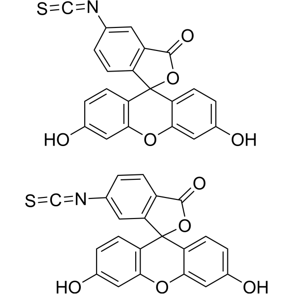
HY-15941
5(6)-FITCFluorescent Dye -
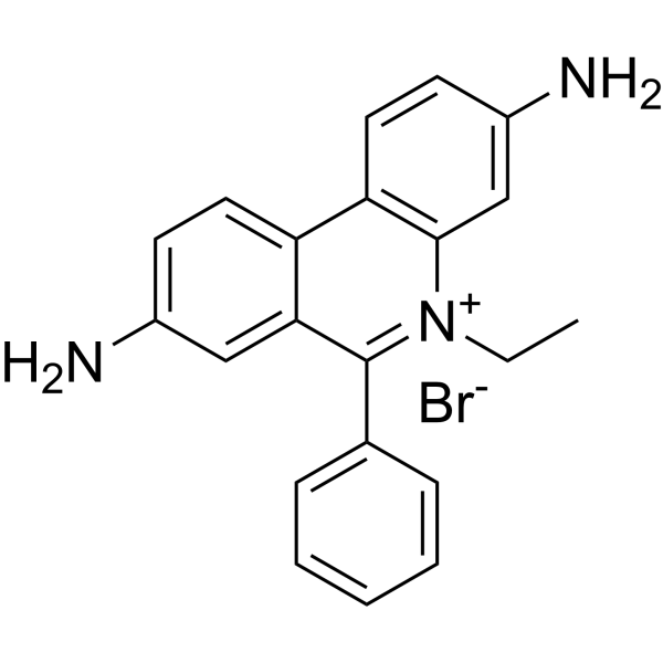
HY-D0021
EthD bromideFluorescent Tag -

HY-DY1050
Ethidium bromide (solution)Fluorescent Tag -
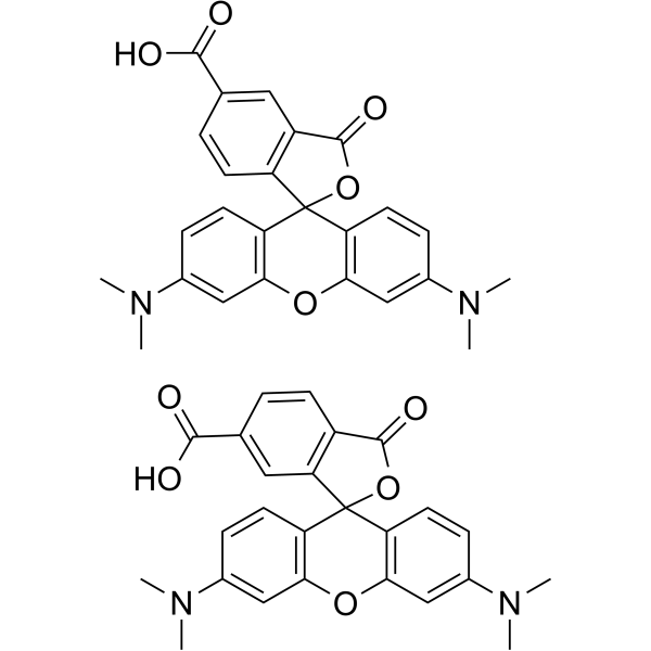
HY-15944
5(6)-TAMRAFluorescent Dye -

HY-DY1058
2′,7′-Dichlorofluorescein diacetate (solution)Fluorescent Probe -
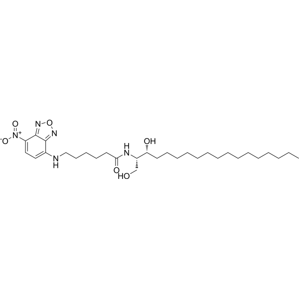
HY-141576
C6-NBD SphinganineFatty Acid Fluorescent Marker -
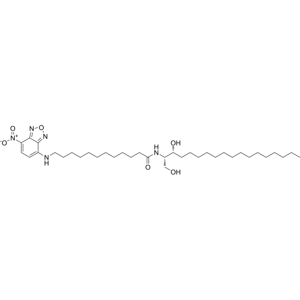
HY-141577
C12-NBD SphinganineCeramidase Substrate -

HY-DY1085
5(6)-TAMRA (solution)Fluorescent Dye


Biological Activity
Description
Solvent and concentration: DMSO: 10 mM
In Vitro
Guide (The following is our recommended protocol. This protocol is only a guide and should be modified according to your specific needs).
1. Fluorescein-5-maleimide for detecting the redox status of thiols in cells
1.1 Prepare PBMCs from venous blood using Ficoll Paque density centrifugation and wash them in d-PBS.
1.2 Wash PBMCs (106cells in 100 μ L) labeled with anti human CD3-APC.
1.3 Stain with 0.1 μ M Fluorescein-5-maleimide and analyze by flow cytometry.
2. Preparation of Fluorescein-5-maleimide Fluorescent Probe[2]
2.1 Preparation of working fluid
Add the working solution (probe: peptide) in a 5:1 molar ratio to a peptide solution (4.06 mM) containing 20.3 mM N - (2-hydroxyethyl) piperazine-N '- ethanesulfonic acid KOH, 50 mM KCl, and 2 mM tris (2-carboxyethyl) phosphine buffer for peptide labeling.
2.2 Incubate the mixture (final volume of 125 μ L) in the dark at room temperature for 2 hours and gently shake.
2.3 Add excess DDT (20 mM) to quench the reaction.
2.4 Further purify the pure labeled peptide mixture by precipitation with acetone (1 mL).
2.5 Add 5 μ L of fluorescent probe to 15 μ L of nanoparticles and 130 μ L of buffer solution to detect their interaction.
2.6 Data analysis: Detecting nanoparticles-peptide interactions through changes in fluorescence intensity of labeled peptides and/or differential light scattering.
3. Fluorescein-5-maleimide is used to detect protein interactions (actin and cardiac myosin binding protein C (cMyBP-C))[3]
3.1 50 μ M G-actin was polymerized by adding 3 M KCl (final concentration of 100 mM) and 0.5 M MgCl2 (final concentration of 2 mM) under the conditions of 20 mM Tris pH 7.5, 0.2 mM CaCl2, and 0.2 mM ATP, and incubated at 23 ° C for 1 hour.
3.2 Add Fluorescein-5-maleimide (final concentration of 1 mM) and label at 23 ° C for 1 hour.
3.3 Add a five fold molar excess of DTT to stop labeling.
3.4 Incubate 0.25 μ M Fluorescein-5-maleimide actin and 0-20 μ M TMR-cC0-C2 (N-terminal fragment of cMyBP-C) together.
3.5 Data analysis (FLT-FRET assay): Firstly, excite Fluorescein-5-maleimide and detect its FLT to measure the changes in fluorescence resonance energy transfer (FRET) from Fluorescein-5-maleimide (donor) to TMR (acceptor), with a decrease in the FLT (and an increase in FRET) of Fluorescein-5-maleimide-actin indicating binding.
MedChemExpress (MCE) has not independently confirmed the accuracy of these methods. They are for reference only. Further protocols information, click here.


Chemical Information
-
CAS No. 75350-46-8
-
Appearance Liquid
-
Molecular Weight 427.36
-
Formula C24H13NO7
-
Color Light yellow to yellow
-
SMILES
O=C(C=C1)N(C2=CC3=C(C4(C5=C(OC6=C4C=CC(O)=C6)C=C(O)C=C5)OC3=O)C=C2)C1=O
-
Synonyms
N-(5-Fluoresceinyl)maleimide (solution)
-
Shipping
Room temperature in continental US; may vary elsewhere.
-
Storage
Please store the product under the recommended conditions in the Certificate of Analysis.

Protocols
-
ROS/oxidative-stress fluorescent staining
ROS/oxidative-stress fluorescent staining uses cell-permeant fluorogenic probes that become fluorescent after oxidation inside cells or tissues; commonly used examples include DCFH-DA/DCFDA for broad cellular oxidant detection, DHE for superoxide-related signal detection, MitoSOX for mitochondrial superoxide-related signal detection, and CellROX probes for oxidative-stress-associated fluorescence readouts. The assay detects probe oxidation rather than a single ROS species unless the probe and analysis method have been chemically validated for that species. DCFH-DA enters cells, is deacetylated by intracellular esterases to DCFH, and produces fluorescent DCF after oxidation, so the readout is used as an operational measure of total cellular oxidative stress rather than a species-specific ROS measurement. DHE and MitoSOX can report superoxide-related oxidation, but red fluorescence alone can include non-specific ethidium-like oxidation products; HPLC or optimized spectral approaches are
-
Research Protocol for Cardiovascular Diseases
Cardiovascular disease can be modeled as maladaptive cardiac remodeling, where ischemic injury or pressure overload activates inflammatory signaling, fibroblast activation, extracellular-matrix deposition, cardiomyocyte hypertrophy, vascular remodeling, and progressive ventricular dysfunction. The TGF-β/SMAD axis is a central profibrotic pathway after myocardial injury and pressure overload, while innate immune and cytokine pathways regulate leukocyte recruitment, scar formation, and adverse remodeling. Key unresolved questions include which inflammatory signals are reparative versus harmful, when fibrosis is protective versus maladaptive, and whether pathway inhibition improves function without weakening necessary infarct healing or compensatory remodeling.


Purity & Documentation
-
Data Sheet (282 KB)
-
SDS (252 KB)
- English - EN (252 KB)
- Français - FR (252 KB)
- Deutsch - DE (252 KB)
- Norwegian - NO (252 KB)
- Español - ES (252 KB)
- Swedish - SV (252 KB)
- Italian - IT (252 KB)
- Korean - KR (252 KB)
- Portuguese - PT (252 KB)
-
Handling Instructions (2659 KB)
References
[1]. Wadley AJ, et al. Detecting intracellular thiol redox state in leukaemia and heterogeneous immune cell populations: An optimised protocol for digital flow cytometers. MethodsX. 2018 Nov 1;5:1473-1483. [Content Brief]
[2]. Akinloye O, et al. Peptide-based fluorescence biosensors for detection/measurement of nanoparticles. Anal Bioanal Chem. 2017 Feb;409(4):903-915. [Content Brief]
[3]. Bunch TA, et al. Drug discovery for heart failure targeting myosin-binding protein C. J Biol Chem. 2023 Dec;299(12):105369. [Content Brief]
[4]. McLachlin DT, et al. A method of screening for mutant proteins containing cysteine residues using fluorescein-5-maleimide. Protein Expr Purif. 1996 May;7(3):275-80. [Content Brief]

Calculators
Concentration (start) × Volume (start) = Concentration (final) × Volume (final)


Fluorescein-5-maleimide (solution) Related Classifications
HY-DY1088 Related Classifications
 Powered by Bioz