



GA-017
Based on 7 publication(s) in Google Scholar
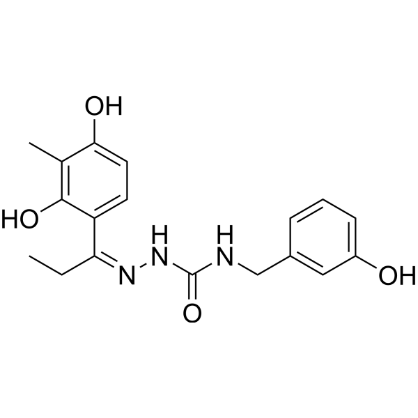
GA-017 is a potent and selective LATS1 and LATS2 (large tumor suppressor kinase 1/2) inhibitor, with IC50 values of 4.10 and 3.92 nM, respectively. GA-017 is an activator of cell proliferation. GA-017 promotes YAP/TAZ activation and nuclear translocation. GA-017 promotes cell growth under 3D culture conditions. GA-017 enhances the ex vivo formation of mouse intestinal organoids.
For research use only. We do not sell to patients.
- Purity : 98.95%
- CAS No.: 2351906-74-4
- Formula: C18H21N3O4
- Molecular Weight:343.38
-
Storage:Powder -20°C, 3 years ; In solvent -80°C, 6 months , -20°C, 1 month

To place orders, for customer services and technical support, please contact: MedChemExpress USA
Tel: 609-228-6898 E-mail: [email protected] [email protected]


-
 Biological Activity
-
Chemical Information
-
Solvent & Solubility
- Protocols
- Purity & Documentation
- References
-
 Help & FAQs


-
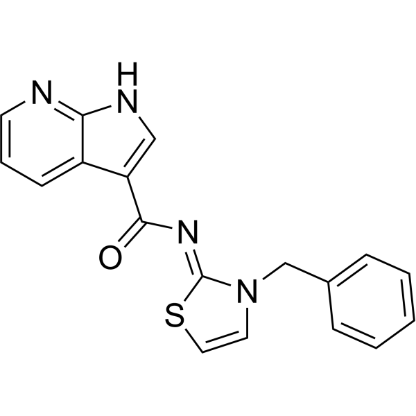
HY-138489
TRULILat1/2 kinases Inhibitor -
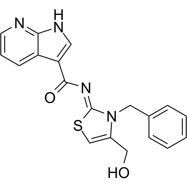
HY-150042
TDI-011536Lats Kinase Inhibitor -
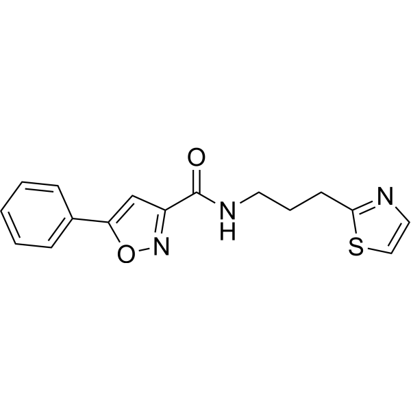
HY-141644
PY-60YAP Activator -
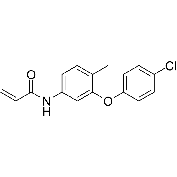
HY-138565
K-975YAP1/TAZ-TEAD Inhibitor -
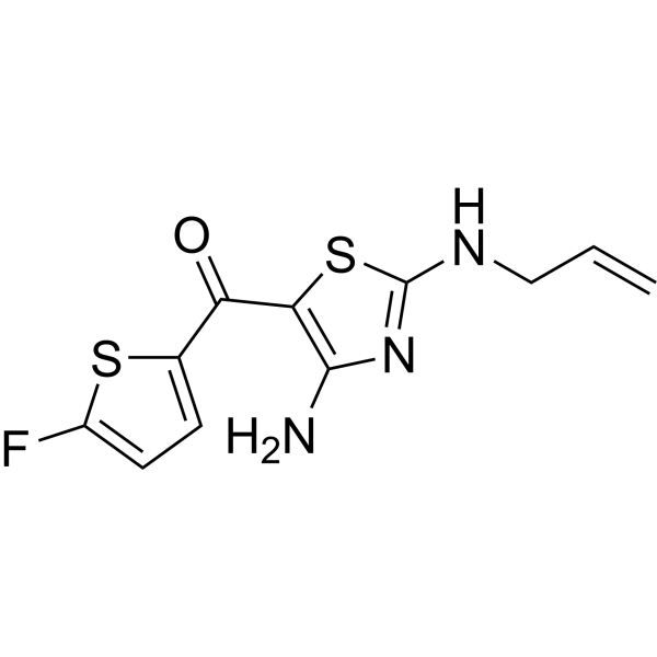
HY-125016
TT-10YAP-TEAD Activator -
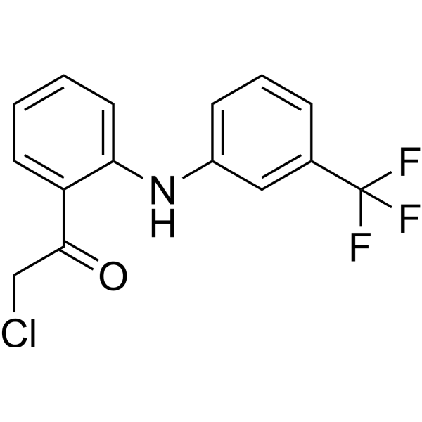
HY-125269
TED-347YAP-TEAD Inhibitor -
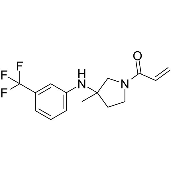
HY-139603
MYF-01-37TEAD Inhibitor -

HY-P1727
Super-TDUYAP Antagonist -
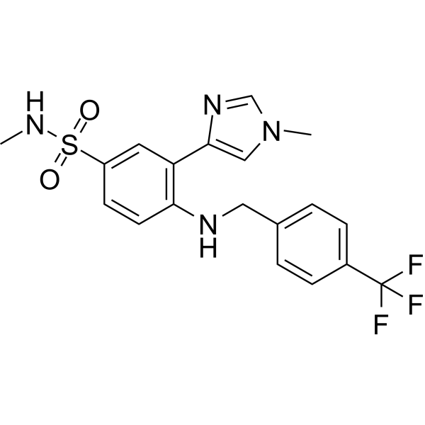
HY-153585
IK-930TEAD Inhibitor -
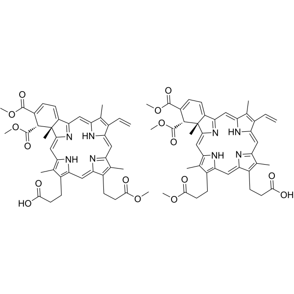
HY-B0146
VerteporfinYAP Inhibitor -
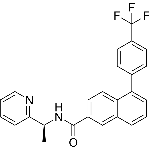
HY-134956
VT104YAP/TAZ Inhibitor -
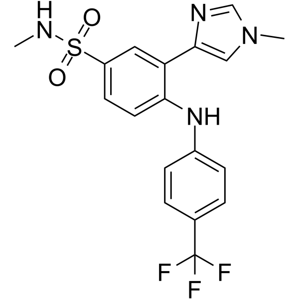
HY-134955
VT103TEAD1 Palmitoylation Inhibitor
Publications Citing Use of MedChemExpress (MCE) GA-017
More- Mol Cell. 2024 Sep 5;84(17):3336-3353.e7. [Abstract]
- Cell Rep Med. 2026 Feb 17;7(2):102622. [Abstract]
- Cell Death Dis. 2026 Mar 26;17(1):381. [Abstract]
- Int Immunopharmacol. 2026 Jan 1;168(Pt 1):115828. [Abstract]
- Cancer Biother Radiopharm. 2025 Mar;40(2):139-150 [Abstract]
- bioRxiv. 2025 April 14.
- SSRN. 2025 Jan 3.
 Customer Validation & Images

-
RT-PCR

-
Cell Migration/Invasion Assay
-
Cell Migration/Invasion Assay
-
RT-PCR
-
WB
All YAP Isoforms
More

Biological Activity
Description
IC50 & Target
|
Lats1 |
Lats2 |
YAP/TAZ |
In Vitro
GA-017 suppresses the cell death of HUVECs and promotes their cell growth[1].
GA-017 facilitates SKOV3 cell growth, with an EC50 of 3.51 ± 0.26 μM[1].
GA-017 competitively inhibited LATS1 and 2 against ATP, with Ki (inhibition constant) values of 0.58 ± 0.11 and 0.25 ± 0.03 nM, respectively[1].
MedChemExpress (MCE) has not independently confirmed the accuracy of these methods. They are for reference only. Further protocols information, click here.


Chemical Information
-
CAS No. 2351906-74-4
-
Appearance Solid
-
Molecular Weight 343.38
-
Formula C18H21N3O4
-
Color Off-white to light yellow
-
SMILES
O=C(NCC1=CC(O)=CC=C1)N/N=C(CC)\C2=CC=C(C(C)=C2O)O
-
Shipping
Room temperature in continental US; may vary elsewhere.
-
Storage
Powder -20°C 3 years In solvent -80°C 6 months -20°C 1 month


Publications (7)
-
Journal Impact Factor
-
Most Recent
-
Mol Cell
Consecutive palmitoylation and phosphorylation orchestrates NLRP3 membrane trafficking and inflammasome activation. [Abstract]2024 Sep 5;84(17):3336-3353.e7. PMID: 39173637
GA-017 purchased from MedChemExpress. Usage Cited in: Mol Cell. 2024 Sep 5;84(17):3336-3353.e7. [Abstract]
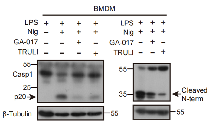
Caspase-1 (p20) and gasdermin D (cleaved N-term) processing upon NLRP3 inflammasome in BMDM cells with or without the treatment of TRULI and GA-017 (10 μM, 5h) .
-
Cell Rep Med
A human patient-derived organoid biobank to model tumor heterogeneity and therapeutic vulnerability for oral squamous cell carcinoma. [Abstract]2026 Feb 17;7(2):102622. PMID: 41707653 -
Cell Death Dis
Generation of proliferative hESC-derived grape-clustered hepatocyte organoids with multipolar architecture as regenerative counterpart via synergy of YAP and IGF2 pathways. [Abstract]2026 Mar 26;17(1):381. PMID: 41888105 -
Int Immunopharmacol
ACTL6A accelerates the progression of NSCLC through hippo/YAP signaling axis and TAMs-mediated immune regulation. [Abstract]2026 Jan 1;168(Pt 1):115828. PMID: 41223612
GA-017 purchased from MedChemExpress. Usage Cited in: Int Immunopharmacol. 2026 Jan 1;168(Pt 1):115828. [Abstract]
ACTL6A-interfered NCI-H520 cells were treated with GA-017 (10 μM) and K-975 for 24 h. Then, qRT-PCR was used to detect the expression of TEAD1 and TEAD4.
GA-017 purchased from MedChemExpress. Usage Cited in: Int Immunopharmacol. 2026 Jan 1;168(Pt 1):115828. [Abstract]
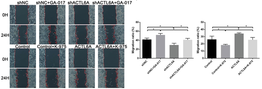
GA-017 (10 μM) for 24 h. Wound healing assay (scale bar =100 μm) was performed to detect the changes of NCI-H520 cell migration. *P < 0.05, **P < 0.01 and ***P < 0.001.
GA-017 purchased from MedChemExpress. Usage Cited in: Int Immunopharmacol. 2026 Jan 1;168(Pt 1):115828. [Abstract]
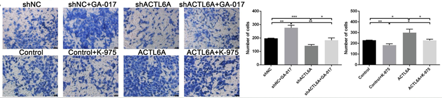
GA-017 (10 μM) for 24 h. Transwell assay (scale bar =50 μm) was performed to detect the changes of cell migration. *P < 0.05, **P < 0.01 and ***P < 0.001.
GA-017 purchased from MedChemExpress. Usage Cited in: Int Immunopharmacol. 2026 Jan 1;168(Pt 1):115828. [Abstract]
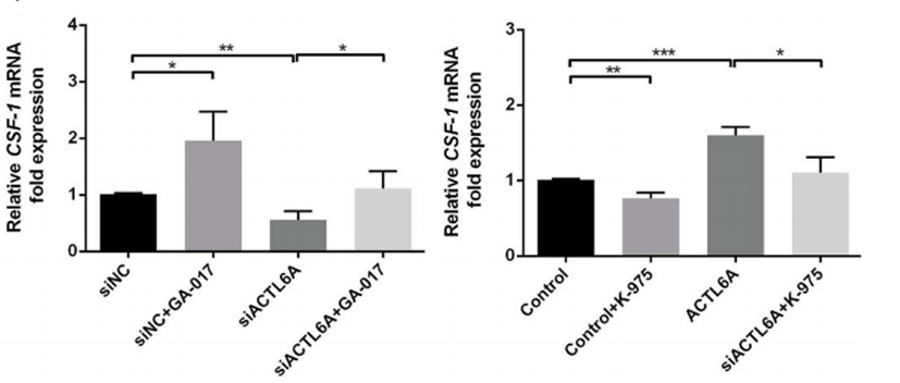
ACTL6A-interfered NCI-H520 cells were treated with GA-017 (10 μM) for 24 h or K-975. Then, qRT-PCR was performed to detect the expression of CSF1.
-
Cancer Biother Radiopharm
Dihydroartemisinin Inhibits Epithelial-Mesenchymal Transition Progression in Medullary Thyroid Carcinoma Through the Hippo Signaling Pathway Regulated by Interleukin-6. [Abstract]2025 Mar;40(2):139-150 PMID: 39286872 -
-


Solvent & Solubility
In Vitro:
DMSO : 20.83 mg/mL (60.66 mM; ultrasonic and warming and heat to 80°C; Hygroscopic DMSO has a significant impact on the solubility of product, please use newly opened DMSO)
Please refer to the solubility information to select the appropriate solvent. Once prepared, please aliquot and store the solution to prevent product inactivation from repeated freeze-thaw cycles.
Storage method and period of stock solution: -80°C, 6 months; -20°C, 1 month. When stored at -80°C, please use it within 6 months. When stored at -20°C, please use it within 1 month.
Please refer to the solubility information to select the appropriate solvent. Once prepared, please aliquot and store the solution to prevent product inactivation from repeated freeze-thaw cycles.
Storage method and period of stock solution: -80°C, 6 months; -20°C, 1 month. When stored at -80°C, please use it within 6 months. When stored at -20°C, please use it within 1 month.
Concentration (start) × Volume (start) = Concentration (final) × Volume (final)

Protocols
-
BrdU Incorporation Assay
Bromodeoxyuridine (BrdU) incorporation assay is based on the principle that BrdU, a thymidine analog, is incorporated into newly synthesized DNA during the S phase of the cell cycle, thereby serving as a marker of DNA replication and cellular proliferation. Incorporated BrdU can be detected using anti-BrdU antibodies following DNA denaturation, enabling visualization or quantification of proliferating cells through immunochemical detection methods such as immunofluorescence or immunohistochemistry.
-
Somatic Cell Culture
A method of simulating the in vivo environment in vitro to maintain the cell growth, differentation and main functions.
-
Organoid Passaging and Mechanical Expansion Protocols
Organoid passaging by mechanical expansion transfers established 3D epithelial organoids from an extracellular matrix dome into fresh matrix after physical fragmentation. The readout is successful re-formation and expansion of organoid fragments into new organoids, reflecting survival of organoid-forming epithelial stem/progenitor cells and continued self-organization in a 3D matrix.
-
ECM-Embedded Organoid (Matrigel/Dome) Culture
ECM-embedded organoid dome culture embeds epithelial stem cells, crypts, organoid fragments, or tumor-derived epithelial cells in a basement-membrane-like hydrogel such as Matrigel, allowing 3D growth, self-organization, lumen formation, budding or cystic morphogenesis, and lineage maintenance under defined niche-factor-containing medium. The primary readouts are organoid establishment efficiency, growth, morphology, passaging capacity, lineage-marker expression, and, when fluorescently labeled lines are used, microscopy- or flow-cytometry-based quantification of population behavior in 3D culture.
-
CFSE Dye Dilution Proliferation Assay
The CFSE (carboxyfluorescein diacetate succinimidyl ester) dye dilution proliferation assay is based on the covalent labeling of intracellular proteins by a cell-permeant fluorescent dye that becomes fluorescent upon intracellular ester cleavage and then is stably retained within cells. As labeled cells divide, the dye is partitioned equally between daughter cells, resulting in a stepwise halving of fluorescence intensity that can be quantified by flow cytometry to determine the number of cell divisions undergone by each cell population. This fluorescence dilution approach enables quantitative tracking of lymphocyte proliferation at the single-cell level over multiple rounds of division. CFSE-based proliferation analysis has been widely applied to measure antigen-driven lymphocyte expansion in vitro, where discrete fluorescence peaks correspond to successive cell divisions and allow reconstruction of proliferative history within heterogeneous populations.
-
Dye-dilution cell tracking and proliferation staining
Dye-dilution cell tracking assays quantify cell proliferation by covalently labeling intracellular proteins with a stable fluorescent dye that is equally partitioned between daughter cells during mitosis, resulting in stepwise halving of fluorescence intensity with each cell division as measured by flow cytometry histograms. Carboxyfluorescein diacetate succinimidyl ester (CFSE) is a prototypical dye that diffuses into cells, is enzymatically converted into a fluorescent compound, and then covalently binds intracellular amine groups, producing long-lived fluorescence suitable for tracking multiple rounds of division in vitro and in vivo. Successive generations of dividing cells form discrete peaks of decreasing fluorescence intensity, enabling estimation of proliferation history, precursor frequency, and division index within heterogeneous populations. Alternative dyes such as CellTrace Violet (CTV) and far-red membrane dyes (e. g. , PKH26) follow the same dilution principle but differ
-
CCK-8/WST-8 Cell Proliferation Assay
The CCK-8/WST-8 assay is based on the reduction of the water-soluble tetrazolium salt WST-8 to a water-soluble formazan product by cellular dehydrogenases in metabolically active cells, where the generated formazan amount is proportional to the number of living cells and is quantified by measuring absorbance in the visible range, providing a colorimetric readout for cell viability and proliferation assessment. This class of tetrazolium-based assays improves upon earlier MTT-based systems by producing a water-soluble formazan, eliminating the need for organic solubilization steps and enabling direct spectrophotometric measurement in culture medium.
-
Protocol for Cell Cycle
Cell-cycle analysis by flow cytometry measures DNA content in single cells to estimate the fraction of cells in G0/G1, S, and G2/M phases. Propidium iodide intercalates into DNA, and after RNA removal with RNase, fluorescence intensity reflects cellular DNA content: 2N cells are assigned to G0/G1, cells between 2N and 4N to S phase, and 4N cells to G2/M. DNA-content analysis alone cannot reliably separate G0 from G1 or G2 from M. Ki-67 can distinguish quiescent G0 cells from cycling cells, EdU or BrdU incorporation marks active DNA synthesis in S phase, and phospho-histone H3 staining identifies mitotic cells within the 4N population.
-
Hanging Drop Spheroid Culture
Hanging drop spheroid culture is a scaffold-free 3D culture method in which a small droplet of cell suspension is inverted so that suspended cells sediment by gravity toward the lowest point of the drop, aggregate, and form a multicellular spheroid with direct cell-cell contact. Spheroids generated by this method are used to study 3D cell cohesion, cell-ECM interactions, drug response, co-culture organization, and tumor-like microenvironmental behavior. The primary readouts are spheroid formation efficiency, spheroid size, circularity or compactness, viability, and treatment response; these can be measured by bright-field microscopy, fluorescence viability staining, ATP-, fluorescence-, or colorimetric-based assays, and image-based diameter or volume calculations.
-
Cell Counting-Based Growth Curve Assay
Cell counting-based growth curve assays quantify cell proliferation by directly measuring changes in viable cell number over time using manual or automated counting methods such as hemocytometer-based counting or instrument-assisted cell enumeration, enabling construction of growth curves that reflect population expansion dynamics in response to culture conditions. A widely used approach is trypan blue exclusion with hemocytometer counting, where membrane-compromised (non-viable) cells take up the dye, allowing discrimination between viable and non-viable cells while simultaneously enabling total cell number quantification. Repeated sampling across time points allows estimation of proliferation rate, growth phases, and comparative growth kinetics between experimental conditions.
-
Colony Formation (Clonogenic) Assay
The clonogenic (colony formation) assay measures the ability of a single cell to retain reproductive viability and form a macroscopic colony, typically defined as a cluster derived from one progenitor cell after a defined growth period. This assay is widely used to evaluate cell survival after exposure to ionizing radiation or cytotoxic treatments and is considered a standard method in radiation biology for generating dose-response relationships of reproductive cell death. Colony formation reflects long-term proliferative capacity rather than short-term metabolic activity, and survival is quantified by comparing treated versus untreated conditions based on colony number and derived survival fractions.
-
Cell Viability Determination by MTT Colorimetric Assay
The following protocol uses the MTT colorimetric assay as a classic literature-established method for assessing cell viability/metabolic activity in cultured mammalian cells. MTT[3-(4,5-dimethylthiazol-2-yl)-2,5-diphenyltetrazolium bromide] is reduced by metabolically active cells to a colored formazan product; the amount of formazan is quantified spectrophotometrically and provides an indirect measure of metabolically active viable cells. Importantly, MTT reduction reflects cellular oxidoreductase/metabolic activity rather than an absolute direct count of living cells, so changes in cellular metabolism can alter the signal independently of cell number.
-
EdU Incorporation Assay (Click Chemistry-Based DNA Synthesis Measurement)
The EdU incorporation assay measures DNA synthesis by adding the thymidine analog 5-ethynyl-2′-deoxyuridine to cells or tissues, where it is incorporated into newly synthesized DNA during S phase. Incorporated EdU is detected by copper-catalyzed azide-alkyne cycloaddition, in which a fluorescent azide covalently reacts with the ethynyl group on EdU, allowing S-phase cells to be detected by fluorescence microscopy, flow cytometry, or high-content imaging. EdU detection does not require DNA denaturation or anti-BrdU antibody access, which preserves sample structure and improves compatibility with immunostaining and multiparameter cytometry compared with BrdU-based detection. EdU can be cytotoxic in a cell-type- and exposure-dependent manner, so pulse duration, concentration, and continuous-labeling designs should be validated for each cell type.
-
Cytoplasmic-Nuclear Fractionated Protein Extraction
Cytoplasmic-nuclear fractionated protein extraction separates soluble cytoplasmic proteins from nuclear-enriched proteins by mild plasma-membrane permeabilization, differential centrifugation, washing of nuclei, and extraction of nuclear proteins for downstream immunoblotting or related molecular analysis. The readout is the relative abundance of a protein in cytoplasmic and nuclear fractions, commonly assessed by western blotting together with compartment markers such as tubulin or pyruvate kinase for cytoplasm and lamin, nucleoporin, hnRNP, H2AX, or Lamin B for nuclear fractions.
-
Ki-67 Immunostaining Proliferation Assay
Ki-67 immunostaining measures the growth fraction of a cell population by detecting Ki-67, a nuclear antigen present in proliferating cells and absent in quiescent G0 cells. The readout is the percentage of Ki-67-positive nuclei among total counted cells, commonly called the Ki-67 labeling index or proliferation index.
-
Cell Cytotoxicity Assay
Cytotoxicity assays are usually based on the assessment of cell membrane damage, which can also be indirectly detected by measuring cell viability. Detection methods include MTT assay, CKK-8 assay, LDH assay and ATP assay, etc.
-
PCNA Immunodetection Proliferation Assay
PCNA immunodetection measures proliferative activity by detecting proliferating cell nuclear antigen, a nuclear protein associated with DNA polymerase δ function and DNA replication. The assay readout is the proportion of PCNA-positive nuclei among total counted cells, but PCNA labeling is not identical to BrdU labeling because PCNA can mark late G1/early S-associated replication competence and may persist beyond active DNA synthesis depending on fixation and extraction conditions.
-
Protocol for Cell Counting and Cell Density Analysis
Cell counting and cell-density analysis estimate the number of cells in a known volume or field area. Manual hemocytometer counting uses a chamber of defined geometry to convert counted cells into cells/mL, while automated counters and image-analysis workflows detect cell objects from optical, brightfield, fluorescence, impedance, or digital-image features. Trypan blue viability counting is based on dye exclusion: viable cells with intact membranes exclude dye, while non-viable cells with compromised membranes stain blue. The readout is total cell density, viable-cell density, dead-cell density, and percent viability. Cell density can also be estimated from microscopy images by counting objects per image area, from flow cytometry using calibrated volume or reference particles, or from in situ microscopy in bioreactors after calibration against reference methods such as hemocytometer or flow cytometry.
-
MTT Cell Proliferation Assay
The MTT assay is a colorimetric endpoint assay for estimating viable cell number, cell growth, cytotoxicity, or cell activation in cultured mammalian cells. Living cells reduce the yellow tetrazolium salt MTT into purple/blue formazan, while dead cells do not generate the same signal; the resulting color can be quantified with a multiwell spectrophotometer. MTT reduction is commonly interpreted as a readout of metabolic activity that often correlates with viable cell number, but it should not be treated as a direct cell-counting method unless the assay is optimized for the cell type and experimental condition. Studies show that MTT reduction can involve mitochondrial and non-mitochondrial reducing systems, and formazan may accumulate in intracellular lipid droplets rather than simply marking mitochondria.


Purity & Documentation
-
Data Sheet (272 KB)
-
SDS (252 KB)
- English - EN (252 KB)
- Français - FR (252 KB)
- Deutsch - DE (252 KB)
- Norwegian - NO (252 KB)
- Español - ES (252 KB)
- Swedish - SV (252 KB)
- Italian - IT (252 KB)
- Korean - KR (252 KB)
- Portuguese - PT (252 KB)
-
Handling Instructions (2659 KB)
References


Complete Stock Solution Preparation Table
Please refer to the solubility information to select the appropriate solvent. Once prepared, please aliquot and store the solution to prevent product inactivation from repeated freeze-thaw cycles.
Storage method and period of stock solution: -80°C, 6 months; -20°C, 1 month. When stored at -80°C, please use it within 6 months. When stored at -20°C, please use it within 1 month.
| Optional Solvent | Concentration Solvent Mass | 1 mg | 5 mg | 10 mg | 25 mg |
|---|---|---|---|---|---|
| DMSO | 1 mM | 2.9122 mL | 14.5611 mL | 29.1223 mL | 72.8056 mL |
| 5 mM | 0.5824 mL | 2.9122 mL | 5.8245 mL | 14.5611 mL | |
| 10 mM | 0.2912 mL | 1.4561 mL | 2.9122 mL | 7.2806 mL | |
| 15 mM | 0.1941 mL | 0.9707 mL | 1.9415 mL | 4.8537 mL | |
| 20 mM | 0.1456 mL | 0.7281 mL | 1.4561 mL | 3.6403 mL | |
| 25 mM | 0.1165 mL | 0.5824 mL | 1.1649 mL | 2.9122 mL | |
| 30 mM | 0.0971 mL | 0.4854 mL | 0.9707 mL | 2.4269 mL | |
| 40 mM | 0.0728 mL | 0.3640 mL | 0.7281 mL | 1.8201 mL | |
| 50 mM | 0.0582 mL | 0.2912 mL | 0.5824 mL | 1.4561 mL | |
| 60 mM | 0.0485 mL | 0.2427 mL | 0.4854 mL | 1.2134 mL |


GA-017 Related Classifications
HY-147082 Related Classifications
 Powered by Bioz