



Dazcapistat
Based on 1 publication(s) in Google Scholar
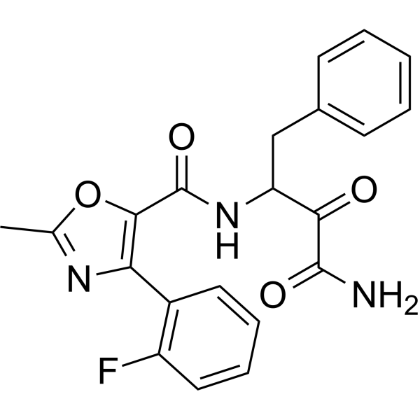
Dazcapistat (BLD-2660) is an orally active small-molecule inhibitor of calpain 1, 2, and 9 (calpain 1, 2, 9). Dazcapistat reduces the expression/production level of IL-6 in injured lung tissue and alleviates fibrosis. Dazcapistat exerts anti-fibrotic effects in various animal models of fibrosis in the skin, liver and lung. Dazcapistat can be used in research related to coronavirus disease 2019 (COVID-19) and idiopathic pulmonary fibrosis.
Para uso exclusivo en investigación. No vendemos a pacientes.
- Pureza : 98.0%
- No. CAS: 2221010-42-8
- Fòrmula: C21H18FN3O4
- Peso molecular:395.38
-
Almacenamiento:Powder -20°C, 3 years , 4°C, 2 years ; In solvent -80°C, 6 months , -20°C, 1 month

To place orders, for customer services and technical support, please contact: MedChemExpress USA
Tel: 609-228-6898 E-mail: [email protected] [email protected]


-
 Actividad biológica
-
Chemical Information
-
Solvente y solubilidad
- Protocolo
- Pureza y Documentación
- Referencias
-
 Help & FAQs


-
HY-109001
AlicapistatCalpains Inhibitor -
HY-18236
MDL-28170Calpain Inhibitor -
HY-18964
MG-101Cysteine Protease Inhibitor -
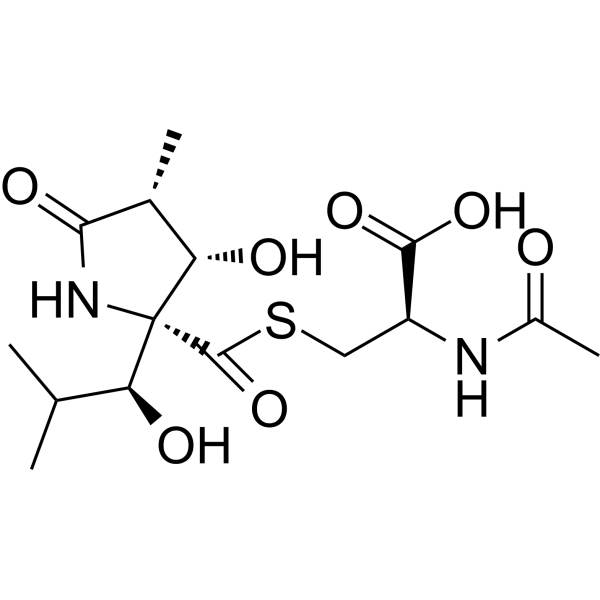
HY-16594
LactacystinProteasome Inhibitor -
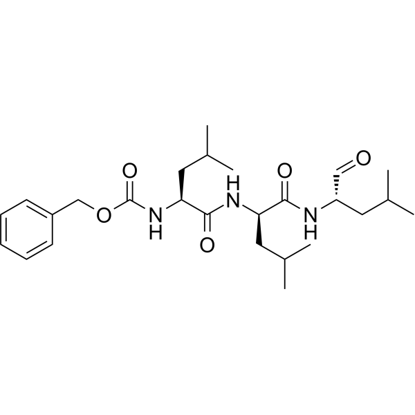
HY-13259C
(R)-MG-132Proteasome Inhibitor -
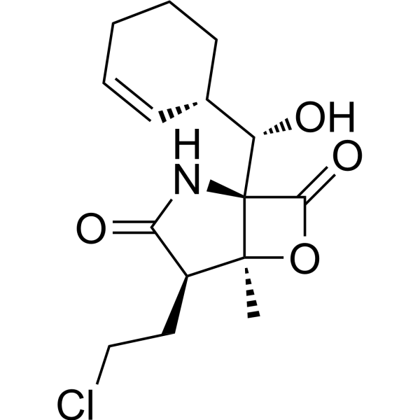
HY-10985
MarizomibProteasome Inhibitor -
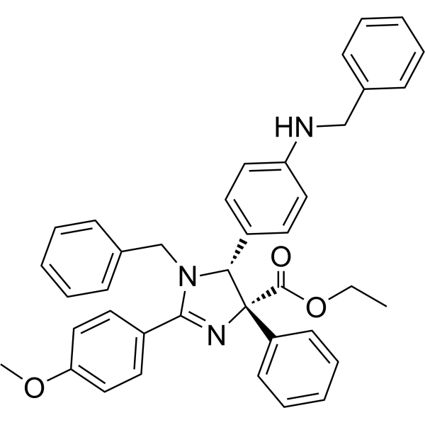
HY-120722
TCH-165Proteasome Assembly Modulator -
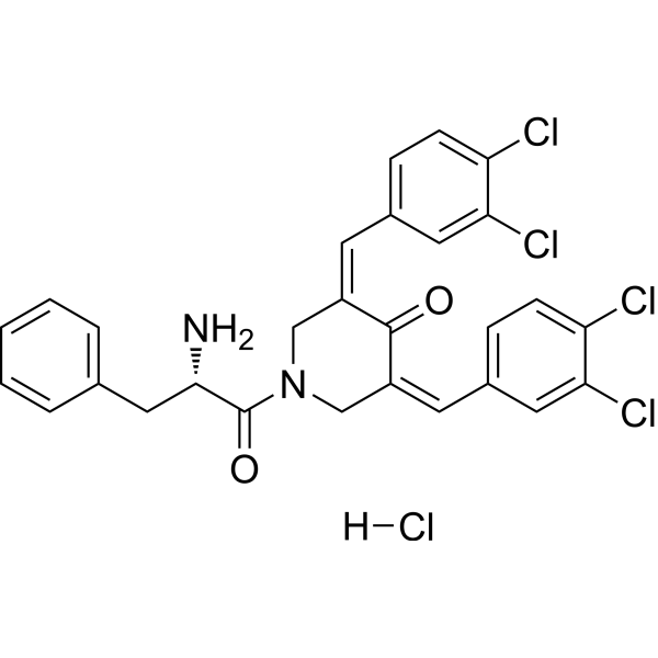
HY-100739
RA190RPN13 Inhibitor -
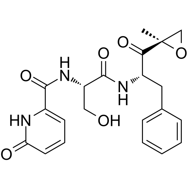
HY-101786
KZR-504LMP2 Inhibitor -
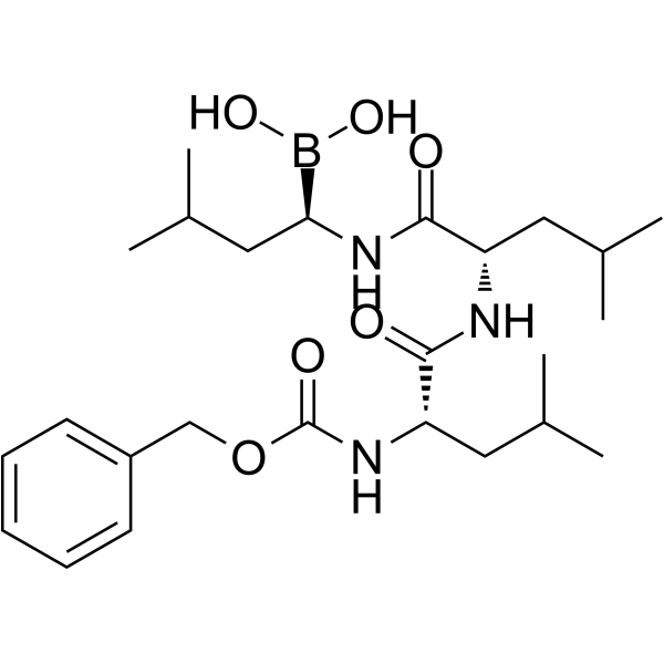
HY-108551
MG-262Proteasome Inhibitor -
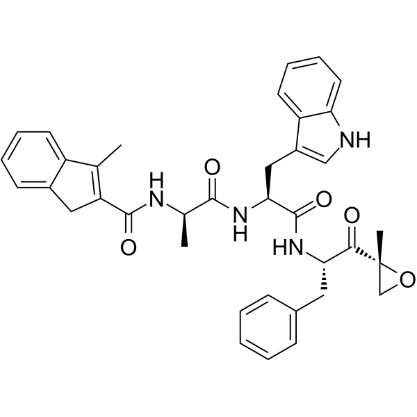
HY-123587
PR-924LMP-7 Inhibitor -
HY-114419
ZetomipzomibImmunoproteasome Inhibitor
Publications Citing Use of MedChemExpress (MCE) Dazcapistat
More

Actividad biológica
Descripciòn
In Vivo
BLD-2660 is shown to reduce the expression/production of IL-6
in injured lungs and to reduce fibrosis[2]。
MedChemExpress (MCE) has not independently confirmed the accuracy of these methods. They are for reference only.


Chemical Information
-
No. CAS 2221010-42-8
-
Appearance Solid
-
Peso molecular 395.38
-
Fòrmula C21H18FN3O4
-
Color White to off-white
-
SMILES
O=C(C(C(CC1=CC=CC=C1)NC(C2=C(C3=C(F)C=CC=C3)N=C(C)O2)=O)=O)N
-
Synonyms
BLD-2660
-
Envío
Room temperature in continental US; may vary elsewhere.
-
Almacenamiento
Powder -20°C 3 years 4°C 2 years In solvent -80°C 6 months -20°C 1 month


Publications (1)
-
Journal Impact Factor
-
Most Recent
-
Cells
Detection of Calpain-Mediated Beclin-1 Cleavage for Drug Discovery in Inflammatory Bowel Diseases. [Abstract]2026 May 18;15(10):917. PMID: 42193926


Solvente y solubilidad
In Vitro:
DMSO : 100 mg/mL (252.92 mM; Need ultrasonic; Hygroscopic DMSO has a significant impact on the solubility of product, please use newly opened DMSO)
Please refer to the solubility information to select the appropriate solvent. Once prepared, please aliquot and store the solution to prevent product inactivation from repeated freeze-thaw cycles.
Storage method and period of stock solution: -80°C, 6 months; -20°C, 1 month. When stored at -80°C, please use it within 6 months. When stored at -20°C, please use it within 1 month.
Please refer to the solubility information to select the appropriate solvent. Once prepared, please aliquot and store the solution to prevent product inactivation from repeated freeze-thaw cycles.
Storage method and period of stock solution: -80°C, 6 months; -20°C, 1 month. When stored at -80°C, please use it within 6 months. When stored at -20°C, please use it within 1 month.
Concentration (start) × Volume (start) = Concentration (final) × Volume (final)
In Vivo:
Select the appropriate dissolution method based on your experimental animal and administration route.
- For the following dissolution methods, please ensure to first prepare a clear stock solution using an In Vitro approach and then sequentially add co-solvents:
- To ensure reliable experimental results, the clarified stock solution can be appropriately stored based on storage conditions. As for the working solution for In Vivo experiments, it is recommended to prepare freshly and use it on the same day.
- The percentages shown for the solvents indicate their volumetric ratio in the final prepared solution. If precipitation or phase separation occurs during preparation, heat and/or sonication can be used to aid dissolution.
Add each solvent one by one: 10% DMSO 40% PEG300 5% Tween-80 45% Saline
Solubility: ≥ 2.5 mg/mL (6.32 mM); Clear solution
This protocol yields a clear solution of ≥ 2.5 mg/mL (saturation unknown).
Taking 1 mL working solution as an example, add 100 μL DMSO stock solution (25.0 mg/mL) to 400 μL PEG300, and mix evenly; then add 50 μL Tween-80 and mix evenly; then add 450 μL Saline to adjust the volume to 1 mL.
Preparation of Saline: Dissolve 0.9 g sodium chloride in ddH₂O and dilute to 100 mL to obtain a clear Saline solution.
Add each solvent one by one: 10% DMSO 90% (20% SBE-β-CD in Saline)
Solubility: ≥ 2.5 mg/mL (6.32 mM); Clear solution
This protocol yields a clear solution of ≥ 2.5 mg/mL (saturation unknown).
Taking 1 mL working solution as an example, add 100 μL DMSO stock solution (25.0 mg/mL) to 900 μL 20% SBE-β-CD in Saline, and mix evenly.
Preparation of 20% SBE-β-CD in Saline (4°C, storage for one week): 2 g SBE-β-CD powder is dissolved in 10 mL Saline, completely dissolve until clear.
In Vivo Dissolution Calculator
Please enter the basic information of animal experiments:
-
-
-
-
Recommended: Prepare an additional quantity of animals to account for potential losses during experiments.
Please enter your animal formula composition:
-
%DMSO +
Recommended: Keep the proportion of DMSO in working solution below 2% if your animal is weak.
-
%+
-
+%Tween-80 + +
-
%Saline +
The co-solvents required include: DMSO, . All of co-solvents are available by MedChemExpress (MCE). , Tween 80. All of co-solvents are available by MedChemExpress (MCE).
Working solution concentration: 0.22 mg/mL
Method for preparing stock solution: mg drug dissolved in μL DMSO. Stock solution concentration: mg/mL.
1. Take μL DMSO stock solution;
2. Add μL .
μL , mix evenly;
3. Then add μL Tween 80, mix evenly;
4. Then add μL
Please ensure that the stock solution in the first step is dissolved to a clear state, and add co-solvents in sequence. You can use ultrasonic heating (ultrasonic cleaner, recommended frequency 20-40 kHz), vortexing, etc. to assist dissolution.


Pureza y Documentación
-
Ficha de datos (281 KB)
-
SDS (251 KB)
- English - EN (251 KB)
- Français - FR (251 KB)
- Deutsch - DE (251 KB)
- Norwegian - NO (251 KB)
- Español - ES (251 KB)
- Swedish - SV (251 KB)
- Italian - IT (251 KB)
- Korean - KR (251 KB)
- Portuguese - PT (251 KB)
-
Instrucciones de manejo (2659 KB)
Referencias
[1]. Oliver JC, et al. Different drug approaches to COVID-19 treatment worldwide: an update of new drugs and drugs repositioning to fight against the novel coronavirus. Ther Adv Vaccines Immunother. 2022;10:25151355221144845. Published 2022 Dec 23. [Content Brief]
[2]. Al-Horani RA, et al. Potential Anti-SARS-CoV-2 Therapeutics That Target the Post-Entry Stages of the Viral Life Cycle: A Comprehensive Review. Viruses. 2020;12(10):1092. Published 2020 Sep 26. [Content Brief]


Complete Stock Solution Preparation Table
Please refer to the solubility information to select the appropriate solvent. Once prepared, please aliquot and store the solution to prevent product inactivation from repeated freeze-thaw cycles.
Storage method and period of stock solution: -80°C, 6 months; -20°C, 1 month. When stored at -80°C, please use it within 6 months. When stored at -20°C, please use it within 1 month.
| Optional Solvent | Concentration Solvent Mass | 1 mg | 5 mg | 10 mg | 25 mg |
|---|---|---|---|---|---|
| DMSO | 1 mM | 2.5292 mL | 12.6461 mL | 25.2921 mL | 63.2303 mL |
| 5 mM | 0.5058 mL | 2.5292 mL | 5.0584 mL | 12.6461 mL | |
| 10 mM | 0.2529 mL | 1.2646 mL | 2.5292 mL | 6.3230 mL | |
| 15 mM | 0.1686 mL | 0.8431 mL | 1.6861 mL | 4.2154 mL | |
| 20 mM | 0.1265 mL | 0.6323 mL | 1.2646 mL | 3.1615 mL | |
| 25 mM | 0.1012 mL | 0.5058 mL | 1.0117 mL | 2.5292 mL | |
| 30 mM | 0.0843 mL | 0.4215 mL | 0.8431 mL | 2.1077 mL | |
| 40 mM | 0.0632 mL | 0.3162 mL | 0.6323 mL | 1.5808 mL | |
| 50 mM | 0.0506 mL | 0.2529 mL | 0.5058 mL | 1.2646 mL | |
| 60 mM | 0.0422 mL | 0.2108 mL | 0.4215 mL | 1.0538 mL | |
| 80 mM | 0.0316 mL | 0.1581 mL | 0.3162 mL | 0.7904 mL | |
| 100 mM | 0.0253 mL | 0.1265 mL | 0.2529 mL | 0.6323 mL |


Dazcapistat Related Classifications
HY-132850 Related Classifications
 Powered by Bioz