



MitoCur-1
Based on 1 publication(s) in Google Scholar
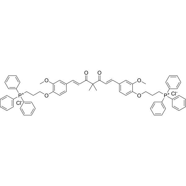
MitoCur-1, a curcumin analogue, is an inhibitor of mitochondrial antioxidative thioredoxin reductase 2 (TrxR2). MitoCur-1 has electrophilic and mitochondrial-targeting properties. MitoCur-1 induces reactive oxygen species (ROS) generation, exerts specifically antitumor efficacy.
Para uso exclusivo en investigación. No vendemos a pacientes.
- Pureza : ≥95.0%
- Fòrmula: C65H64Cl2O6P2
- Peso molecular:1074.05
-
Almacenamiento:
4°C, sealed storage, away from moisture and light
* In solvent : -80°C, 6 months; -20°C, 1 month (sealed storage, away from moisture and light)

To place orders, for customer services and technical support, please contact: MedChemExpress USA
Tel: 609-228-6898 E-mail: [email protected] [email protected]


-
 Actividad biológica
-
Chemical Information
- Protocolo
- Pureza y Documentación
- Referencias
-
 Help & FAQs


Publications Citing Use of MedChemExpress (MCE) MitoCur-1
More

Actividad biológica
Descripciòn
IC50 & Target
Thioredoxin reductase 2 (TrxR2)[1]
In Vitro
MitoCur-1 exhibits selective cytotoxicity against HepG2 and L02 cells with IC50s of 1.4 μM and 9.1 μM, respectively[1].
MitoCur-1 (10 μM; 12 h) generates ROS and depletes GSH selectively in HepG2 cells over L02 cells[1].
MitoCur-1 (10 μM; 24 h) arrests cell cycle at G0/G1 phase, inhibits cyclin protein expression[1].
MedChemExpress (MCE) has not independently confirmed the accuracy of these methods. They are for reference only. Further protocols information, click here.
-
Cell Line:HepG2 cells
-
Concentration:2.5 μM, 5 μM, and 10 μM
-
Incubation Time:24 h, 36 h, and 48 h
-
Result:Decreased the protein level of CDK2 and CDK4, Cyclin E1/D1/A2.
-
Cell Line:HepG2 and L02 cells
-
Concentration:10 μM
-
Incubation Time:12 h
-
Result:Showed selectivity on HepG2 cells over L02 cells to generates ROS. Promoted mitochondrial O2- production.
-
Cell Line:HepG2 cells
-
Concentration:1.25 μM, 2.5 μM, 5 μM, and 10 μM
-
Incubation Time:48 h; pretreated with 400 μM DTT or not
-
Result:Inhibited HepG2 cells viability. Showed significant reversion with DTT for 1 h pretreatment.
In Vivo
MedChemExpress (MCE) has not independently confirmed the accuracy of these methods. They are for reference only.
-
Animal Model:BALB/c nude mice bearing HepG2 tumor[1]
-
Dosage:5 mg/kg, 15 mg/kg
-
Administration:Intraperitoneal injection; every other day for 4 weeks
-
Result:Decreased the tumor growth in vivo.


Chemical Information
-
Appearance Solid
-
Peso molecular 1074.05
-
Fòrmula C65H64Cl2O6P2
-
Color Light yellow to orange
-
SMILES
O=C(C(C(/C=C/C1=CC=C(OCCC[P+](C2=CC=CC=C2)(C3=CC=CC=C3)C4=CC=CC=C4)C(OC)=C1)=O)(C)C)/C=C/C5=CC(OC)=C(OCCC[P+](C6=CC=CC=C6)(C7=CC=CC=C7)C8=CC=CC=C8)C=C5.[Cl-].[Cl-]
-
Envío
Room temperature in continental US; may vary elsewhere.
-
Almacenamiento
4°C, sealed storage, away from moisture and light
* In solvent : -80°C, 6 months; -20°C, 1 month (sealed storage, away from moisture and light)


Publications (1)
-
Journal Impact Factor
-
Most Recent
-
Adv Sci (Weinh)
TrxR2 Lactylation Facilitates Mitochondrial Protection and Endothelial Ferroptosis Resistance in Diabetic Cardiomyopathy. [Abstract]2026 Apr;13(22):e21997. PMID: 41704008

Protocolo
-
ROS/oxidative-stress fluorescent staining
ROS/oxidative-stress fluorescent staining uses cell-permeant fluorogenic probes that become fluorescent after oxidation inside cells or tissues; commonly used examples include DCFH-DA/DCFDA for broad cellular oxidant detection, DHE for superoxide-related signal detection, MitoSOX for mitochondrial superoxide-related signal detection, and CellROX probes for oxidative-stress-associated fluorescence readouts. The assay detects probe oxidation rather than a single ROS species unless the probe and analysis method have been chemically validated for that species. DCFH-DA enters cells, is deacetylated by intracellular esterases to DCFH, and produces fluorescent DCF after oxidation, so the readout is used as an operational measure of total cellular oxidative stress rather than a species-specific ROS measurement. DHE and MitoSOX can report superoxide-related oxidation, but red fluorescence alone can include non-specific ethidium-like oxidation products; HPLC or optimized spectral approaches are
-
Patient-Derived Xenograft (PDX)
Patient-derived xenograft (PDX) models are generated by engrafting primary human tumor tissue directly into immunodeficient mice, allowing in vivo propagation of patient tumor biology without initial in vitro adaptation. These models are used to preserve key histopathological and molecular characteristics of the original tumor and enable assessment of tumor growth dynamics and therapeutic response in a living organism. The biological readout is tumor engraftment and subsequent growth in the murine host, which reflects the ability of human tumor cells to survive, vascularize, and expand in an immunocompromised microenvironment.


Pureza y Documentación
-
Ficha de datos (271 KB)
-
SDS (252 KB)
- English - EN (252 KB)
- Français - FR (252 KB)
- Deutsch - DE (252 KB)
- Norwegian - NO (252 KB)
- Español - ES (252 KB)
- Swedish - SV (252 KB)
- Italian - IT (252 KB)
- Korean - KR (252 KB)
- Portuguese - PT (252 KB)
-
Instrucciones de manejo (2659 KB)
Referencias

Calculators
Concentration (start) × Volume (start) = Concentration (final) × Volume (final)


MitoCur-1 Related Classifications
HY-150228 Related Classifications
 Powered by Bioz