



Gly-His-Lys acetate

Based on 1 Customer Validation
Gly-His-Lys acetate is a natural, circulating regulatory and antimicrobial tripeptide derived from extracellular matrix proteins. Gly-His-Lys acetate binds Cu2+ to support copper enzyme activation, antioxidant processes, cellular bioenergetics, and the synthesis of elastin, collagen and catecholamines. Gly-His-Lys acetate regulates cell growth, differentiation and tissue repair, and exerts regenerative, anxiolytic, anti-inflammatory, analgesic and immunosuppressive activities. Gly-His-Lys acetate induces liver degenerative changes. Gly-His-Lys acetate can be used for the research of infections, anxiety, pain-related behaviors and immune-associated liver diseases.
For research use only. We do not sell to patients.
- Purity : 99.68%
- CAS No.: 72957-37-0
- Formula: C16H28N6O6
- Molecular Weight:400.43
-
Storage:
4°C, protect from light
* In solvent : -80°C, 6 months; -20°C, 1 month (protect from light)


To place orders, for customer services and technical support, please contact: MedChemExpress USA
Tel: 609-228-6898 E-mail: [email protected] [email protected]


-
 Biological Activity
-
Chemical Information
-
Solvent & Solubility
- Protocols
- Purity & Documentation
- References
-
 Help & FAQs


-
HY-W013494
L-CarnosineFree Radical Scavenger -

HY-W013494R
L-Carnosine (Standard) -
HY-W129161
Acetyl tetrapeptide-3ECM Protein Stimulant -

HY-66018
Palmitoyl tripeptide-5 diTFAAnti-aging Peptide -
HY-P0064
Palmitoyl Tetrapeptide-3IL-6 Inhibitor -
HY-125616
Biotinoyl tripeptide-1Peptide -
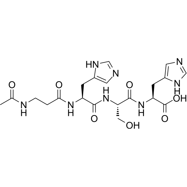
HY-P0095
Acetyl tetrapeptide-5ACE-1 Inhibitor -
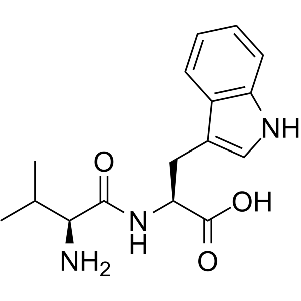
HY-118060
Dipeptide 2ACE Inhibitor -
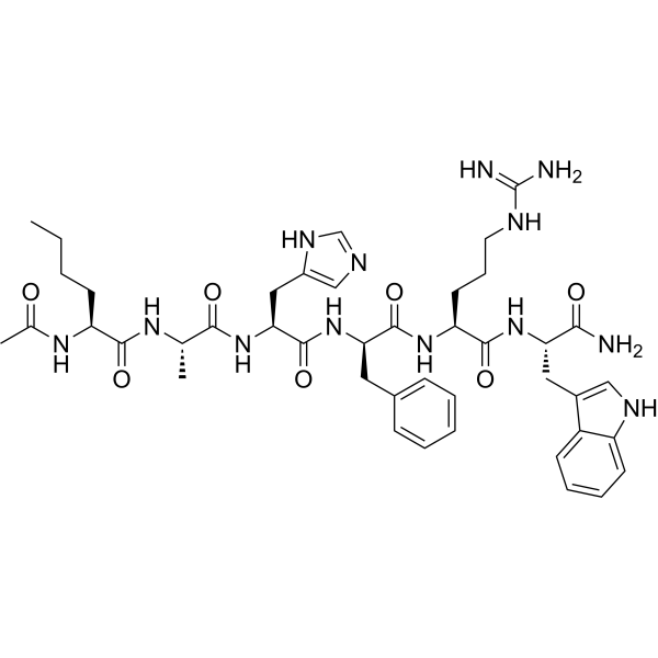
HY-P5220
Acetyl hexapeptide-1Cosmetic Peptide -
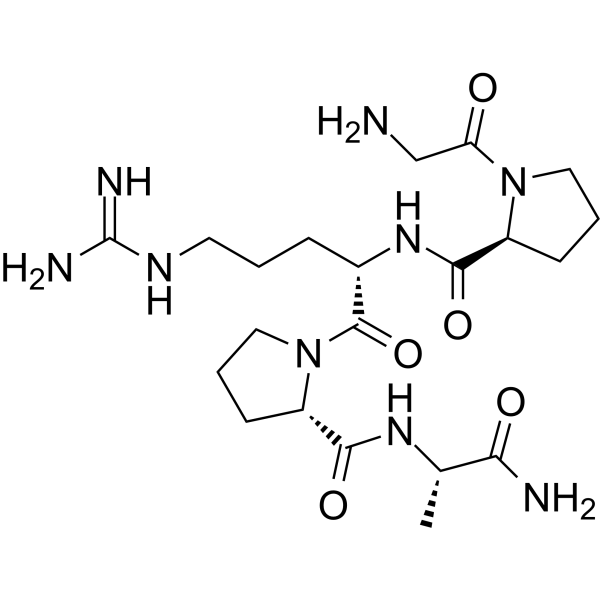
HY-P0099
Pentapeptide-3Acetylcholine Receptor Antagonist -
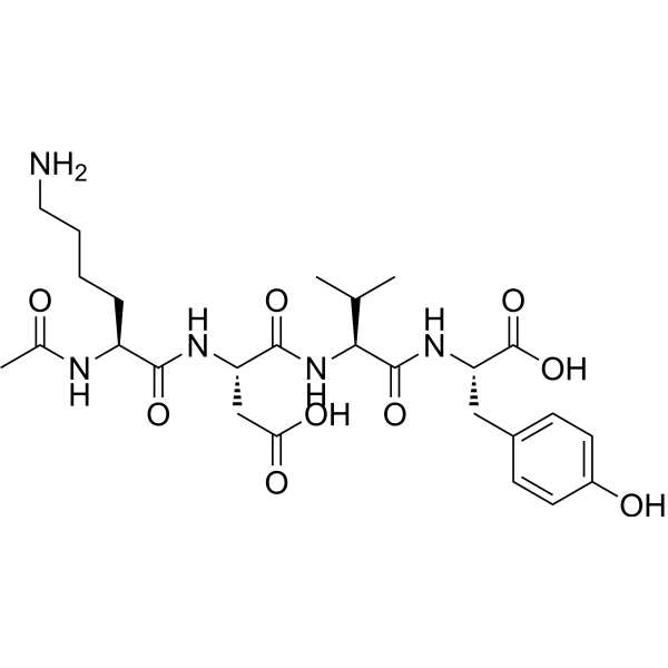
HY-P2663
Acetyl tetrapeptide-2Anti-aging Peptide -

HY-118060R
Dipeptide 2 (Standard)Anti-aging Peptide


Biological Activity
Description
In Vitro
Gly-His-Lys acetate display potent antimicrobial activity against Staphylococcus aureus spp., Escherichia coli spp., Pseudomonas aeruginosa spp. and Candida albicans spp., and exert rapid bactericidal and fungicidal effects[1].
MedChemExpress (MCE) has not independently confirmed the accuracy of these methods. They are for reference only. Further protocols information, click here.
In Vivo
Gly-His-Lys (5-150 μg/kg; i.p.; single dose, 12 min pre-stimulation) acetate exhibits dose-dependent analgesic and antiaggresogenic effects in rats with pain-induced aggressive-defensive behavior[3].
Gly-His-Lys (0.5-450 mg/kg; i.p.; daily; 10 days) acetate stimulates hepatocyte mitotic index in a dose-independent manner at ≥ 1.5 mg/kg and exerts a dose-dependent suppression of humoral immune response in CBA mice[4].
Gly-His-Lys (0.5-450 mg/kg; i.p.; daily; 10 days) acetate exerts a dose-dependent suppression of delayed-type hypersensitivity in Wistar rats at ≥ 1.5 mg/kg[4].
MedChemExpress (MCE) has not independently confirmed the accuracy of these methods. They are for reference only.
-
Animal Model:Wistar (male, 180-220 g)[2]
-
Dosage:0.5 μg/kg; 5 μg/kg; 50 μg/kg
-
Administration:i.p.; single dose
-
Result:Increased time spent in open arms, entries into open arms, and time on the central platform, while decreasing time in closed arms at 0.5 and 5 μg/kg; at 50 μg/kg, it still elevated central platform time and closed-arm entries but showed no significant difference in open-arm time versus control.
-
Animal Model:Wistar rats (male, 180-200 g)[3]
-
Dosage:5 μg/kg; 15 μg/kg; 50 μg/kg; 150 μg/kg
-
Administration:i.p.; single dose, 12 min pre-stimulation
-
Result:Increased flinching, vocalization, rearing, running, and fighting thresholds while reducing fight frequency at 5, 15, 50, and 150 μg/kg; the effects were most prominent at 150 μg/kg.
-
Animal Model:CBA (weight 20-25 g)[4]
-
Dosage:0.5 mg/kg; 1.5 mg/kg; 5 mg/kg; 50 mg/kg; 150 mg/kg; 450 mg/kg
-
Administration:i.p.; daily; 10 days
-
Result:Increased hepatocyte mitotic index significantly at 1.5-450 mg/kg with no obvious dose dependence, dose-dependently reduced antibody-producing spleen cell number, and induced liver degeneration at 150 mg/kg and 450 mg/kg with more severe lesions at 450 mg/kg.
-
Animal Model:Wistar (weight 180-220 g)[4]
-
Dosage:0.5 mg/kg; 1.5 mg/kg; 5 mg/kg; 50 mg/kg; 150 mg/kg; 450 mg/kg
-
Administration:i.p.; daily; 10 days
-
Result:Reduced the regional-versus-contralateral popliteal lymph node weight difference and nucleated cell count difference dose-dependently at 1.5-450 mg/kg, significantly inhibited delayed-type hypersensitivity, and induced obvious liver degeneration at 150 mg/kg and 450 mg/kg with more severe injury at 450 mg/kg.


Chemical Information
-
CAS No. 72957-37-0
-
Appearance Solid
-
Molecular Weight 400.43
-
Formula C16H28N6O6
-
Color White to off-white
-
SMILES
NCCCC[C@H](NC([C@@H](NC(CN)=O)CC1=CNC=N1)=O)C(O)=O.CC(O)=O
-
Shipping
Room temperature in continental US; may vary elsewhere.
-
Storage
4°C, protect from light
* In solvent : -80°C, 6 months; -20°C, 1 month (protect from light)


Solvent & Solubility
In Vitro:
H2O : 100 mg/mL (249.73 mM; Need ultrasonic)
Please refer to the solubility information to select the appropriate solvent. Once prepared, please aliquot and store the solution to prevent product inactivation from repeated freeze-thaw cycles.
Storage method and period of stock solution: -80°C, 6 months; -20°C, 1 month (protect from light). When stored at -80°C, please use it within 6 months. When stored at -20°C, please use it within 1 month.
* Note: If you choose water as the stock solution, please dilute it to the working solution, then filter and sterilize it with a 0.22 μm filter before use.
Please refer to the solubility information to select the appropriate solvent. Once prepared, please aliquot and store the solution to prevent product inactivation from repeated freeze-thaw cycles.
Storage method and period of stock solution: -80°C, 6 months; -20°C, 1 month (protect from light). When stored at -80°C, please use it within 6 months. When stored at -20°C, please use it within 1 month.
* Note: If you choose water as the stock solution, please dilute it to the working solution, then filter and sterilize it with a 0.22 μm filter before use.
Concentration (start) × Volume (start) = Concentration (final) × Volume (final)

Protocols
-
Research Protocol for Infectious Diseases
Infectious-disease experiments test how pathogens interact with host barriers, innate immune receptors, inflammatory signaling, pathogen replication, and tissue injury; pattern-recognition receptors such as TLRs, RIG-I-like receptors, NOD-like receptors, and inflammasomes detect microbial molecules and activate NF-κB, interferon, and cytokine responses. The central hypothesis is that infection severity reflects the balance between pathogen burden and host response: protective inflammation restricts pathogen growth, whereas excessive or mislocalized inflammation contributes to tissue damage and disease phenotype. Unresolved questions include which host pathways are protective versus pathogenic, why some infection models fail to translate to human disease, and which combined readouts best predict clinically relevant infection outcomes.
-
Collagen: Sirius Red Staining
Sirius Red or picrosirius red staining is a histochemical method for visualizing collagen-rich extracellular matrix in tissue sections, and collagen fibers are detected as red-stained structures under bright-field microscopy with enhanced birefringence under polarized light. Picrosirius red is useful for assessing total collagen organization, distribution, and fibrosis burden, but polarized color should not be interpreted as a definitive collagen type I versus type III readout because color is affected by fiber orientation, thickness, and packing.
-
Somatic Cell Culture
A method of simulating the in vivo environment in vitro to maintain the cell growth, differentation and main functions.
-
ECM-Embedded Organoid (Matrigel/Dome) Culture
ECM-embedded organoid dome culture embeds epithelial stem cells, crypts, organoid fragments, or tumor-derived epithelial cells in a basement-membrane-like hydrogel such as Matrigel, allowing 3D growth, self-organization, lumen formation, budding or cystic morphogenesis, and lineage maintenance under defined niche-factor-containing medium. The primary readouts are organoid establishment efficiency, growth, morphology, passaging capacity, lineage-marker expression, and, when fluorescently labeled lines are used, microscopy- or flow-cytometry-based quantification of population behavior in 3D culture.
-
Fibers: Elastic Fiber Staining
Elastic fiber staining is a histochemical technique used to selectively visualize elastin-rich structures such as elastic fibers in connective tissues (e. g. , blood vessels, lung, dermis) based on the affinity of specific dyes or oxidation products for elastin-associated amino acid residues and cross-linked elastic matrix components. Classical methods such as Verhoeff-Van Gieson (VVG), resorcin-fuchsin (Weigert-type stains), or aldehyde fuchsin rely on differential binding of dye complexes to elastic fibers, allowing them to be distinguished from collagen and other extracellular matrix components by contrast staining (typically black or deep purple elastic fibers against red collagen counterstain). These methods are widely used in histopathology to evaluate elastic fiber integrity, fragmentation, or remodeling in vascular diseases, pulmonary pathology, and connective tissue disorders.
-
CCK-8/WST-8 Cell Proliferation Assay
The CCK-8/WST-8 assay is based on the reduction of the water-soluble tetrazolium salt WST-8 to a water-soluble formazan product by cellular dehydrogenases in metabolically active cells, where the generated formazan amount is proportional to the number of living cells and is quantified by measuring absorbance in the visible range, providing a colorimetric readout for cell viability and proliferation assessment. This class of tetrazolium-based assays improves upon earlier MTT-based systems by producing a water-soluble formazan, eliminating the need for organic solubilization steps and enabling direct spectrophotometric measurement in culture medium.
-
Cell Counting-Based Growth Curve Assay
Cell counting-based growth curve assays quantify cell proliferation by directly measuring changes in viable cell number over time using manual or automated counting methods such as hemocytometer-based counting or instrument-assisted cell enumeration, enabling construction of growth curves that reflect population expansion dynamics in response to culture conditions. A widely used approach is trypan blue exclusion with hemocytometer counting, where membrane-compromised (non-viable) cells take up the dye, allowing discrimination between viable and non-viable cells while simultaneously enabling total cell number quantification. Repeated sampling across time points allows estimation of proliferation rate, growth phases, and comparative growth kinetics between experimental conditions.
-
Research Protocol for Inflammation-related Diseases
The NLRP3 inflammasome is a cytosolic innate immune signaling platform that integrates priming signals and danger-signal activation to promote caspase-1 activation, maturation of IL-1β and IL-18, and gasdermin D-mediated pyroptotic cell death. The core experimental logic is to determine whether inflammatory disease phenotypes are driven by increased NLRP3 expression, ASC-containing inflammasome assembly, caspase-1 cleavage, GSDMD cleavage, and extracellular release of IL-1β/IL-18 rather than by nonspecific cell injury alone. The pathway is strongly linked to inflammation-related disease phenotypes because monosodium urate crystals activate NALP3/NLRP3 inflammasome signaling in gout-like crystal inflammation, cholesterol crystals activate NLRP3 inflammasomes in atherogenesis models, and DSS-induced intestinal inflammation has been reported to involve NLRP3 inflammasome activity. However, experimental colitis studies also show context-dependent protective effects of NLRP3 inflammasome co
-
Matrigel Transwell/Boyden Chamber Invasion Assay
Matrigel Transwell/Boyden chamber invasion assay measures the ability of cells to degrade or traverse an extracellular matrix-coated porous membrane and move from an upper chamber toward a chemoattractant in a lower chamber. Invasion is distinguished from migration by coating the membrane with Matrigel or basement membrane matrix; uncoated inserts measure migration, while coated inserts require cells to cross an ECM barrier before reaching the underside of the membrane.
-
Cell Viability Determination by MTT Colorimetric Assay
The following protocol uses the MTT colorimetric assay as a classic literature-established method for assessing cell viability/metabolic activity in cultured mammalian cells. MTT[3-(4,5-dimethylthiazol-2-yl)-2,5-diphenyltetrazolium bromide] is reduced by metabolically active cells to a colored formazan product; the amount of formazan is quantified spectrophotometrically and provides an indirect measure of metabolically active viable cells. Importantly, MTT reduction reflects cellular oxidoreductase/metabolic activity rather than an absolute direct count of living cells, so changes in cellular metabolism can alter the signal independently of cell number.
-
Fibrosis/Collagen Morphometry
Fibrosis and collagen morphometry is based on the quantitative visualization of fibrillar collagen deposition in tissue sections using histochemical stains such as Sirius Red (Picrosirius Red) or Masson's trichrome, followed by image-based or polarization-enhanced analysis to estimate collagen proportional area as a surrogate of extracellular matrix accumulation during fibrotic remodeling. Sirius Red combined with polarized light microscopy enhances detection of collagen fibers due to birefringence properties, enabling more specific visualization of collagen type I and III fibrils compared to conventional bright-field histology, while whole-section or region-restricted digital morphometry reduces field-selection bias in fibrosis assessment. Alternative quantitative approaches include second harmonic generation (SHG) and two-photon excited fluorescence microscopy, which enable label-free detection of fibrillar collagen and have been validated against histological staining and biochemica
-
Protocol for Cell Counting and Cell Density Analysis
Cell counting and cell-density analysis estimate the number of cells in a known volume or field area. Manual hemocytometer counting uses a chamber of defined geometry to convert counted cells into cells/mL, while automated counters and image-analysis workflows detect cell objects from optical, brightfield, fluorescence, impedance, or digital-image features. Trypan blue viability counting is based on dye exclusion: viable cells with intact membranes exclude dye, while non-viable cells with compromised membranes stain blue. The readout is total cell density, viable-cell density, dead-cell density, and percent viability. Cell density can also be estimated from microscopy images by counting objects per image area, from flow cytometry using calibrated volume or reference particles, or from in situ microscopy in bioreactors after calibration against reference methods such as hemocytometer or flow cytometry.
-
Matrigel/ECM Transwell Invasion Assay
The Matrigel/ECM Transwell invasion assay measures the ability of cells to move toward a chemoattractant while crossing an extracellular-matrix barrier placed on a porous membrane; therefore, the readout reflects both chemotactic motility and matrix invasion rather than migration alone. Matrigel is a basement-membrane-rich matrix derived from Engelbreth-Holm-Swarm mouse sarcoma and has been used as a reconstituted basement membrane barrier in chemoinvasion assays. The assay readout is generated by quantifying cells that reach the underside of the insert membrane or lower compartment after incubation, commonly by staining and counting invaded cells or by fluorescence-based quantification.
-
MTT Cell Proliferation Assay
The MTT assay is a colorimetric endpoint assay for estimating viable cell number, cell growth, cytotoxicity, or cell activation in cultured mammalian cells. Living cells reduce the yellow tetrazolium salt MTT into purple/blue formazan, while dead cells do not generate the same signal; the resulting color can be quantified with a multiwell spectrophotometer. MTT reduction is commonly interpreted as a readout of metabolic activity that often correlates with viable cell number, but it should not be treated as a direct cell-counting method unless the assay is optimized for the cell type and experimental condition. Studies show that MTT reduction can involve mitochondrial and non-mitochondrial reducing systems, and formazan may accumulate in intracellular lipid droplets rather than simply marking mitochondria.


Purity & Documentation
-
Data Sheet (277 KB)
-
SDS (394 KB)
- English - EN (394 KB)
- Français - FR (394 KB)
- Deutsch - DE (394 KB)
- Norwegian - NO (394 KB)
- Español - ES (394 KB)
- Swedish - SV (394 KB)
- Italian - IT (394 KB)
- Korean - KR (394 KB)
- Portuguese - PT (394 KB)
-
Handling Instructions (2659 KB)
References


Complete Stock Solution Preparation Table
Please refer to the solubility information to select the appropriate solvent. Once prepared, please aliquot and store the solution to prevent product inactivation from repeated freeze-thaw cycles.
Storage method and period of stock solution: -80°C, 6 months; -20°C, 1 month (protect from light). When stored at -80°C, please use it within 6 months. When stored at -20°C, please use it within 1 month.
| Optional Solvent | Concentration Solvent Mass | 1 mg | 5 mg | 10 mg | 25 mg |
|---|---|---|---|---|---|
| H2O | 1 mM | 2.4973 mL | 12.4866 mL | 24.9732 mL | 62.4329 mL |
| 5 mM | 0.4995 mL | 2.4973 mL | 4.9946 mL | 12.4866 mL | |
| 10 mM | 0.2497 mL | 1.2487 mL | 2.4973 mL | 6.2433 mL | |
| 15 mM | 0.1665 mL | 0.8324 mL | 1.6649 mL | 4.1622 mL | |
| 20 mM | 0.1249 mL | 0.6243 mL | 1.2487 mL | 3.1216 mL | |
| 25 mM | 0.0999 mL | 0.4995 mL | 0.9989 mL | 2.4973 mL | |
| 30 mM | 0.0832 mL | 0.4162 mL | 0.8324 mL | 2.0811 mL | |
| 40 mM | 0.0624 mL | 0.3122 mL | 0.6243 mL | 1.5608 mL | |
| 50 mM | 0.0499 mL | 0.2497 mL | 0.4995 mL | 1.2487 mL | |
| 60 mM | 0.0416 mL | 0.2081 mL | 0.4162 mL | 1.0405 mL | |
| 80 mM | 0.0312 mL | 0.1561 mL | 0.3122 mL | 0.7804 mL | |
| 100 mM | 0.0250 mL | 0.1249 mL | 0.2497 mL | 0.6243 mL |
* Note: If you choose water as the stock solution, please dilute it to the working solution, then filter and sterilize it with a 0.22 μm filter before use.


Gly-His-Lys acetate Related Classifications
HY-W010989 Related Classifications
 Powered by Bioz