



HER2-IN-9
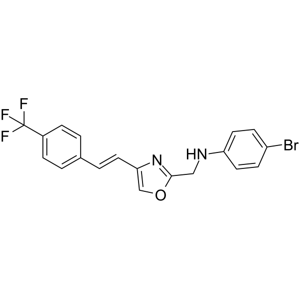
HER2-IN-9 is an orally active HER2 inhibitor, with an IC50 value of 0.03 μM. HER2-IN-9 inhibits HER-2 positive breast cancer cells proliferation and migration. HER2-IN-9 can be used in the research of breast cancers.
For research use only. We do not sell to patients.
- Formula: C19H14BrF3N2O
- Molecular Weight:423.23
-
Storage:
Please store the product under the recommended conditions in the Certificate of Analysis.

To place orders, for customer services and technical support, please contact: MedChemExpress USA
Tel: 609-228-6898 E-mail: [email protected] [email protected]


-
 Biological Activity
-
Chemical Information
- Protocols
- Purity & Documentation
- References
-
 Help & FAQs


All EGFR Isoforms
More

Biological Activity
Description
IC50 & Target
[1]|
HER2 0.03 μM (IC50) |
In Vitro
HER2-IN-9 (24 h) shows anti-proliferation activities against cancer cells (A549, HEPG2, MCF7, SKBR3)[1].
HER2-IN-9 (0-60 nM, 24 h) inhibits the migration of SKBR3 cells[1].
HER2-IN-9 (0-60 nM, 24 h) increases E-cadherin levels and decreases N-cadherin levels in SKBR3 cells[1].
HER2-IN-9 (0-60 nM, 24 h) suppresses the expression of p-HER-2, further inhibits the activation of the EMT signal pathway to inhibit the migration of SKBR3 cells[1].
MedChemExpress (MCE) has not independently confirmed the accuracy of these methods. They are for reference only.
-
Cell Line:Cancer cells (A549, HEPG2, MCF7, SKBR3), normal cells (Beas2b, LO2, MCF-0A)
-
Concentration:0.05-30 μM approximately
-
Incubation Time:24 h
-
Result:Inhibited cancer cell proliferation with IC50s of 0.05-12.17 μM.
Inhibited noamal cell proliferation with IC50s of 15.4- 26.95 μM.
-
Cell Line:SKBR3 cells
-
Concentration:0, 20, 40, 60 nM
-
Incubation Time:24 h
-
Result:Inhibited HER-2 phosphorylation with no significant change in total HER-2 protein levels.
Down-regulated β-catenin, snail, and Vimentin level.
In Vivo
MedChemExpress (MCE) has not independently confirmed the accuracy of these methods. They are for reference only.
-
Animal Model:SKBR3 orthotopic xenograft model[1]
-
Dosage:30 mg/kg
-
Administration:Oral administration, every two days.
-
Result:Inhibited the growth of cancer cells in vivo without noticeable toxic effects.
Increased the level of cleaved-caspase 3 implicated in cell death pathways (immunohistochemistry assay in tumor).


Chemical Information
-
Molecular Weight 423.23
-
Formula C19H14BrF3N2O
-
SMILES
FC(F)(C1=CC=C(C=C1)/C=C/C2=COC(CNC3=CC=C(C=C3)Br)=N2)F
-
Shipping
Room temperature in continental US; may vary elsewhere.
-
Storage
Please store the product under the recommended conditions in the Certificate of Analysis.


Purity & Documentation
References

Calculators
Concentration (start) × Volume (start) = Concentration (final) × Volume (final)


HER2-IN-9 Related Classifications
HY-143323 Related Classifications
 Powered by Bioz