



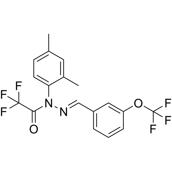
CAD031

Based on 1 Customer Validation
CAD031 is an orally active AMPK/ACC1 signaling pathway activator and a derivative of the Alzheimer's disease (AD) targeted agent J147 (HY-13779) (more active than J147 in human neural stem cell assays). CAD031 can cross the blood-brain barrier, activate AMPK and inhibit ACC1, thereby increasing ac-CoA levels, improving mitochondrial function and reducing free fatty acid synthesis. CAD031 has neuroprotective, neurogenesis-promoting and memory-improving activities and can be used in the study of Alzheimer's disease and aging-related neurodegenerative diseases. CAD031 effectively enhances the memory of mice, improves dendritic structure, and stimulates cell division in the germinal zone of the brain of elderly mice.
For research use only. We do not sell to patients.
- Purity : 99.39%
- CAS No.: 2071209-49-7
- Formula: C18H14F6N2O2
- Molecular Weight:404.31
-
Storage:Powder -20°C, 3 years , 4°C, 2 years ; In solvent -80°C, 6 months , -20°C, 1 month

To place orders, for customer services and technical support, please contact: MedChemExpress USA
Tel: 609-228-6898 E-mail: [email protected] [email protected]


-
 Biological Activity
-
Chemical Information
-
Solvent & Solubility
- Protocols
- Purity & Documentation
- References
-
 Help & FAQs


All AMPK Isoforms
More

Biological Activity
Description
In Vitro
CAD031 (100 nM; 7 d) significantly induces the mRNA expression of neurogenesis markers Nestin, Pax6, DCX, and Ki67 in human neural precursor cells (hNPCs) experiments; and 1 μM CAD031 inhibits Aβ-induced cell death in MC65 cells and glutamate-induced cytotoxicity in HT22 cells, exerting neuroprotective activity[1].
MedChemExpress (MCE) has not independently confirmed the accuracy of these methods. They are for reference only.
-
Cell Line:Human embryonic stem cell-derived neural progenitor cells (Hues6 and H9)
-
Concentration:100 nM
-
Incubation Time:7 days
-
Result:Increased transcript levels of neurogenesis-related genes (Nestin, Pax6, DCX, Ki67) compared to control, while showing no significant effect on early stem cell markers (Sox1, Sox2). The effect was normalized to fibroblast growth factor (FGF).
In Vivo
CAD031 (200 ppm; dietary administration; daily; 4 months) extends median lifespan, maintained mitochondrial gene expression and protein levels, reduces plasma free fatty acids, and activates the hepatic AMPK/ACC1 pathway in rapidly aging SAMP8 mice[2].
MedChemExpress (MCE) has not independently confirmed the accuracy of these methods. They are for reference only.
-
Animal Model:APPswe/PS1ΔE9 Transgenic AD Mouse Model** Female APPswe/PS1ΔE9 transgenic mice (3 months old, weight not specified) + wild-type (WT) C57BL/6 controls[2]
-
Dosage:200 ppm (premixed in diet)
-
Administration:Dietary administration, daily for 6 months
-
Result:Rescued context-dependent fear memory deficits in AD mice (increased freezing time) and restored the recognition index in a spatial pattern separation test.
Increased the number of BrdU+ cells in the dentate gyrus and BrdU+/DCX+ neural progenitors in the subventricular zone, while having no effect on Aβ plaque number or size.
- Mitochondrial membrane-potential fluorescent assay
- hPSC directed neural induction/NPC culture
- Mitochondrial membrane-potential and mitochondrial mass staining
- SH-SY5Y neuronal-like differentiation
- CFSE Dye Dilution Proliferation Assay
- Dye-dilution cell tracking and proliferation staining
- Cell differentiation
- Alzheimer’s Disease Modeling


Chemical Information
-
CAS No. 2071209-49-7
-
Appearance Solid
-
Molecular Weight 404.31
-
Formula C18H14F6N2O2
-
Color Light yellow to yellow
-
SMILES
O=C(N(C1=CC=C(C)C=C1C)/N=C/C2=CC=CC(OC(F)(F)F)=C2)C(F)(F)F
-
Shipping
Room temperature in continental US; may vary elsewhere.
-
Storage
Powder -20°C 3 years 4°C 2 years In solvent -80°C 6 months -20°C 1 month


Solvent & Solubility
In Vitro:
DMSO : 100 mg/mL (247.33 mM; Need ultrasonic and warming; Hygroscopic DMSO has a significant impact on the solubility of product, please use newly opened DMSO)
Please refer to the solubility information to select the appropriate solvent. Once prepared, please aliquot and store the solution to prevent product inactivation from repeated freeze-thaw cycles.
Storage method and period of stock solution: -80°C, 6 months; -20°C, 1 month. When stored at -80°C, please use it within 6 months. When stored at -20°C, please use it within 1 month.
Please refer to the solubility information to select the appropriate solvent. Once prepared, please aliquot and store the solution to prevent product inactivation from repeated freeze-thaw cycles.
Storage method and period of stock solution: -80°C, 6 months; -20°C, 1 month. When stored at -80°C, please use it within 6 months. When stored at -20°C, please use it within 1 month.
Concentration (start) × Volume (start) = Concentration (final) × Volume (final)
In Vivo:
Select the appropriate dissolution method based on your experimental animal and administration route.
- For the following dissolution methods, please ensure to first prepare a clear stock solution using an In Vitro approach and then sequentially add co-solvents:
- To ensure reliable experimental results, the clarified stock solution can be appropriately stored based on storage conditions. As for the working solution for In Vivo experiments, it is recommended to prepare freshly and use it on the same day.
- The percentages shown for the solvents indicate their volumetric ratio in the final prepared solution. If precipitation or phase separation occurs during preparation, heat and/or sonication can be used to aid dissolution.
Add each solvent one by one: 10% DMSO 40% PEG300 5% Tween-80 45% Saline
Solubility: 2.5 mg/mL (6.18 mM); Clear solution; Need ultrasonic
This protocol yields a clear solution of 2.5 mg/mL.
Taking 1 mL working solution as an example, add 100 μL DMSO stock solution (25.0 mg/mL) to 400 μL PEG300, and mix evenly; then add 50 μL Tween-80 and mix evenly; then add 450 μL Saline to adjust the volume to 1 mL.
Preparation of Saline: Dissolve 0.9 g sodium chloride in ddH₂O and dilute to 100 mL to obtain a clear Saline solution.
In Vivo Dissolution Calculator
Please enter the basic information of animal experiments:
-
-
-
-
Recommended: Prepare an additional quantity of animals to account for potential losses during experiments.
Please enter your animal formula composition:
-
%DMSO +
Recommended: Keep the proportion of DMSO in working solution below 2% if your animal is weak.
-
%+
-
+%Tween-80 + +
-
%Saline +
The co-solvents required include: DMSO, . All of co-solvents are available by MedChemExpress (MCE). , Tween 80. All of co-solvents are available by MedChemExpress (MCE).
Working solution concentration: 0.22 mg/mL
Method for preparing stock solution: mg drug dissolved in μL DMSO. Stock solution concentration: mg/mL.
1. Take μL DMSO stock solution;
2. Add μL .
μL , mix evenly;
3. Then add μL Tween 80, mix evenly;
4. Then add μL
Please ensure that the stock solution in the first step is dissolved to a clear state, and add co-solvents in sequence. You can use ultrasonic heating (ultrasonic cleaner, recommended frequency 20-40 kHz), vortexing, etc. to assist dissolution.


Purity & Documentation
-
Data Sheet (275 KB)
-
SDS (251 KB)
- English - EN (251 KB)
- Français - FR (251 KB)
- Deutsch - DE (251 KB)
- Norwegian - NO (251 KB)
- Español - ES (251 KB)
- Swedish - SV (251 KB)
- Italian - IT (251 KB)
- Korean - KR (251 KB)
- Portuguese - PT (251 KB)
-
Handling Instructions (2659 KB)
References
[1]. Prior M, et al. Selecting for neurogenic potential as an alternative for Alzheimer's disease drug discovery. Alzheimers Dement. 2016 Jun;12(6):678-86. [Content Brief]
[2]. Currais A, et al. A chemical biology approach to identifying molecular pathways associated with aging. Geroscience. 2021 Feb;43(1):353-365. [Content Brief]
[3]. Kepchia D, et al. The Alzheimer's disease drug candidate J147 decreases blood plasma fatty acid levels via modulation of AMPK/ACC1 signaling in the liver. Biomed Pharmacother. 2022 Mar;147:112648. [Content Brief]


Complete Stock Solution Preparation Table
Please refer to the solubility information to select the appropriate solvent. Once prepared, please aliquot and store the solution to prevent product inactivation from repeated freeze-thaw cycles.
Storage method and period of stock solution: -80°C, 6 months; -20°C, 1 month. When stored at -80°C, please use it within 6 months. When stored at -20°C, please use it within 1 month.
| Optional Solvent | Concentration Solvent Mass | 1 mg | 5 mg | 10 mg | 25 mg |
|---|---|---|---|---|---|
| DMSO | 1 mM | 2.4733 mL | 12.3667 mL | 24.7335 mL | 61.8337 mL |
| 5 mM | 0.4947 mL | 2.4733 mL | 4.9467 mL | 12.3667 mL | |
| 10 mM | 0.2473 mL | 1.2367 mL | 2.4733 mL | 6.1834 mL | |
| 15 mM | 0.1649 mL | 0.8244 mL | 1.6489 mL | 4.1222 mL | |
| 20 mM | 0.1237 mL | 0.6183 mL | 1.2367 mL | 3.0917 mL | |
| 25 mM | 0.0989 mL | 0.4947 mL | 0.9893 mL | 2.4733 mL | |
| 30 mM | 0.0824 mL | 0.4122 mL | 0.8244 mL | 2.0611 mL | |
| 40 mM | 0.0618 mL | 0.3092 mL | 0.6183 mL | 1.5458 mL | |
| 50 mM | 0.0495 mL | 0.2473 mL | 0.4947 mL | 1.2367 mL | |
| 60 mM | 0.0412 mL | 0.2061 mL | 0.4122 mL | 1.0306 mL | |
| 80 mM | 0.0309 mL | 0.1546 mL | 0.3092 mL | 0.7729 mL | |
| 100 mM | 0.0247 mL | 0.1237 mL | 0.2473 mL | 0.6183 mL |


CAD031 Related Classifications
HY-124609 Related Classifications
 Powered by Bioz