



Resistomycin

Based on 1 Customer Validation
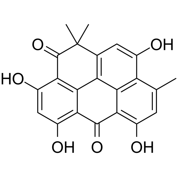
Resistomycin, a pentacyclic polyketide antibiotic, possesses anticancer activity and induces apoptosis.
For research use only. We do not sell to patients.
- Purity : 99.9%
- CAS No.: 20004-62-0
- Formula: C22H16O6
- Molecular Weight:376.36
-
Storage:Powder -20°C, 3 years ; In solvent -80°C, 6 months , -20°C, 1 month

To place orders, for customer services and technical support, please contact: MedChemExpress USA
Tel: 609-228-6898 E-mail: [email protected] [email protected]


-
 Biological Activity
-
Chemical Information
- Protocols
- Purity & Documentation
-
 Help & FAQs


All Antibiotic Isoforms
More

Biological Activity
Description
Cellular Effect
|
Cell Line

|
Type | Value | Description | References |
|---|---|---|---|---|
| CCRF-CEM | IC50 |
0.5 μM
Compound: 1
|
Antiproliferative activity against human CEM cells after 72 hrs by MTT assay
Antiproliferative activity against human CEM cells after 72 hrs by MTT assay
|
[PMID: 29137865] |
| CCRF-CEM | IC50 |
0.5 μM
Compound: 5
|
Antiproliferative activity against human CEM cells assessed as growth inhibition after 72 hrs by MTT assay
Antiproliferative activity against human CEM cells assessed as growth inhibition after 72 hrs by MTT assay
|
[PMID: 33974416] |
| HCT-116 | IC50 |
0.1 μM
Compound: 1
|
Antiproliferative activity against p53 deficient human HCT116 cells after 72 hrs by MTT assay
Antiproliferative activity against p53 deficient human HCT116 cells after 72 hrs by MTT assay
|
[PMID: 29137865] |
| HCT-116 | IC50 |
0.3 μM
Compound: 1
|
Antiproliferative activity against human HCT116 cells after 72 hrs by MTT assay
Antiproliferative activity against human HCT116 cells after 72 hrs by MTT assay
|
[PMID: 29137865] |
| HeLa | IC50 |
0.1 μM
Compound: 1
|
Antiproliferative activity against human HeLa cells after 72 hrs by MTT assay
Antiproliferative activity against human HeLa cells after 72 hrs by MTT assay
|
[PMID: 29137865] |
| HeLa | IC50 |
0.1 μM
Compound: 5
|
Antiproliferative activity against human HeLa cells assessed as growth inhibition after 72 hrs by MTT assay
Antiproliferative activity against human HeLa cells assessed as growth inhibition after 72 hrs by MTT assay
|
[PMID: 33974416] |
| HMEC-1 | IC50 |
0.5 μM
Compound: 1
|
Antiproliferative activity against HMEC1 cells after 72 hrs by MTT assay
Antiproliferative activity against HMEC1 cells after 72 hrs by MTT assay
|
[PMID: 29137865] |
| HMEC-1 | IC50 |
0.5 μM
Compound: 5
|
Cytotoxicity against human HMEC-1 cells assessed as growth inhibition after 72 hrs by MTT assay
Cytotoxicity against human HMEC-1 cells assessed as growth inhibition after 72 hrs by MTT assay
|
[PMID: 33974416] |
| K562 | IC50 |
0.3 μM
Compound: 1
|
Antiproliferative activity against human K562 cells after 72 hrs by MTT assay
Antiproliferative activity against human K562 cells after 72 hrs by MTT assay
|
[PMID: 29137865] |
| L1210 | IC50 |
0.7 μM
Compound: 1
|
Antiproliferative activity against mouse L1210 cells after 72 hrs by MTT assay
Antiproliferative activity against mouse L1210 cells after 72 hrs by MTT assay
|
[PMID: 29137865] |
| L1210 | IC50 |
0.7 μM
Compound: 5
|
Antiproliferative activity against mouse L1210 cells assessed as growth inhibition after 72 hrs by MTT assay
Antiproliferative activity against mouse L1210 cells assessed as growth inhibition after 72 hrs by MTT assay
|
[PMID: 33974416] |
| T-24 | IC50 |
26 μM
Compound: 1
|
Growth inhibition of human T24 cells after 72 hrs by cell impedance method
Growth inhibition of human T24 cells after 72 hrs by cell impedance method
|
[PMID: 29137865] |
In Vitro
Resistomycin attenuates triple-negative breast cancer progression by inhibiting E3 ligase Pellino-1 and inducing SNAIL/SLUG degradation[1].
Resistomycin displays a high affinity for GST-Pellino-1 (KD = 2.58 μM) as indicated by surface plasmon resonance[1].
Resistomycin treatment reduced the expression of SNAIL and SLUG in TNBC cells[1].
Resistomycin induced apoptosis and cycle arrest in human hepatocellular carcinoma cells by activating p38 MAPK pathway in vitro and in vivo[3].
MedChemExpress (MCE) has not independently confirmed the accuracy of these methods. They are for reference only.


Chemical Information
-
CAS No. 20004-62-0
-
Appearance Solid
-
Molecular Weight 376.36
-
Formula C22H16O6
-
Color Yellow to orange
-
SMILES
OC1=C(C(C2=C(O)C=C(C)C(C(O)=C3)=C2C4=C3C(C)(C)C5=O)=O)C4=C5C(O)=C1
-
Structure Classification
-
Initial Source
Streptomyces sulphureus
-
Shipping
Room temperature in continental US; may vary elsewhere.
-
Storage
Powder -20°C 3 years In solvent -80°C 6 months -20°C 1 month


Purity & Documentation
-
Data Sheet (248 KB)
-
SDS (557 KB)
- English - EN (557 KB)
- Français - FR (557 KB)
- Deutsch - DE (557 KB)
- Norwegian - NO (557 KB)
- Español - ES (557 KB)
- Swedish - SV (557 KB)
- Italian - IT (557 KB)
- Korean - KR (557 KB)
- Portuguese - PT (557 KB)
-
Handling Instructions (2659 KB)

Calculators
Concentration (start) × Volume (start) = Concentration (final) × Volume (final)


Resistomycin Related Classifications
HY-N10449 Related Classifications
 Powered by Bioz