



Veligrotug

Based on 1 Customer Validation
Veligrotug (AVE1642) is a selective, fully human IGF-1R antagonist antibody with a Kd value of 0.55 nM for hIGF-1R. Veligrotug blocks the phosphorylation of downstream AKT. Veligrotug is applicable to research related to thyroid eye disease.
For research use only. We do not sell to patients.
- Purity : 99.25%
- CAS No.: 2728655-31-8
- Molecular Weight:147.7 kDa
-
Storage:
Please store the product under the recommended conditions in the Certificate of Analysis.


To place orders, for customer services and technical support, please contact: MedChemExpress USA
Tel: 609-228-6898 E-mail: [email protected] [email protected]


-
 Biological Activity
-
Chemical Information
- Protocols
- Purity & Documentation
- References
-
 Help & FAQs


-

HY-P99294
GanitumabAnti-IGF1R Antibody -

HY-P99197
FigitumumabAnti-IGF1R Antibody -

HY-P99165
TeprotumumabIGF-1R Inhibitor -

HY-P99218
RobatumumabAnti-human IGF-1R Antibody -

HY-P2093
S961Insulin Receptor Antagonist -
HY-135680
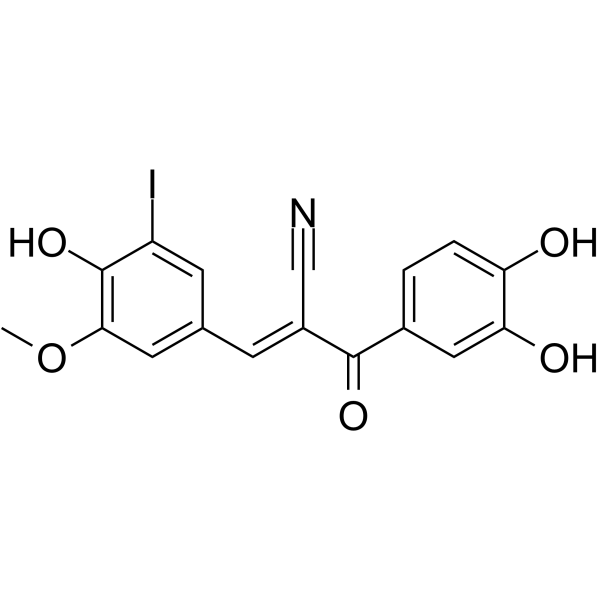
I-OMe-Tyrphostin AG 538IGF-1R/PI5P4Kα Inhibitor -
HY-18785
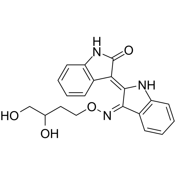
Indirubin Derivative E804IGF-1R Inhibitor -

HY-P99867
DusigitumabIGF Antibody


Biological Activity
Description
Isotype
Human IgG1 kappa
Recommend Isotype Controls
Species Reactivity
Human
IC50 & Target
IGF1R/CD221
In Vitro
Veligrotug binds to recombinant hIGF-1R ECD protein with high affinity, with a mean Kd value of 0.55 nM[2].
Veligrotug (30 min) binds with high affinity to HOCF cells endogenously expressing IGF-1R, with a mean EC50 of 2.41 nM and a mean Kd of 1.85 nM[2].
Veligrotug (0.46-1000 nM; 1 h) does not inhibit insulin-mediated insulin receptor phosphorylation in HepG2 cells even at concentrations as high as 1000 nM[2].
Veligrotug (0.008-500 nM; 30 min preincubation on ice) almost completely inhibits the binding of IGF-1 to IGF-1R on FreeStyle 293-F cells at concentrations ≥50 nM[2].
Veligrotug (0.0001-333 nM; 1 h) completely inhibits IGF-1-mediated AKT phosphorylation in primary HOCF cells at concentrations of 10-30 nM[2].
MedChemExpress (MCE) has not independently confirmed the accuracy of these methods. They are for reference only.
-
Cell Line:HepG2 cells
-
Concentration:0.46-1000 nM
-
Incubation Time:1 h preincubation at 37°C and 5% CO2
-
Result:Showed no significant antagonism of insulin-mediated IR phosphorylation in HepG2 cells at concentrations up to 1000 nM.
Parmacokinetics
In Vivo
MedChemExpress (MCE) has not independently confirmed the accuracy of these methods. They are for reference only.
-
Animal Model:Cynomolgus monkeys (male and female, 2.0 to 2.9 years old, 1.6 to 2.2 kg)[2]
-
Dosage:2 mg/kg; 10 mg/kg; 50 mg/kg
-
Administration:i.v.; weekly 30-minute infusion; 13 weeks
-
Result:Induced an approximately 2.5-fold increase in IGF-1 concentrations for all dose groups following the first dose (Days 0-7).
Sustained elevated IGF-1 serum concentrations throughout the 13-week dosing period for the 10 and 50 mg/kg dose groups.
Maintained elevated IGF-1 concentrations during the recovery period for the 50 mg/kg dose group.
Saw IGF-1 concentrations return to baseline in 5 of 6 animals in the 2 mg/kg group, correlating with anti-drug antibody development.
Clinical Trial
| NCT Number | Sponsor | Condition | Start Date |
Phase

|
|---|---|---|---|---|
| NCT01329991 | Plexxikon| | 2011-05 | PHASE1 |
Gene ID
Accession
Conjugated
Unconjugated
Reconsititution
The product can be reconstituted/diluted with sterile PBS or saline.
Format
-
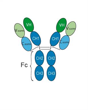
Human IgG1 kappa
Application
ELISA, FACS, Functional assay
Verified Bioactivity
-
 Flow cytometric analysis of 1X10⁶ MCF-7 cells labeling Veligrotug (HY-P990084, red). Cells were fixed with 4% paraformaldehyde. Then stained with the primary antibody at 1/200 dilution for an hour at 4℃. Alexa Fluor 488-conjugated AffiniPure Goat Anti-Human IgG H&L (AF488) (HY-P83776) was used as the secondary antibody at 1/1,000 dilution for 30 minutes at 4℃. Human IgG1 kappa (HY-P99001, blue) was used as the isotype control, cells without incubation with primary antibody were used as the unlabeled control (black).
 -
 Immobilized IGF1R/IGF-I R Protein, Human (HEK293, HY-P72606) can bind Veligrotug. The ED50 for this effect is 3.569 ng/mL.


Chemical Information
-
CAS No. 2728655-31-8
-
Appearance Liquid
-
Molecular Weight 147.7 kDa
-
Color Colorless to light yellow
-
SMILES
[Veligrotug]
-
Synonyms
AVE1642; VRDN-001
-
Shipping
Shipping with dry ice.
-
Formulation
Please refer to the lot-specific COA for specific buffer information.
-
Storage
Please store the product under the recommended conditions in the Certificate of Analysis.


Purity & Documentation
-
Data Sheet (281 KB)
-
SDS (252 KB)
- English - EN (252 KB)
- Français - FR (252 KB)
- Deutsch - DE (252 KB)
- Norwegian - NO (252 KB)
- Español - ES (252 KB)
- Swedish - SV (252 KB)
- Italian - IT (252 KB)
- Korean - KR (252 KB)
- Portuguese - PT (252 KB)
-
Inhibitory Antibodies User Guide (603 KB)
References

Calculators
Concentration (start) × Volume (start) = Concentration (final) × Volume (final)


Veligrotug Related Classifications
HY-P990084 Related Classifications
 Powered by Bioz