



JLK-6
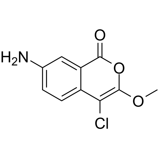
JLK-6 is a rhomboid protease GlpG inhibitor. JLK-6 reduces the production of Amyloid β-peptide by altering the cleavage of β-amyloid precursor protein by γ-secretase, with no effect on the cleavage of Notch receptors. JLK-6 can be used in research related to Alzheimer's disease.
For research use only. We do not sell to patients.
- CAS No.: 62252-26-0
- Formula: C10H8ClNO3
- Molecular Weight:225.63
-
Storage:
Please store the product under the recommended conditions in the Certificate of Analysis.

To place orders, for customer services and technical support, please contact: MedChemExpress USA
Tel: 609-228-6898 E-mail: [email protected] [email protected]


-
 Biological Activity
-
Chemical Information
- Protocols
- Purity & Documentation
- References
-
 Help & FAQs


-
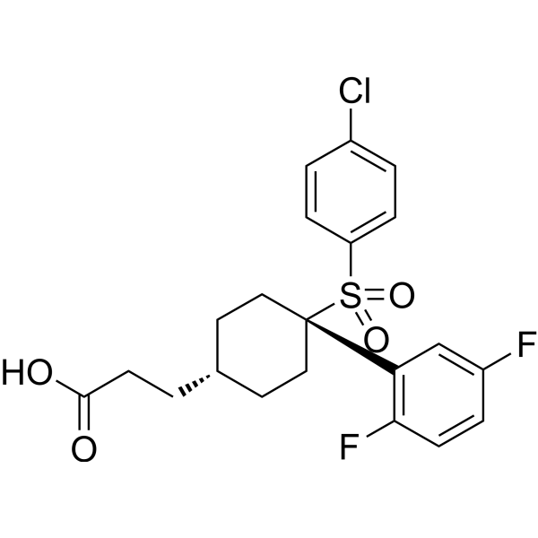
HY-10974
MK-0752 -
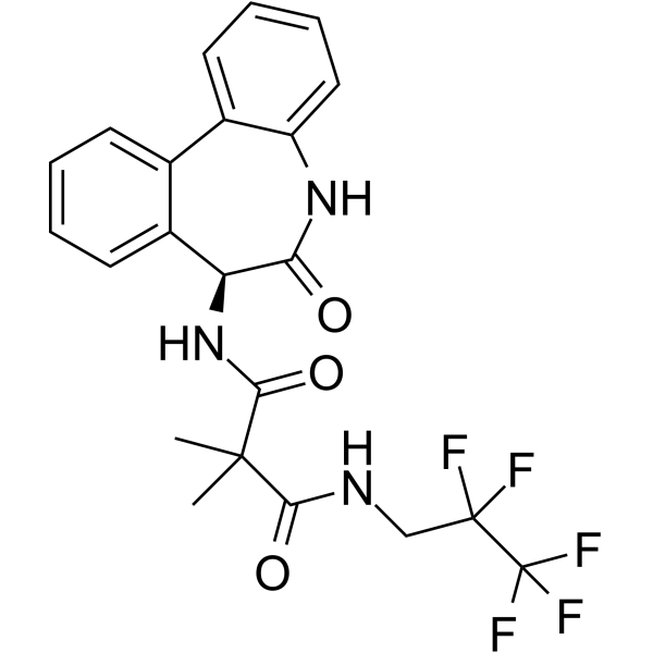
HY-11102
RO4929097γ-secretase Inhibitor -
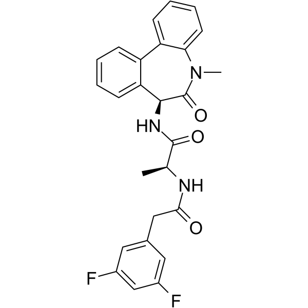
HY-13526
YO-01027γ-secretase Inhibitor -
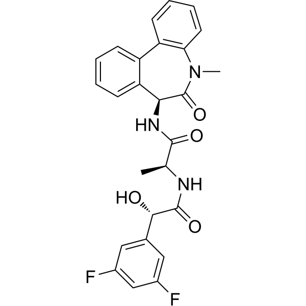
HY-50752
LY-411575γ-secretase Inhibitor -
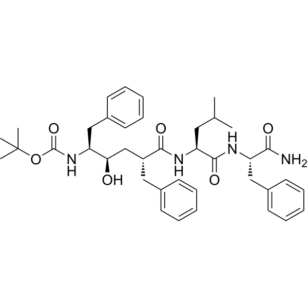
HY-19369
L-685458γ-Secretase Inhibitor -
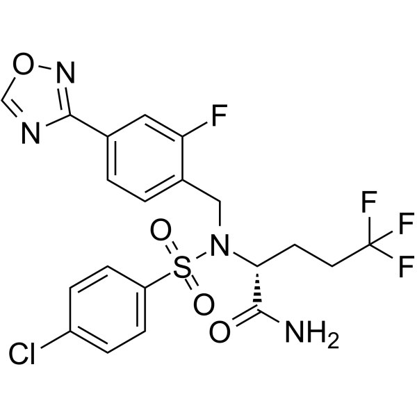
HY-50845
Avagacestatγ-secretase Inhibitor -
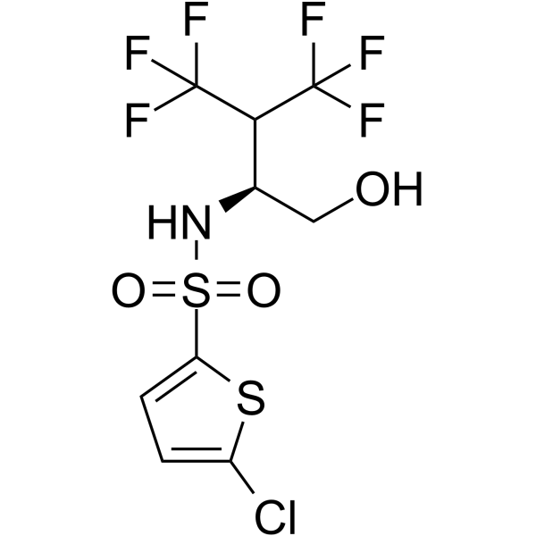
HY-14175
Begacestatγ-secretase Inhibitor -
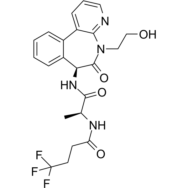
HY-12449
CrenigacestatNotch/γ-secretase Inhibitor -
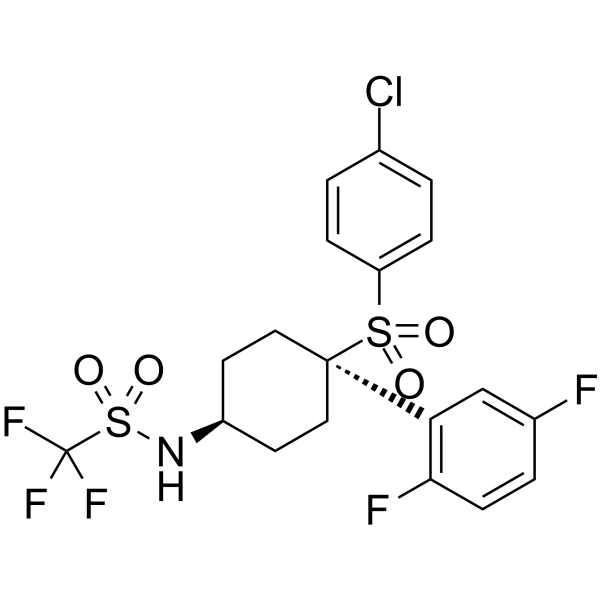
HY-14174
MRK-560γ-Secretase Inhibitor -
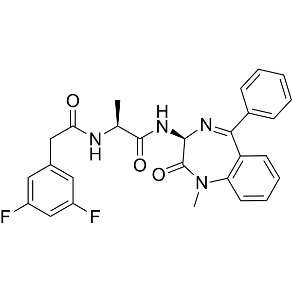
HY-14176
Compound Eγ-secretase Inhibitor -
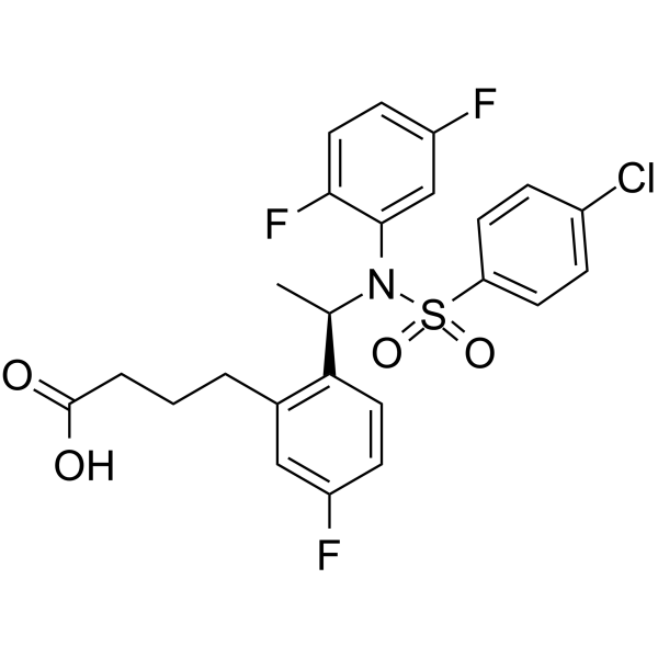
HY-50883
BMS 299897γ-secretase Inhibitor -
HY-10009
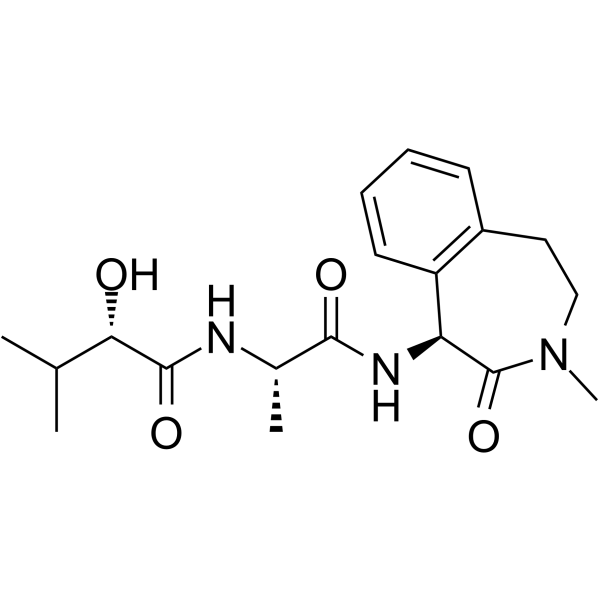
Semagacestatγ-secretase Inhibitor


Biological Activity
Description
Cellular Effect
|
Cell Line

|
Type | Value | Description | References |
|---|---|---|---|---|
| HEK293 | IC50 |
30 μM
Compound: 24; JLK6
|
Inhibition of gamma secretase mediated amyloid beta level reduction in HEK293 cells expressing APP after 72 hrs by chemiluminescence assay
Inhibition of gamma secretase mediated amyloid beta level reduction in HEK293 cells expressing APP after 72 hrs by chemiluminescence assay
|
[PMID: 27045975] |
In Vitro
JLK-6 (3-60 μM; 7 h) potently reduces Aβ production in bAPPswe-overexpressing HEK293 cells with an IC50 of 30-60 μM, while sparing Notch signaling and other protease pathways without inducing cell toxicity at 100 μM[1].
JLK-6 (10 μM; 1 h) irreversibly inhibits E. coli GlpG rhomboid protease, as no enzyme activity recovery is observed after dilution of the inhibitor-enzyme complex[2].
JLK-6 forms a double-bonded end product with E. coli GlpG rhomboid protease, resulting in a detectable mobility shift on SDS-PAGE[2].
MedChemExpress (MCE) has not independently confirmed the accuracy of these methods. They are for reference only. Further protocols information, click here.
-
Cell Line:HEK293 cells stably overexpressing Swedish mutant β-amyloid precursor protein (bAPPswe)
-
Concentration:3 μM, 10 μM, 30 μM, 60 μM
-
Incubation Time:7 h
-
Result:Reduced Aβ production in a concentration-dependent manner: 3 μM and 10 μM caused partial reduction, 30 μM caused further reduction, and 60 μM resulted in near-complete inhibition of Aβ production.
Exhibited an IC50 of approximately 30-60 μM in this assay.
Did not interfere with the processing of Notch receptor, cadherins, CD44, α-secretase, β-secretase, or GSK3β kinase.
Showed no cell toxicity at 100 μM.


Chemical Information
-
CAS No. 62252-26-0
-
Molecular Weight 225.63
-
Formula C10H8ClNO3
-
SMILES
O=C1C2=CC(N)=CC=C2C(Cl)=C(OC)O1
-
Shipping
Room temperature in continental US; may vary elsewhere.
-
Storage
Please store the product under the recommended conditions in the Certificate of Analysis.

Protocols
-
Notch Pathway Solutions
The Notch pathway is a contact-dependent signaling pathway that controls cell-fate decisions, differentiation, proliferation, and tissue patterning through interactions between membrane-bound Notch receptors and membrane-bound ligands on neighboring cells. Canonical Notch signaling is activated when ligand engagement triggers proteolytic release of the Notch intracellular domain, which enters the nucleus and regulates transcription together with DNA-binding transcriptional complexes. In the canonical mechanism, ligand-dependent Notch activation leads to release of the intracellular Notch domain, and presenilin-dependent γ-secretase activity is required for production of the active intracellular signaling fragment. The released intracellular domain functions as a nuclear signal that converts Notch receptor activation at the membrane into transcriptional regulation of target programs such as HES/HEY-family genes and other context-dependent downstream targets. The literature links Notch p
-
Amyloid: Congo Red Amyloid Staining
Congo red amyloid staining is a histochemical method used to detect extracellular amyloid deposits in tissue sections based on the affinity of Congo red dye for β-pleated sheet-rich protein aggregates. When bound to amyloid, Congo red produces characteristic apple-green birefringence under polarized light microscopy, which is widely regarded as a diagnostic feature of amyloid deposition in histopathology. The diagnostic principle relies on the combination of dye binding (congophilia) and optical anisotropy under polarized illumination, which distinguishes amyloid from most non-amyloid eosinophilic extracellular deposits in routine histological evaluation. Amyloid identification by Congo red staining remains a cornerstone in diagnostic pathology despite the availability of adjunct methods such as immunohistochemistry and mass spectrometry, particularly because of its ability to localize deposits directly within tissue architecture. The specificity of Congo red-positive deposits is incre
-
Alzheimer’s Disease Modeling
Alzheimer’s Disease (AD) is a neurodegenerative disorder characterized by a progressive decline in cognitive functions and loss of specific types of neurons and synapses. Alzheimer's symptoms can be simulated in mice by injecting drugs (such as Aβ) or genetically modified.


Purity & Documentation
References
[1]. Peuchmaur M, et al. Further characterization of a putative serine protease contributing to the γ-secretase cleavage of β-amyloid precursor protein. Bioorganic & medicinal chemistry. 2013 Feb 15;21(4):1018-29. [Content Brief]
[2]. Goel P, et al. Discovery and Biological Evaluation of Potent and Selective N-Methylene Saccharin-Derived Inhibitors for Rhomboid Intramembrane Proteases. Biochemistry. 2017 Dec 26;56(51):6713-6725. [Content Brief]

Calculators
Concentration (start) × Volume (start) = Concentration (final) × Volume (final)


JLK-6 Related Classifications
HY-103538 Related Classifications
 Powered by Bioz