



Pomstafib-2

Based on 1 Customer Validation
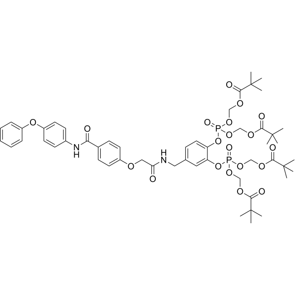
Pomstafib-2 is a potent and selective STAT5b inhibitor. Pomstafib-2 decreases the expression of pSTAT5b and induces Apoptosis.
For research use only. We do not sell to patients.
- Purity : 99.37%
- CAS No.: 2332841-83-3
- Formula: C52H66N2O20P2
- Molecular Weight:1101.03
-
Storage:
4°C, sealed storage, away from moisture
* In solvent : -80°C, 6 months; -20°C, 1 month (sealed storage, away from moisture)

To place orders, for customer services and technical support, please contact: MedChemExpress USA
Tel: 609-228-6898 E-mail: [email protected] [email protected]


-
 Biological Activity
-
Chemical Information
-
Solvent & Solubility
- Protocols
- Purity & Documentation
- References
-
 Help & FAQs




Biological Activity
Description
In Vitro
Pomstafib-2 (compound 8) (0-10 µM; 4 h) decreases the expression of pSTAT5b in a dose-dependent manner in STAT5b-GFP-transfected K562 cells[1].
Pomstafib-2 (0, 10, 20, 40, 60, 80 µM; 48 h) induces apoptosis in a dose-dependent manner in STAT5-dependent K562 cells[1].
MedChemExpress (MCE) has not independently confirmed the accuracy of these methods. They are for reference only.
-
Cell Line:K562 leukaemia cells
-
Concentration:0, 0.3, 1, 3, 10 µM
-
Incubation Time:4 h
-
Result:Decreased the expression of pSTAT5b in a dose-dependent manner.
-
Cell Line:K562, MDA-MB-231 cells
-
Concentration:0, 10, 20, 40, 60, 80 µM
-
Incubation Time:48 h
-
Result:Induced apoptosis in a dose-dependent manner in K562 cells, does not increased the apoptotic rate of STAT5-independent MDA-MB-231 cells.


Chemical Information
-
CAS No. 2332841-83-3
-
Appearance Solid
-
Molecular Weight 1101.03
-
Formula C52H66N2O20P2
-
Color White to light yellow
-
SMILES
O=C(NCC1=CC=C(OP(OCOC(C(C)(C)C)=O)(OCOC(C(C)(C)C)=O)=O)C(OP(OCOC(C(C)(C)C)=O)(OCOC(C(C)(C)C)=O)=O)=C1)COC2=CC=C(C(NC3=CC=C(OC4=CC=CC=C4)C=C3)=O)C=C2
-
Shipping
Room temperature in continental US; may vary elsewhere.
-
Storage
4°C, sealed storage, away from moisture
* In solvent : -80°C, 6 months; -20°C, 1 month (sealed storage, away from moisture)


Solvent & Solubility
In Vitro:
DMSO : 175 mg/mL (158.94 mM; Need ultrasonic; Hygroscopic DMSO has a significant impact on the solubility of product, please use newly opened DMSO)
Please refer to the solubility information to select the appropriate solvent. Once prepared, please aliquot and store the solution to prevent product inactivation from repeated freeze-thaw cycles.
Storage method and period of stock solution: -80°C, 6 months; -20°C, 1 month (sealed storage, away from moisture). When stored at -80°C, please use it within 6 months. When stored at -20°C, please use it within 1 month.
Please refer to the solubility information to select the appropriate solvent. Once prepared, please aliquot and store the solution to prevent product inactivation from repeated freeze-thaw cycles.
Storage method and period of stock solution: -80°C, 6 months; -20°C, 1 month (sealed storage, away from moisture). When stored at -80°C, please use it within 6 months. When stored at -20°C, please use it within 1 month.
Concentration (start) × Volume (start) = Concentration (final) × Volume (final)


Purity & Documentation
-
Data Sheet (272 KB)
-
SDS (251 KB)
- English - EN (251 KB)
- Français - FR (251 KB)
- Deutsch - DE (251 KB)
- Norwegian - NO (251 KB)
- Español - ES (251 KB)
- Swedish - SV (251 KB)
- Italian - IT (251 KB)
- Korean - KR (251 KB)
- Portuguese - PT (251 KB)
-
Handling Instructions (2659 KB)
References
[1]. Elumalai N, et al. Rational development of Stafib-2: a selective, nanomolar inhibitor of the transcription factor STAT5b. Sci Rep. 2017 Apr 11;7(1):819. [Content Brief]
[2]. Gräb J, et al. The STAT5b Linker Domain Mediates the Selectivity of Catechol Bisphosphates for STAT5b over STAT5a. ACS Chem Biol. 2019 Apr 19;14(4):796-805. [Content Brief]


Complete Stock Solution Preparation Table
Please refer to the solubility information to select the appropriate solvent. Once prepared, please aliquot and store the solution to prevent product inactivation from repeated freeze-thaw cycles.
Storage method and period of stock solution: -80°C, 6 months; -20°C, 1 month (sealed storage, away from moisture). When stored at -80°C, please use it within 6 months. When stored at -20°C, please use it within 1 month.
| Optional Solvent | Concentration Solvent Mass | 1 mg | 5 mg | 10 mg | 25 mg |
|---|---|---|---|---|---|
| DMSO | 1 mM | 0.9082 mL | 4.5412 mL | 9.0824 mL | 22.7060 mL |
| 5 mM | 0.1816 mL | 0.9082 mL | 1.8165 mL | 4.5412 mL | |
| 10 mM | 0.0908 mL | 0.4541 mL | 0.9082 mL | 2.2706 mL | |
| 15 mM | 0.0605 mL | 0.3027 mL | 0.6055 mL | 1.5137 mL | |
| 20 mM | 0.0454 mL | 0.2271 mL | 0.4541 mL | 1.1353 mL | |
| 25 mM | 0.0363 mL | 0.1816 mL | 0.3633 mL | 0.9082 mL | |
| 30 mM | 0.0303 mL | 0.1514 mL | 0.3027 mL | 0.7569 mL | |
| 40 mM | 0.0227 mL | 0.1135 mL | 0.2271 mL | 0.5677 mL | |
| 50 mM | 0.0182 mL | 0.0908 mL | 0.1816 mL | 0.4541 mL | |
| 60 mM | 0.0151 mL | 0.0757 mL | 0.1514 mL | 0.3784 mL | |
| 80 mM | 0.0114 mL | 0.0568 mL | 0.1135 mL | 0.2838 mL | |
| 100 mM | 0.0091 mL | 0.0454 mL | 0.0908 mL | 0.2271 mL |


Pomstafib-2 Related Classifications
HY-112649 Related Classifications
 Powered by Bioz