



TM5441
Based on 10 publication(s) in Google Scholar
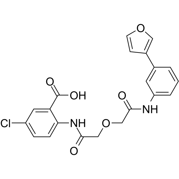
TM5441 is an orally bioavailable inhibitor of plasminogen activator inhibitor-1 (PAI-1), has IC50 values between 13.9 and 51.1 μM and induces intrinsic apoptosis in several human cancer cell lines. TM5441 attenuates Nω-nitro-l-arginine methyl ester-induced cardiac hypertension and vascular senescence.
商品は「研究用試薬」です。人や動物の医療用・臨床診断用・食品用の製品ではありません。
研究用途以外に使用した場合、当社は一切の責任を負いかねます。
- 純度 : 99.97%
- CAS 番号: 1190221-43-2
- 分子式: C21H17ClN2O6
- 分子量:428.82
-
保管条件:Powder -20°C, 3 years , 4°C, 2 years ; In solvent -80°C, 2 years , -20°C, 1 year

To place orders, for customer services and technical support, please contact: MedChemExpress USA
Tel: 609-228-6898 E-mail: [email protected] [email protected]


-
 生物活性
生物活性
-
化学情報
-
溶剤 & 溶解度
- プロトコル
- 純度とドキュメンテーション
- 参考文献
-
 Help & FAQs


-
HY-15253
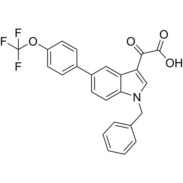
TiplaxtininApoptosis Inducer -
HY-100447
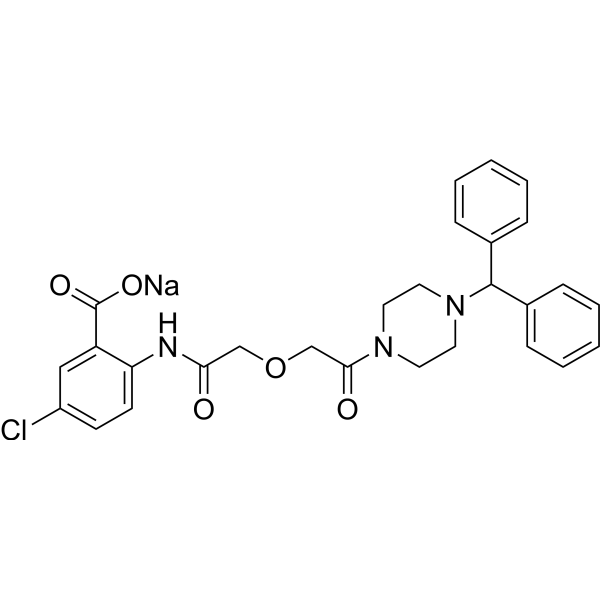
TM5275 sodiumPAI-1 Inhibitor -
HY-116868
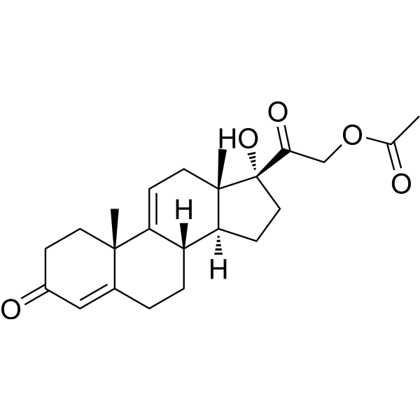
Anecortave acetateAngiostatic Agent -

HY-101761R
TM5441 (Standard)Reference Standard -

HY-P71077
Serpin E1 Protein, Human (HEK293, His) -
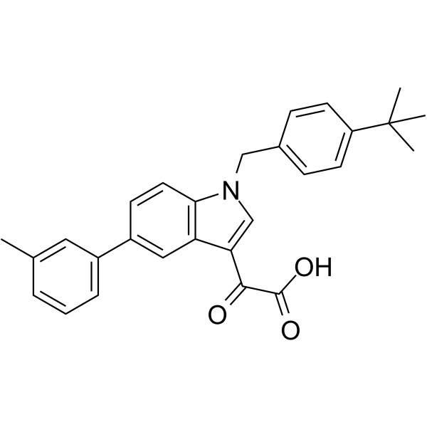
HY-14759
AleplasininPAI-1 Inhibitor -
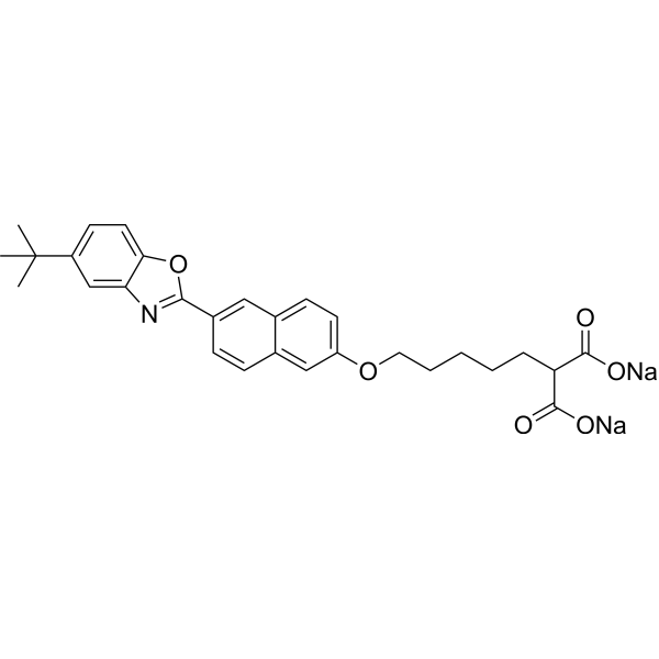
HY-122714
SK-216PAI-1 Inhibitor -
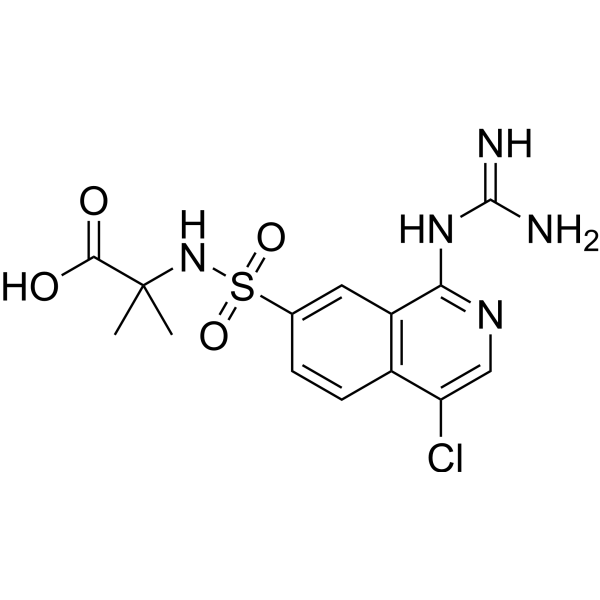
HY-101214
UK-371804uPA Inhibitor -
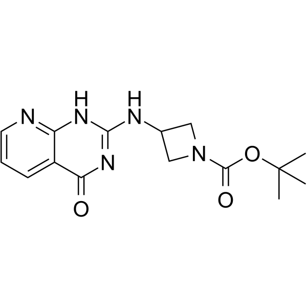
HY-117724
AZ3976PAI-1 Inhibitor -
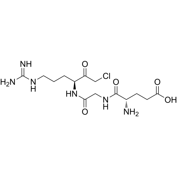
HY-137495
GGACKuPA Inhibitor -
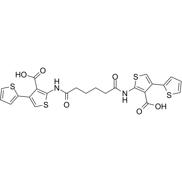
HY-119160
TM5007PAI-1 Inhibitor
MedChemExpress(MCE)の使用を引用している文献 TM5441
More- Kidney Int. 2023 Sep;104(3):508-525. [Abstract]
- Sci Adv. 2025 Jun 6;11(23):eadu2695. [Abstract]
- Eur J Pharmacol. 2024 Oct 5:980:176843. [Abstract]
- Eur J Pharm Sci. 2020 Feb 15:143:105195. [Abstract]
- Sci Rep. 2023 Sep 27;13(1):16210. [Abstract]
- Biol Pharm Bull. 2023 Oct 10;46(12):1753-1760. [Abstract]
- bioRxiv. 2025 May 12.
- bioRxiv. 2024 October 03.
- bioRxiv. 2023 Nov 26.
- Res Sq. 2023 Jun 5:rs.3.rs-2986934. [Abstract]
 Customer Validation & Images

-
Bio/Physico-chemical Assay
-
RT-PCR

-
RT-PCR
-
WB
-
WB


生物活性
製品説明
IC50 & Target
IC50: 13.9~51.1 μM (Tumor cell lines)[1]
体外実験
TM5441 dose-dependently decreases HT1080, HCT116, Daoy, MDA-MB-231 and Jurkat cells with an IC50 ranging between 13.9 and 51.1 μM[1].
TM5441 increases caspase 3/7 activity for both HT1080 and HCT116 cells in a dose dependant manner. TM5441 increases apoptosis in HT1080 and HCT116 cells[1].
TM5441 induces mitochondrial depolarization[1].
In mouse proximal tubular epithelial cells, TM5441 effectively inhibits PAI-1-induced mRNA expression of fibrosis and inflammation markers and also reverses PAI-1-induced inhibition of plasmin activity[2].
MedChemExpress (MCE) has not independently confirmed the accuracy of these methods. They are for reference only.
体内実験
TM5441 attenuates Nω-nitro-l-arginine methyl ester-induced cardiac hypertension and vascular senescence, prolongs lifespan in klotho null mice and elicits anti-tumorigenic and anti-angiogenic activities in cancer[3].
MedChemExpress (MCE) has not independently confirmed the accuracy of these methods. They are for reference only.


化学情報
-
CAS 番号 1190221-43-2
-
性状 Solid
-
分子量 428.82
-
分子式 C21H17ClN2O6
-
Color White to off-white
-
SMILES
ClC1=CC=C(NC(COCC(NC2=CC=CC(C3=COC=C3)=C2)=O)=O)C(C(O)=O)=C1
-
輸送条件
Room temperature in continental US; may vary elsewhere.
-
保管条件
Powder -20°C 3 years 4°C 2 years In solvent -80°C 2 years -20°C 1 year


Publications (10)
-
Journal Impact Factor
-
Most Recent
-
Kidney Int
Dual deletion of guanylyl cyclase-A and p38 mitogen-activated protein kinase in podocytes with aldosterone administration causes glomerular intra-capillary thrombi. [Abstract]2023 Sep;104(3):508-525. PMID: 37356621
TM5441 purchased from MedChemExpress. Usage Cited in: Kidney Int. 2023 Sep;104(3):508-525. [Abstract]
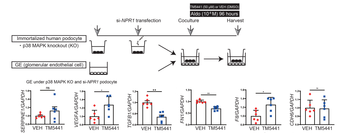
TM5441 decreased SERPINE1, TGFB1 and CDH6.
TM5441 purchased from MedChemExpress. Usage Cited in: Kidney Int. 2023 Sep;104(3):508-525. [Abstract]
TM5441 (PAI-1 inhibitor) or vehicle (DMSO) to the previous transwell systems. TM5441 treatment in the podocytes decreased TGFB1 and FN1.
-
Sci Adv
Determining sex differences in aortic valve myofibroblast responses to drug combinations identified using a digital medicine platform. [Abstract]2025 Jun 6;11(23):eadu2695. PMID: 40479052
TM5441 purchased from MedChemExpress. Usage Cited in: Sci Adv. 2025 Jun 6;11(23):eadu2695. [Abstract]
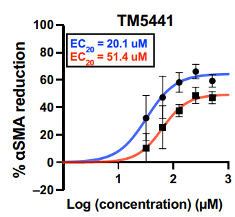
Percent αSMA reduction in male VICs (blue) and female VICs (red) cultured on TCPS with indicated doses of TM5441 (0-1000 μM).
-
Eur J Pharmacol
Carbon monoxide attenuates cellular senescence-mediated pulmonary fibrosis via modulating p53/PAI-1 pathway. [Abstract]2024 Oct 5:980:176843. PMID: 39068977 -
Eur J Pharm Sci
Metabolic activation of TM5441 in vitro and in vivo: Formation of reactive metabolites and human enzymes involved. [Abstract]2020 Feb 15:143:105195. PMID: 31852629 -
Sci Rep
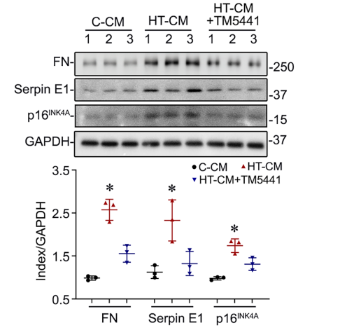
Serpin E1 mediates the induction of renal tubular degeneration and premature senescence upon diabetic insult. [Abstract]2023 Sep 27;13(1):16210. PMID: 37758806
TM5441 purchased from MedChemExpress. Usage Cited in: Sci Rep. 2023 Sep 27;13(1):16210. [Abstract]
Paracrine senescence effects were largely abolished by the addition of the Serpin E1 inhibitor TM5441 (10 μM, pre-incubate for 1 hour) to the conditioned medium.
-
Biol Pharm Bull
Arid5a/IL-6/PAI-1 Signaling Is Involved in the Pathogenesis of Lipopolysaccharide-Induced Kidney Injury. [Abstract]2023 Oct 10;46(12):1753-1760. PMID: 38044094 -
-
-
-
Res Sq
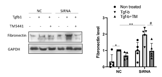
Hypermethylation suppresses microRNA-219a-2 to activate the ALDH1L2/GSH/PAI-1 pathway for fibronectin degradation in renal fibrosis. [Abstract]2023 Jun 5:rs.3.rs-2986934. PMID: 37333081
TM5441 purchased from MedChemExpress. Usage Cited in: Res Sq. 2023 Jun 5:rs.3.rs-2986934. [Abstract]
HEK293 cells with NC or ALDH1L2 siRNA were treated with PAI-1 inhibitor TM5441 and then subjected to TGF-b1 treatment.


溶剤 & 溶解度
体外:
DMSO : 100 mg/mL (233.20 mM; Need ultrasonic; Hygroscopic DMSO has a significant impact on the solubility of product, please use newly opened DMSO)
Please refer to the solubility information to select the appropriate solvent. Once prepared, please aliquot and store the solution to prevent product inactivation from repeated freeze-thaw cycles.
Storage method and period of stock solution: -80°C, 2 years; -20°C, 1 year. When stored at -80°C, please use it within 2 years. When stored at -20°C, please use it within 1 year.
Please refer to the solubility information to select the appropriate solvent. Once prepared, please aliquot and store the solution to prevent product inactivation from repeated freeze-thaw cycles.
Storage method and period of stock solution: -80°C, 2 years; -20°C, 1 year. When stored at -80°C, please use it within 2 years. When stored at -20°C, please use it within 1 year.
濃度 (開始) × 体積 (開始) = 濃度 (終了) × 体積 (終了)
体内:
Select the appropriate dissolution method based on your experimental animal and administration route.
- For the following dissolution methods, please ensure to first prepare a clear stock solution using an In Vitro approach and then sequentially add co-solvents:
- To ensure reliable experimental results, the clarified stock solution can be appropriately stored based on storage conditions. As for the working solution for In Vivo experiments, it is recommended to prepare freshly and use it on the same day.
- The percentages shown for the solvents indicate their volumetric ratio in the final prepared solution. If precipitation or phase separation occurs during preparation, heat and/or sonication can be used to aid dissolution.
Add each solvent one by one: 10% DMSO 40% PEG300 5% Tween-80 45% Saline
Solubility: ≥ 2.5 mg/mL (5.83 mM); Clear solution
This protocol yields a clear solution of ≥ 2.5 mg/mL (saturation unknown).
Taking 1 mL working solution as an example, add 100 μL DMSO stock solution (25.0 mg/mL) to 400 μL PEG300, and mix evenly; then add 50 μL Tween-80 and mix evenly; then add 450 μL Saline to adjust the volume to 1 mL.
Preparation of Saline: Dissolve 0.9 g sodium chloride in ddH₂O and dilute to 100 mL to obtain a clear Saline solution.
In Vivo Dissolution Calculator
Please enter the basic information of animal experiments:
-
-
-
-
Recommended: Prepare an additional quantity of animals to account for potential losses during experiments.
Please enter your animal formula composition:
-
%DMSO +
Recommended: Keep the proportion of DMSO in working solution below 2% if your animal is weak.
-
%+
-
+%Tween-80 + +
-
%Saline +
The co-solvents required include: DMSO, . All of co-solvents are available by MedChemExpress (MCE). , Tween 80. All of co-solvents are available by MedChemExpress (MCE).
Working solution concentration: 0.22 mg/mL
Method for preparing stock solution: mg drug dissolved in μL DMSO. Stock solution concentration: mg/mL.
1. Take μL DMSO stock solution;
2. Add μL .
μL , mix evenly;
3. Then add μL Tween 80, mix evenly;
4. Then add μL
Please ensure that the stock solution in the first step is dissolved to a clear state, and add co-solvents in sequence. You can use ultrasonic heating (ultrasonic cleaner, recommended frequency 20-40 kHz), vortexing, etc. to assist dissolution.


純度とドキュメンテーション
-
データシート (280 KB)
-
SDS (396 KB)
- English - EN (396 KB)
- Français - FR (396 KB)
- Deutsch - DE (396 KB)
- Norwegian - NO (396 KB)
- Español - ES (396 KB)
- Swedish - SV (396 KB)
- Italian - IT (396 KB)
- Korean - KR (396 KB)
- Portuguese - PT (396 KB)
-
取扱説明書 (2659 KB)
参考文献
[1]. Placencio VR, et al. Small Molecule Inhibitors of Plasminogen Activator Inhibitor-1 Elicit Anti-Tumorigenic and Anti-Angiogenic Activity. PLoS One. 2015 Jul 24;10(7):e0133786. [Content Brief]
[2]. Jeong BY, et al. Novel Plasminogen Activator Inhibitor-1 Inhibitors Prevent Diabetic Kidney Injury in a Mouse Model. PLoS One. 2016 Jun 3;11(6):e0157012. [Content Brief]


Complete Stock Solution Preparation Table
Please refer to the solubility information to select the appropriate solvent. Once prepared, please aliquot and store the solution to prevent product inactivation from repeated freeze-thaw cycles.
Storage method and period of stock solution: -80°C, 2 years; -20°C, 1 year. When stored at -80°C, please use it within 2 years. When stored at -20°C, please use it within 1 year.
| Optional Solvent | Concentration Solvent Mass | 1 mg | 5 mg | 10 mg | 25 mg |
|---|---|---|---|---|---|
| DMSO | 1 mM | 2.3320 mL | 11.6599 mL | 23.3198 mL | 58.2995 mL |
| 5 mM | 0.4664 mL | 2.3320 mL | 4.6640 mL | 11.6599 mL | |
| 10 mM | 0.2332 mL | 1.1660 mL | 2.3320 mL | 5.8300 mL | |
| 15 mM | 0.1555 mL | 0.7773 mL | 1.5547 mL | 3.8866 mL | |
| 20 mM | 0.1166 mL | 0.5830 mL | 1.1660 mL | 2.9150 mL | |
| 25 mM | 0.0933 mL | 0.4664 mL | 0.9328 mL | 2.3320 mL | |
| 30 mM | 0.0777 mL | 0.3887 mL | 0.7773 mL | 1.9433 mL | |
| 40 mM | 0.0583 mL | 0.2915 mL | 0.5830 mL | 1.4575 mL | |
| 50 mM | 0.0466 mL | 0.2332 mL | 0.4664 mL | 1.1660 mL | |
| 60 mM | 0.0389 mL | 0.1943 mL | 0.3887 mL | 0.9717 mL | |
| 80 mM | 0.0291 mL | 0.1457 mL | 0.2915 mL | 0.7287 mL | |
| 100 mM | 0.0233 mL | 0.1166 mL | 0.2332 mL | 0.5830 mL |


TM5441 関連分類
HY-101761 関連分類
 Powered by Bioz