



GE 2270A
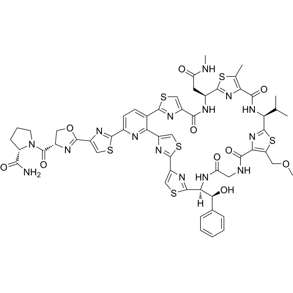
GE 2270A (MDL 62879) is an antibiotic. GE 2270A inhibits gram-positive bacteria and anaerobes by inhibiting protein synthesis. GE 2270A can be used for the research of infection.
商品は「研究用試薬」です。人や動物の医療用・臨床診断用・食品用の製品ではありません。
研究用途以外に使用した場合、当社は一切の責任を負いかねます。
- CAS 番号: 134861-34-0
- 分子式: C56H55N15O10S6
- 分子量:1290.52
-
保管条件:
Please store the product under the recommended conditions in the Certificate of Analysis.

To place orders, for customer services and technical support, please contact: MedChemExpress USA
Tel: 609-228-6898 E-mail: [email protected] [email protected]


-
 生物活性
生物活性
-
化学情報
- プロトコル
- 純度とドキュメンテーション
- 参考文献
-
 Help & FAQs


Antibiotic アイソフォーム固有の製品をすべて表示
More

生物活性
製品説明
IC50 & Target
IC50: 0.4 μM (poly(U)-directed poly(Phe) synthesis)[2]
体外実験
GE 2270A (0.01-100 mg/L) inhibits poly(U)-directed poly(Phe) synthesis in cell-free systems with an IC50 value of 0.12 mg/L[1]. GE 2270A (0-0.1 mM; 10 min) is a strong inhibitor of protein biosynthesis in vitro as determined in a minimal E. coli purified system for poly(U)-directed poly(Phe) synthesis with an IC50 value of 0.4 μM[2].
MedChemExpress (MCE) has not independently confirmed the accuracy of these methods. They are for reference only. Further protocols information, click here.


化学情報
-
CAS 番号 134861-34-0
-
分子量 1290.52
-
分子式 C56H55N15O10S6
-
別名
MDL 62879
-
輸送条件
Room temperature in continental US; may vary elsewhere.
-
保管条件
Please store the product under the recommended conditions in the Certificate of Analysis.

プロトコル
-
Research Protocol for Infectious Diseases
Infectious-disease experiments test how pathogens interact with host barriers, innate immune receptors, inflammatory signaling, pathogen replication, and tissue injury; pattern-recognition receptors such as TLRs, RIG-I-like receptors, NOD-like receptors, and inflammasomes detect microbial molecules and activate NF-κB, interferon, and cytokine responses. The central hypothesis is that infection severity reflects the balance between pathogen burden and host response: protective inflammation restricts pathogen growth, whereas excessive or mislocalized inflammation contributes to tissue damage and disease phenotype. Unresolved questions include which host pathways are protective versus pathogenic, why some infection models fail to translate to human disease, and which combined readouts best predict clinically relevant infection outcomes.
-
Gram Staining of Tissue Sections
Gram staining of tissue sections is a histochemical technique used to differentiate Gram-positive and Gram-negative bacteria within histological specimens based on differences in bacterial cell wall structure and dye retention, adapted from classical bacteriological Gram staining into tissue-compatible “histological Gram stain” variants. In tissue applications, modifications of the Brown-Hopps and Brown-Brenn methods are commonly used to improve differentiation of microorganisms embedded within host connective tissue and to reduce overstaining or loss of Gram-negative signal, which are known limitations of earlier approaches. The principle relies on crystal violet-iodine complex retention in Gram-positive organisms and subsequent decolorization and counterstaining steps that allow contrast visualization of Gram-negative organisms against tissue background.


純度とドキュメンテーション
参考文献
[1]. Landini P, B et al. Inhibition of bacterial protein synthesis by elongation-factor-Tu-binding antibiotics MDL 62,879 and efrotomycin. Biochem J. 1992 May 1;283 ( Pt 3)(Pt 3):649-52. [Content Brief]
[2]. Anborgh PH, Parmeggiani A. New antibiotic that acts specifically on the GTP-bound form of elongation factor Tu. EMBO J. 1991 Apr;10(4):779-84. [Content Brief]

Calculators
濃度 (開始) × 体積 (開始) = 濃度 (終了) × 体積 (終了)


GE 2270A 関連分類
HY-106882 関連分類
 Powered by Bioz