



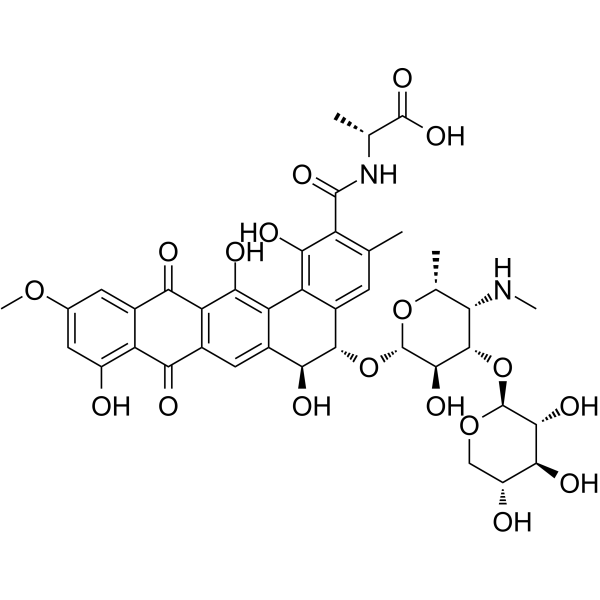
Pradimicin A
Pradimicin A is a potent antifungal agent with a minimum inhibitory concentration (MIC) of 4 μg/mL against Candida rugosa. Pradimicin A exhibits antiviral activity against SARS-CoV, human immunodeficiency virus (HIV) and other enveloped viruses. Pradimicin A exerts its fungicidal activity through a Ca2+-dependent mechanism, which induces fungal lysis. Pradimicin A can be used in research related to fungal infections, HIV infections and COVID-19.
商品は「研究用試薬」です。人や動物の医療用・臨床診断用・食品用の製品ではありません。
研究用途以外に使用した場合、当社は一切の責任を負いかねます。
- CAS 番号: 117704-65-1
- 分子式: C40H44N2O18
- 分子量:840.78
-
保管条件:
Please store the product under the recommended conditions in the Certificate of Analysis.

To place orders, for customer services and technical support, please contact: MedChemExpress USA
Tel: 609-228-6898 E-mail: [email protected] [email protected]


-
 生物活性
生物活性
-
化学情報
- プロトコル
- 純度とドキュメンテーション
- 参考文献
-
 Help & FAQs




生物活性
製品説明
IC50 & Target
[1]|
HIV |
Cellular Effect
|
Cell Line

|
Type | Value | Description | References |
|---|---|---|---|---|
| C8166 | CC50 |
>50 μM
Compound: PRM-A
|
Cytotoxicity against human C8166 cells after 3 to 4 days
Cytotoxicity against human C8166 cells after 3 to 4 days
|
[PMID: 20047920] |
| C8166 | EC50 |
2.6 μM
Compound: PRM-A
|
Antiviral activity against HIV-1 3B infected in human C8166 cells assessed as inhibition of virus-induced cytopathicity after 3 to 4 days by microscopic analysis
Antiviral activity against HIV-1 3B infected in human C8166 cells assessed as inhibition of virus-induced cytopathicity after 3 to 4 days by microscopic analysis
|
[PMID: 20047920] |
| Caco-2 | CC50 |
>50 μM
Compound: PRM-A
|
Cytotoxicity against human Caco2 cells after 3 to 4 days
Cytotoxicity against human Caco2 cells after 3 to 4 days
|
[PMID: 20047920] |
| CCRF-CEM | CC50 |
>100 μM
Compound: Pradimicin A
|
Cytotoxicity against human CEM cells assessed as cell count after 3 days
Cytotoxicity against human CEM cells assessed as cell count after 3 days
|
[PMID: 26513643] |
| CCRF-CEM | CC50 |
>100 μM
Compound: PRM-A
|
Cytotoxicity against human CEM cells after 7 days by trypan blue staining
Cytotoxicity against human CEM cells after 7 days by trypan blue staining
|
[PMID: 20047920] |
| CCRF-CEM | CC50 |
>100 μM
Compound: Pradimicin
|
Cytotoxicity against human CEM cells assessed as inhibition of cell proliferation
Cytotoxicity against human CEM cells assessed as inhibition of cell proliferation
|
[PMID: 25617695] |
| CCRF-CEM | CC50 |
>50 μM
Compound: PRM-A
|
Cytotoxicity against human CD4+ T-lymphocyte CEM cells
Cytotoxicity against human CD4+ T-lymphocyte CEM cells
|
[PMID: 26540494] |
| CCRF-CEM | EC50 |
>50 μM
Compound: PRM-A
|
Antiviral activity against HIV-1 3B harboring wild type gp120 infected in human CEM cells assessed as inhibition of virus-induced cytopathicity after 3 to 4 days by microscopic analysis
Antiviral activity against HIV-1 3B harboring wild type gp120 infected in human CEM cells assessed as inhibition of virus-induced cytopathicity after 3 to 4 days by microscopic analysis
|
[PMID: 20047920] |
| CCRF-CEM | EC50 |
>50 μM
Compound: PRM-A
|
Cytotoxicity against human CEM cells after 4 days by by Coulter counter analysis
Cytotoxicity against human CEM cells after 4 days by by Coulter counter analysis
|
[PMID: 21749165] |
| CCRF-CEM | EC50 |
3.3 μM
Compound: Pradimicin
|
Antiviral activity against HIV1 3B infected in human CEM cells assessed as reduction in virus-induced cytopathicity after 4 to 5 days by microscopy based syncytium cell formation assay
Antiviral activity against HIV1 3B infected in human CEM cells assessed as reduction in virus-induced cytopathicity after 4 to 5 days by microscopy based syncytium cell formation assay
|
[PMID: 25617695] |
| CCRF-CEM | EC50 |
3.3 μM
Compound: Pradimicin A
|
Antiviral activity against HIV1 3B infected in human CEM cells assessed as inhibition of virus-induced giant cell formation after 4 to 5 days by microscopic analysis
Antiviral activity against HIV1 3B infected in human CEM cells assessed as inhibition of virus-induced giant cell formation after 4 to 5 days by microscopic analysis
|
[PMID: 26513643] |
| CCRF-CEM | EC50 |
3.4 μM
Compound: PRM-A
|
Antiviral activity against HIV1 3B infected in human CD4+ T-lymphocyte CEM cells assessed as inhibition of virus-induced cytopathicity after 4 to 5 days by microscopic analysis
Antiviral activity against HIV1 3B infected in human CD4+ T-lymphocyte CEM cells assessed as inhibition of virus-induced cytopathicity after 4 to 5 days by microscopic analysis
|
[PMID: 26540494] |
| CCRF-CEM | EC50 |
4.3 μM
Compound: PRM-A
|
Antiviral activity against SIV mac251 infected in human HUT78 cells assessed as prevention of syncytium formation between persistently SIV mac251-infected HUT-78 cells and uninfected CEM cells after 3 to 4 days by ELISA
Antiviral activity against SIV mac251 infected in human HUT78 cells assessed as prevention of syncytium formation between persistently SIV mac251-infected HUT-78 cells and uninfected CEM cells after 3 to 4 days by ELISA
|
[PMID: 20047920] |
| CCRF-CEM | EC50 |
5.2 μM
Compound: PRM-A
|
Antiviral activity against compound pretreated HIV-1 3B isolate 42 harboring gp120 mutant infected in human CEM cells assessed as inhibition of virus-induced cytopathicity after 3 to 4 days by microscopic analysis
Antiviral activity against compound pretreated HIV-1 3B isolate 42 harboring gp120 mutant infected in human CEM cells assessed as inhibition of virus-induced cytopathicity after 3 to 4 days by microscopic analysis
|
[PMID: 20047920] |
| CCRF-CEM | EC50 |
5.2 μM
Compound: PRM-A
|
Antiviral activity against HIV-2 ROD infected in human CEM cells assessed as inhibition of virus-induced cytopathicity after 3 to 4 days by microscopic analysis
Antiviral activity against HIV-2 ROD infected in human CEM cells assessed as inhibition of virus-induced cytopathicity after 3 to 4 days by microscopic analysis
|
[PMID: 20047920] |
| CCRF-CEM | EC50 |
5.2 μM
Compound: PRM-A
|
Antiviral activity against HIV1 3B infected in human CEM cells assessed as inhibition of virus-induced cytopathicity after 4 days
Antiviral activity against HIV1 3B infected in human CEM cells assessed as inhibition of virus-induced cytopathicity after 4 days
|
[PMID: 21749165] |
| CCRF-CEM | EC50 |
5.9 μM
Compound: Pradimicin
|
Antiviral activity against HIV2 ROD infected in human CEM cells assessed as reduction in virus-induced cytopathicity after 4 to 5 days by microscopy based syncytium cell formation assay
Antiviral activity against HIV2 ROD infected in human CEM cells assessed as reduction in virus-induced cytopathicity after 4 to 5 days by microscopy based syncytium cell formation assay
|
[PMID: 25617695] |
| CCRF-CEM | EC50 |
5.9 μM
Compound: Pradimicin A
|
Antiviral activity against HIV2 ROD infected in human CEM cells assessed as inhibition of virus-induced giant cell formation after 4 to 5 days by microscopic analysis
Antiviral activity against HIV2 ROD infected in human CEM cells assessed as inhibition of virus-induced giant cell formation after 4 to 5 days by microscopic analysis
|
[PMID: 26513643] |
| CCRF-CEM | EC50 |
5.9 μM
Compound: PRM-A
|
Antiviral activity against HIV-1 3B infected in human CEM cells assessed as inhibition of virus-induced cytopathicity after 3 to 4 days by microscopic analysis
Antiviral activity against HIV-1 3B infected in human CEM cells assessed as inhibition of virus-induced cytopathicity after 3 to 4 days by microscopic analysis
|
[PMID: 20047920] |
| CCRF-CEM | EC50 |
5.9 μM
Compound: PRM-A
|
Antiviral activity against HIV2 ROD infected in human CEM cells assessed as inhibition of virus-induced cytopathicity after 4 days
Antiviral activity against HIV2 ROD infected in human CEM cells assessed as inhibition of virus-induced cytopathicity after 4 days
|
[PMID: 21749165] |
| HeLa | CC50 |
>50 μM
Compound: PRM-A
|
Cytotoxicity against human HeLa cells after 3 to 4 days
Cytotoxicity against human HeLa cells after 3 to 4 days
|
[PMID: 20047920] |
| L1210 | CC50 |
>50 μM
Compound: PRM-A
|
Cytotoxicity against mouse L1210 cells after 3 to 4 days
Cytotoxicity against mouse L1210 cells after 3 to 4 days
|
[PMID: 20047920] |
| MOLT-4 | CC50 |
>50 μM
Compound: PRM-A
|
Cytotoxicity against human Molt4 cells after 3 to 4 days
Cytotoxicity against human Molt4 cells after 3 to 4 days
|
[PMID: 20047920] |
| MT4 | CC50 |
>100 μM
Compound: PRM-A
|
Cytotoxicity against human MT4 cells assessed as reduction in cell viability measured after 5 days by MTS/PES assay
Cytotoxicity against human MT4 cells assessed as reduction in cell viability measured after 5 days by MTS/PES assay
|
[PMID: 31809045] |
| MT4 | CC50 |
>100 μM
Compound: PRM-A
|
Cytotoxicity against human MT4 cells assessed as reduction in cell viability measured after 5 days by MTS/PES assay
Cytotoxicity against human MT4 cells assessed as reduction in cell viability measured after 5 days by MTS/PES assay
|
[PMID: 34229438] |
| MT4 | CC50 |
>100 μM
Compound: PRM-A
|
Cytotoxicity against human MT4 cells assessed as cell viability measured after 5 days by MTS assay
Cytotoxicity against human MT4 cells assessed as cell viability measured after 5 days by MTS assay
|
[PMID: 35512567] |
| MT4 | CC50 |
>50 μM
Compound: PRM-A
|
Cytotoxicity against human MT4 cells after 7 days by trypan blue staining
Cytotoxicity against human MT4 cells after 7 days by trypan blue staining
|
[PMID: 20047920] |
| MT4 | EC50 |
5 μM
Compound: PRM-A
|
Antiviral activity against HIV-2 ROD infected in human MT4 cells assessed as inhibition of virus-induced cytopathicity after 7 days by trypan blue staining
Antiviral activity against HIV-2 ROD infected in human MT4 cells assessed as inhibition of virus-induced cytopathicity after 7 days by trypan blue staining
|
[PMID: 20047920] |
| MT4 | EC50 |
5.2 μM
Compound: PRM-A
|
Antiviral activity against HIV-1 3B infected in human MT4 cells assessed as inhibition of virus-induced cytopathicity after 7 days by trypan blue staining
Antiviral activity against HIV-1 3B infected in human MT4 cells assessed as inhibition of virus-induced cytopathicity after 7 days by trypan blue staining
|
[PMID: 20047920] |
| MT4 | EC50 |
5.5 μM
Compound: PRM-A
|
Antiviral activity against SIV mac251 infected in human MT4 cells assessed as inhibition of virus-induced cytopathicity after 7 days by trypan blue staining
Antiviral activity against SIV mac251 infected in human MT4 cells assessed as inhibition of virus-induced cytopathicity after 7 days by trypan blue staining
|
[PMID: 20047920] |
| OST | CC50 |
>50 μM
Compound: PRM-A
|
Cytotoxicity against human thymidine kinase-negative OST cells after 3 to 4 days
Cytotoxicity against human thymidine kinase-negative OST cells after 3 to 4 days
|
[PMID: 20047920] |
| PBMC | CC50 |
>250 μM
Compound: PRM-A
|
Cytotoxicity against human PBMC after 7 days by trypan blue staining
Cytotoxicity against human PBMC after 7 days by trypan blue staining
|
[PMID: 20047920] |
| PBMC | CC50 |
>50 μM
Compound: PRM-A
|
Cytotoxicity against human PBMC cells after 3 to 4 days
Cytotoxicity against human PBMC cells after 3 to 4 days
|
[PMID: 20047920] |
| PBMC | EC50 |
>50 μM
Compound: PRM-A
|
Antiviral activity against HIV-1 3B cells infected in human PBMC cells assessed as inhibition of virus-induced cytopathicity after 3 to 4 days by microscopic analysis
Antiviral activity against HIV-1 3B cells infected in human PBMC cells assessed as inhibition of virus-induced cytopathicity after 3 to 4 days by microscopic analysis
|
[PMID: 20047920] |
| Raji | EC50 |
3.4 μM
Compound: PRM-A
|
Antiviral activity against compound pretreated X4 tropic HIV-1 3B infected in human Raji cells expressing DC-SIGN gene assessed as prevention of syncytium formation between persistently HIV-1 3B-infected Raji cells and uninfected human C8166 cells after 4
Antiviral activity against compound pretreated X4 tropic HIV-1 3B infected in human Raji cells expressing DC-SIGN gene assessed as prevention of syncytium formation between persistently HIV-1 3B-infected Raji cells and uninfected human C8166 cells after 4
|
[PMID: 20047920] |
| Raji | EC50 |
5.4 μM
Compound: PRM-A
|
Antiviral activity against X4 tropic HIV-1 3B infected in human Raji cells expressing DC-SIGN gene assessed as prevention of syncytium formation between persistently HIV-1 3B-infected Raji cells and uninfected human C8166 cells after 36 to 42 hrs by micro
Antiviral activity against X4 tropic HIV-1 3B infected in human Raji cells expressing DC-SIGN gene assessed as prevention of syncytium formation between persistently HIV-1 3B-infected Raji cells and uninfected human C8166 cells after 36 to 42 hrs by micro
|
[PMID: 20047920] |
| Raji | IC50 |
16 μM
Compound: PRM-A
|
Antiviral activity against SIV mac251 infected in human Raji cells expressing DC-SIGN gene assessed as inhibition of virus capture by Raji cells after 60 mins by ELISA
Antiviral activity against SIV mac251 infected in human Raji cells expressing DC-SIGN gene assessed as inhibition of virus capture by Raji cells after 60 mins by ELISA
|
[PMID: 20047920] |
| Raji | IC50 |
16 μM
Compound: PRM-A
|
Antiviral activity against SIV mac251 infected in human Raji cells expressing DC-SIGN gene assessed as prevention of syncytium formation between persistently HIV-1 3B-infected Raji cells and uninfected human C8166 cells after 60 mins by ELISA
Antiviral activity against SIV mac251 infected in human Raji cells expressing DC-SIGN gene assessed as prevention of syncytium formation between persistently HIV-1 3B-infected Raji cells and uninfected human C8166 cells after 60 mins by ELISA
|
[PMID: 20047920] |
| Raji | IC50 |
21 μM
Compound: PRM-A
|
Antiviral activity against R5 tropic HIV-1 Bal infected in human Raji cells expressing DC-SIGN gene assessed as inhibition of virus capture by Raji cells after 60 mins by ELISA
Antiviral activity against R5 tropic HIV-1 Bal infected in human Raji cells expressing DC-SIGN gene assessed as inhibition of virus capture by Raji cells after 60 mins by ELISA
|
[PMID: 20047920] |
| Raji | IC50 |
3.5 μM
Compound: PRM-A
|
Antiviral activity against X4 tropic HIV-1 3B infected in human Raji cells expressing DC-SIGN gene assessed as inhibition of virus capture by Raji cells after 60 mins by ELISA
Antiviral activity against X4 tropic HIV-1 3B infected in human Raji cells expressing DC-SIGN gene assessed as inhibition of virus capture by Raji cells after 60 mins by ELISA
|
[PMID: 20047920] |
| SUP-T1 | EC50 |
3.4 μM
Compound: PRM-A
|
Antiviral activity against HIV-1 3B infected in human HUT78 cells assessed as prevention of syncytium formation between persistently HIV-1 3B-infected HUT-78 cells and uninfected SupT1 cells after 3 to 4 days by ELISA
Antiviral activity against HIV-1 3B infected in human HUT78 cells assessed as prevention of syncytium formation between persistently HIV-1 3B-infected HUT-78 cells and uninfected SupT1 cells after 3 to 4 days by ELISA
|
[PMID: 20047920] |
| U-87MG ATCC | CC50 |
>50 μM
Compound: PRM-A
|
Cytotoxicity against human U87 cells after 3 to 4 days
Cytotoxicity against human U87 cells after 3 to 4 days
|
[PMID: 20047920] |
体外実験
Pradimicin A (48 h) inhibits the growth of Candida rugosa with a minimum inhibitory concentration (MIC) of 4 μg/mL, and completely inhibits Candida rugosa growth while showing preferential binding to branched oligomannose motifs 6 and 7 (MAC = 1 mM) relative to other tested glycan structures[1][3].
Pradimicin A (0.3-100 μM; 1 h pre-incubation, 3 days post challenge) inhibits SARS-CoV-2 infection of Calu-3 cells with an IC50 of 1.2 μM, shows no cytotoxicity at concentrations up to 100 μM, and its antiviral activity is dependent on binding to branched oligomannose motifs on viral spike proteins[1].
Pradimicin A (1 mM) shows Ca2+‑dependent specific binding to Man-OMe (100 mM) with two distinct binding sites in ITC assays (40 injections, 30 °C). The primary binding (Ka = 10400 M⁻¹) is approximately 40‑fold stronger than the secondary binding (Ka = 263 M⁻¹)[2].
Pradimicin A (1 mM) binds methyl α-D-mannopyranoside in a cell-free buffer system with a primary dissociation constant (Kd1) of 96 μM and a secondary dissociation constant (Kd2) of 3800 μM[3].
Pradimicin A (0.8-12.5 μg/mL) exhibits broad-spectrum antifungal activity against diverse fungal species including Candida albicans A9540[5].
Pradimicin A exhibits antiviral activity against coronaviruses and HIV-1, targeting viral glycoprotein mannose moieties and demonstrating a high genetic barrier to HIV-1 resistance[5].
MedChemExpress (MCE) has not independently confirmed the accuracy of these methods. They are for reference only.


化学情報
-
CAS 番号 117704-65-1
-
分子量 840.78
-
分子式 C40H44N2O18
-
SMILES
OC1=C2C3=C(O)C(C(N[C@H](C)C(O)=O)=O)=C(C)C=C3[C@@H]([C@H](C2=CC4=C1C(C5=CC(OC)=CC(O)=C5C4=O)=O)O)O[C@H]6[C@@H]([C@H]([C@H]([C@H](O6)C)NC)O[C@H]7[C@@H]([C@H]([C@@H](CO7)O)O)O)O
-
輸送条件
Room temperature in continental US; may vary elsewhere.
-
保管条件
Please store the product under the recommended conditions in the Certificate of Analysis.


純度とドキュメンテーション
参考文献
[1]. Nakagawa Y, et al. Molecular basis of N-glycan recognition by pradimicin a and its potential as a SARS-CoV-2 entry inhibitor. Bioorg Med Chem. 2024 May 1;105:117732. [Content Brief]
[2]. Nakagawa Y, et al. Mapping of the primary mannose binding site of pradimicin A. J Am Chem Soc. 2011;133(43):17485-17493. [Content Brief]
[3]. Miyanishi W, et al. d-Mannose binding, aggregation property, and antifungal activity of amide derivatives of pradimicin A. Bioorg Med Chem. 2022;55:116590. [Content Brief]
[4]. Nakagawa Y, et al. Molecular Basis of Mannose Recognition by Pradimicins and their Application to Microbial Cell Surface Imaging. Cell Chem Biol. 2019;26(7):950-959.e8. [Content Brief]
[5]. Guo Y, et al. Isolation, biosynthesis, and biological activity of pradimicins from actinomycetes. Bioorg Chem. 2025;164:108884. [Content Brief]

Calculators
濃度 (開始) × 体積 (開始) = 濃度 (終了) × 体積 (終了)


Pradimicin A 関連分類
HY-132191 関連分類
 Powered by Bioz